. 2012;7(6):e39520.
doi: 10.1371/journal.pone.0039520. Epub 2012 Jun 25.

Antagonism of miR-21 reverses epithelial-mesenchymal transition and cancer stem cell phenotype through AKT/ERK1/2 inactivation by targeting PTEN

Affiliations

Antagonism of miR-21 reverses epithelial-mesenchymal transition and cancer stem cell phenotype through AKT/ERK1/2 inactivation by targeting PTEN

Mingli Han et al. PLoS One. 2012.

Abstract

Background: Accumulating evidence suggested that epithelial-mesenchymal transition (EMT) and cancer stem cell (CSC) characteristics, both of which contribute to tumor invasion and metastasis, are interrelated with miR-21. MiR-21 is one of the important microRNAs associated with tumor progression and metastasis, but the molecular mechanisms underlying EMT and CSC phenotype during miR-21 contributes to migration and invasion of breast cancer cells remain to be elucidated.

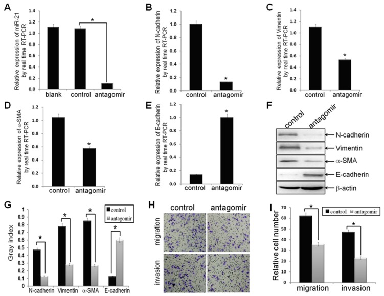
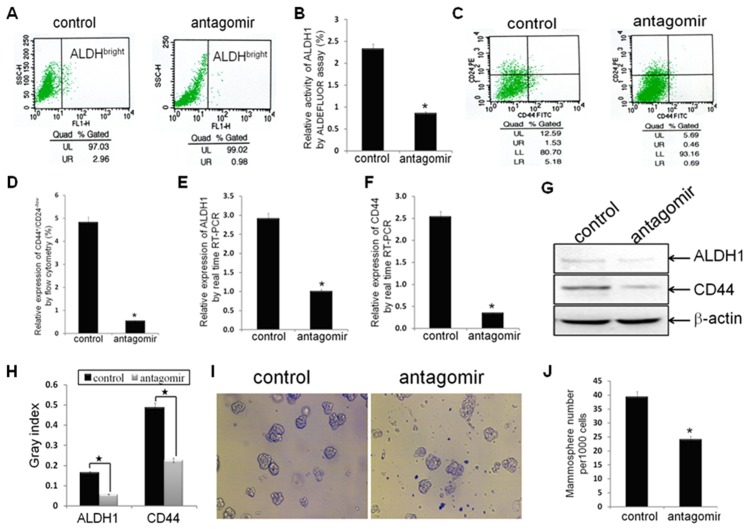
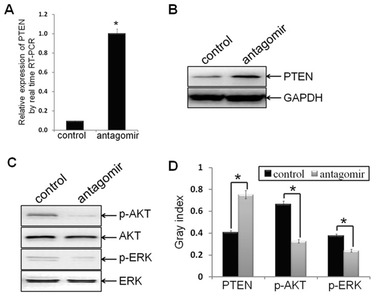
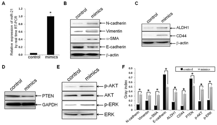
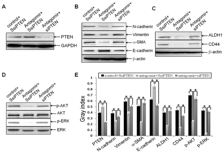
Methodology/principal findings: In this study, MDA-MB-231/anti-miR-21 cells were established by transfected hsa-miR-21 antagomir into breast cancer MDA-MB-231 cells. EMT was evaluated by the changes of mesenchymal cell markers (N-cadherin, Vimentin, and alpha-SMA), epithelial cell marker (E-cadherin), as well as capacities of cell migration and invasion; CSC phenotype was measured using the changes of CSC surface markers (ALDH1 and CD44), and the capacity of sphereforming (mammospheres). We found that antagonism of miR-21 reversed EMT and CSC phenotype, accompanied with PTEN up-regulation and AKT/ERK1/2 inactivation. Interestingly, down-regulation of PTEN by siPTEN suppressed the effects of miR-21 antagomir on EMT and CSC phenotype, confirming that PTEN is a target of miR-21 in reversing EMT and CSC phenotype. The inhibitors of PI3K-AKT and ERK1/2 pathways, LY294002 and U0126, both significantly suppressed EMT and CSC phenotype, indicating that AKT and ERK1/2 pathways are required for miR-21 mediating EMT and CSC phenotype.

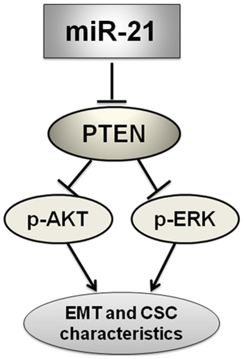
Conclusions/significance: In conclusion, our results demonstrated that antagonism of miR-21 reverses EMT and CSC phenotype through targeting PTEN, via inactivation of AKT and ERK1/2 pathways, and showed a novel mechanism of which might relieve the malignant biological behaviors of breast cancer.


Conflict of interest statement

Competing Interests: The authors have declared that no competing interests exist.

Figures

Figure 1
Figure 1. Antagonism of miR-21 reversed EMT phenotype, as well as decreased cell migration and invasion.
MDA-MB-231 cells were transfected with hsa-miR-21 antagomir or hsa-miR-21 antagomir control at a final concentration of 50 nmol for 48 h. (A) MDA-MB-231 cells were treated with hsa-miR-21 antagomir decreased the expression of miR-21, as compared to control groups (n1 = n2 = 3; p = 0.0015), by real-time RT-PCR analysis. (B-E) The mRNA levels of mesenchymal biomarkers (N-cadherin, Vimentin and alpha-SMA) and epithelial biomarker (E-cadherin) in MDA-MB-231/anti-miR-21 cells and MDA-MB-231/control cells, as measured by real-time RT-PCR analysis. The real-time RT-PCR reactions were performed in a 20 µl reaction volume in triplicate, simultaneously. (F, G) The relative protein levels of EMT markers in indicated cells were shown by Western blot analysis, and bands were semi-quantified using ImageJ software. Beta-actin was used as loading control. (H, I) The migratory and invasive properties of indicated cells were tested in migration and invasion assay in Transwell inserts. Penetrated cells were counted and analyzed in histogram. Data represent at least three experiments done in triplicate. (*indicates p<0.05; indicates p<0.001).
Figure 2
Figure 2. Antagonism of miR-21 reversed CSC phenotype.
(A, B) ALDH1 enzymatic activity (ALDHbright) in established breast cancer MDA-MB-231/anti-miR-21 cells and MDA-MB-231/control cells (n1 = n2 = 3) were detected using the ALDEFLUOR assay. (C, D) The CD44+/CD24−/low phenotype in indicated cells (n1 = n2 = 3) were detected by FACS analysis. (E, F) The relative mRNA levels of ALDH1 and CD44 were detected by real time RT-PCR assay. (G, H) The relative protein levels of ALDH1 and CD44 were detected by Western blot analysis. Beta-actin was used as loading control. (I, J) The number of mammospheres from 1000 MDA-MB-231/anti-miR-21 cells or MDA-MB-231/control cells was counted under microscope. All the data represent at least three experiments done in triplicate. (*indicates p<0.05; indicates p<0.001).
Figure 3
Figure 3. Antagonism of miR-21 induced the expression of PTEN, as well as inactivated AKT and ERK1/2.
(A) Ectopic expression of PTEN mRNA in MDA-MB-231/anti-miR-21 cells and MDA-MB-231/control cells were verified by real time RT-PCR assay (p = 0.003). (B, C, D) Protein levels of PTEN, p-AKT, AKT, p-ERK1/2, and ERK1/2 in indicated cells were detected by Western blot analysis, and bands were semi-quantified using ImageJ software. GAPDH was used as loading control. (*indicates p<0.05).
Figure 4
Figure 4. Hsa-miR-21 mimics induced EMT and CSC phenotype, accompanied with PTEN down-regulation and AKT/ERK1/2 activation.
Established MDA-MB-231/anti-miR-21 cells were transfected with hsa-miR-21 mimics at a concentration of 40 nmol for 72 h. (A) MDA-MB-231/anti-miR-21 cells were treated with hsa-miR-21 mimics elevated the expression of miR-21, as compared to control groups (n1 = n2 = 3; p = 0.00373), by real-time RT-PCR analysis. (B-F) Protein levels of mesenchymal markers (N-cadherin, Vimentin and alpha-SMA) (B), epithelial marker (E-cadherin) (B), CSC markers (ALDH1 and CD44) (C), PTEN (D), p-AKT and AKT (E), as well as p-ERK1/2 and ERK1/2 (E) in indicated cells were measured by Western blot analysis, and bands were semi-quantified using ImageJ software (F). Beta-actin or GAPDH was used as loading control. (*indicates p<0.05; indicates p<0.001).
Figure 5
Figure 5. PTEN was the downstream target of miR-21 during reversing EMT and CSC phenotype
. MDA-MB-231 cells were transfected with siPTEN or the scrambled control SsiPTEN at a final concentration of 50 nmol for 24 h. Then the cells were transfected with hsa-miR-21 antagomir or negative control at a final concentration of 50 nmol for 72 h. Cells were trypsinized, and the relative protein levels of PTEN (A), EMT markers (B), CSC surface markers (C), p-AKT and AKT (D), as well as p-ERK and ERK (D) were measured and semi-quantified (E) as stated before. The representative plugs from treatments of SsiPTEN plus miR-21 antagomir control, SsiPTEN plus miR-21 antagomir, and siPTEN plus miR-21 antagomir were shown in the picture. Beta-actin or GAPDH was used as loading control. (*indicates p<0.05; indicates p<0.001).
Figure 6
Figure 6. MiR-21 regulated EMT and CSC phenotype through mediating AKT and ERK1/2 activation.
Established MDA-MB-231/anti-miR-21 cells were transfected with hsa-miR-21 mimics at a concentration of 40 nmol for 72 h. Then the cells were trypsinized, and treated with LY294002 (20 µmol/l) or U0126 (10 µmol/l) for 24 h. (A-E) The relative protein levels of p-AKT and AKT (A), p-ERK and ERK (A), EMT markers (B), CSC surface markers (C), as well as PTEN (D) from blank control, LY294002, and U0126 treatment groups were shown, and semi-quantified (E) as stated before. Beta-actin or GAPDH was used as loading control. (*indicates p<0.05; indicates p<0.001; indicates p>0.05). (F) The effects of antagonism of miR-21, re-expression of miR-21, LY294002, and U0126 on cell proliferation by cell count (A vs. B, p = 0.0077; B+C vs. B+D, p = 0.0091; B+D vs. B+D+E, p = 0.0109; B+D vs. B+D+F, p = 0.0031).
Figure 7
Figure 7. Schematic diagram of the proposed model: possible mechanism of miR-21 regulates EMT and CSC phenotype.
Antagonism of miR-21 could inactivate AKT and ERK1/2 pathways presumably through PTEN up-regulation, and finally reverse EMT and CSC phenotype in MDA-MB-231 cells.

References

    1. Thiery JP. Epithelial-mesenchymal transitions in tumour progression. Nat Rev Cancer. 2002;2:442–454. - PubMed
    1. Thiery JP, Sleeman JP. Complex networks orchestrate epithelial-mesenchymal transitions. Nat Rev Mol Cell Biol. 2006;7:131–142. - PubMed
    1. Christiansen JJ, Rajasekaran AK. Reassessing epithelial to mesenchymal transition as a prerequisite for carcinoma invasion and metastasis. Cancer Res. 2006;66:8319–8326. - PubMed
    1. Thiery JP. Epithelial–mesenchymal transitions in development and pathologies. Curr Opin Cell Biol. 2000;15:740–746. - PubMed
    1. Polakis P. Wnt signaling and cancer. Genes Dev. 2000;14:1837–1851. - PubMed

Publication types

MeSH terms