

Initiation of resuscitation with high tidal volumes causes cerebral hemodynamic disturbance, brain inflammation and injury in preterm lambs
- PMID: 22761816
- PMCID: PMC3382197
- DOI: 10.1371/journal.pone.0039535
Initiation of resuscitation with high tidal volumes causes cerebral hemodynamic disturbance, brain inflammation and injury in preterm lambs
Abstract
Aims: Preterm infants can be inadvertently exposed to high tidal volumes (V(T)) in the delivery room, causing lung inflammation and injury, but little is known about their effects on the brain. The aim of this study was to compare an initial 15 min of high V(T) resuscitation strategy to a less injurious resuscitation strategy on cerebral haemodynamics, inflammation and injury.
Methods: Preterm lambs at 126 d gestation were surgically instrumented prior to receiving resuscitation with either: 1) High V(T) targeting 10-12 mL/kg for the first 15 min (n = 6) or 2) a protective resuscitation strategy (Prot V(T)), consisting of prophylactic surfactant, a 20 s sustained inflation and a lower initial V(T) (7 mL/kg; n = 6). Both groups were subsequently ventilated with a V(T) 7 mL/kg. Blood gases, arterial pressures and carotid blood flows were recorded. Cerebral blood volume and oxygenation were assessed using near infrared spectroscopy. The brain was collected for biochemical and histologic assessment of inflammation, injury, vascular extravasation, hemorrhage and oxidative injury. Unventilated controls (UVC; n = 6) were used for comparison.
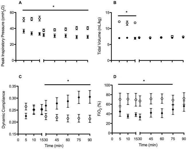
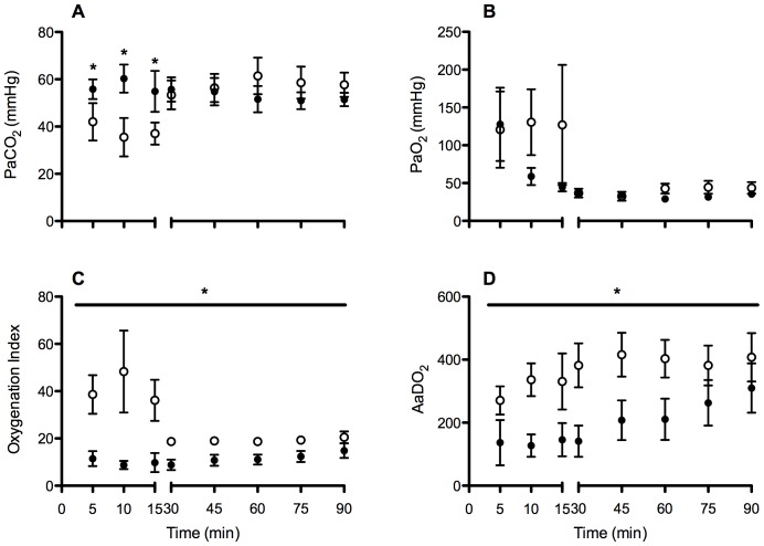
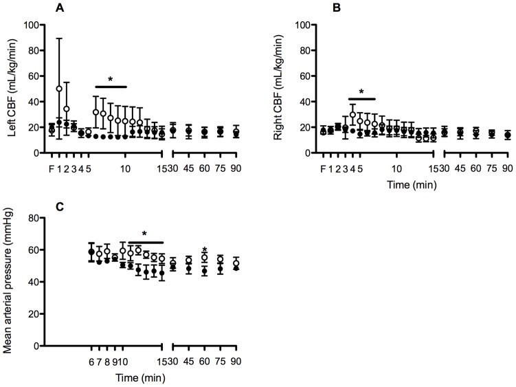
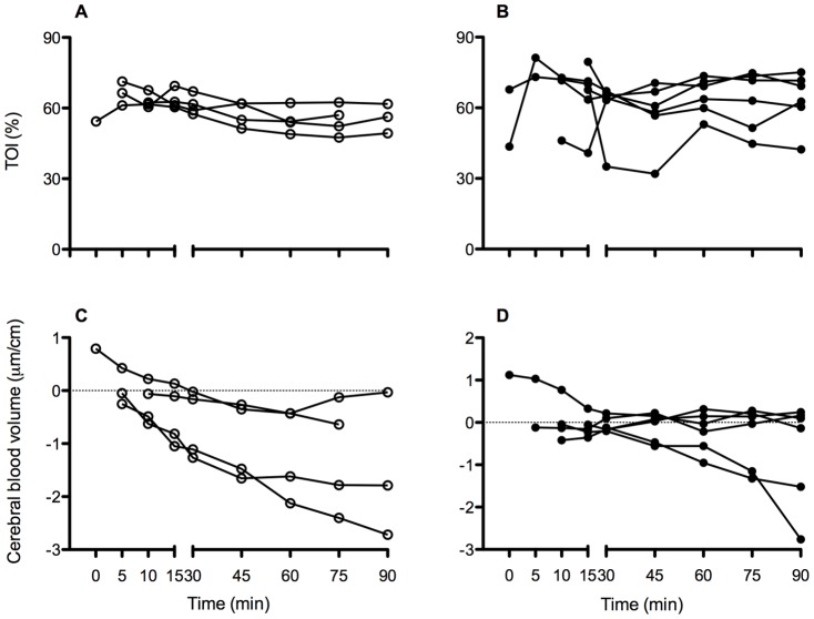
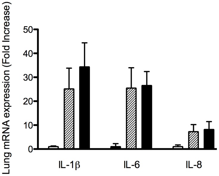
Results: High V(T) lambs had worse oxygenation and required greater ventilatory support than Prot V(T) lambs. High V(T) resulted in cerebral haemodynamic instability during the initial 15 min, adverse cerebral tissue oxygenation index and cerebral vasoparalysis. While both resuscitation strategies increased lung and brain inflammation and oxidative stress, High V(T) resuscitation significantly amplified the effect (p = 0.014 and p<0.001). Vascular extravasation was evident in the brains of 60% of High V(T) lambs, but not in UVC or Prot V(T) lambs.
Conclusion: High V(T) resulted in greater cerebral haemodynamic instability, increased brain inflammation, oxidative stress and vascular extravasation than a Prot V(T) strategy. The initiation of resuscitation targeting Prot V(T) may reduce the severity of brain injury in preterm neonates.
Conflict of interest statement
Figures
References
-
- Volpe JJ. Brain injury in the premature infant: overview of clinical aspects, neuropathology, and pathogenesis. Semin Pediatr Neurol. 1998;5:135–151. - PubMed
-
- Del Toro J, Louis PT, Goddard-Finegold J. Cerebrovascular regulation and neonatal brain injury. Pediatr Neurol. 1991;7:3–12. - PubMed
-
- Lou HC, Lassen NA, Friis-Hansen B. Impaired autoregulation of cerebral blood flow in the distressed newborn infant. J Pediatr. 1979;94:118–121. - PubMed
-
- Schmolzer GM, Kamlin OC, O'Donnell CP, Dawson JA, Morley CJ, et al. Assessment of tidal volume and gas leak during mask ventilation of preterm infants in the delivery room. Arch Dis Child Fetal Neonatal Ed. 2010;95:F393–397. - PubMed
Publication types
MeSH terms
LinkOut - more resources
Full Text Sources
Medical
