. 2012;7(6):e39766.
doi: 10.1371/journal.pone.0039766. Epub 2012 Jun 27.

Myoferlin depletion in breast cancer cells promotes mesenchymal to epithelial shape change and stalls invasion

Affiliations

Myoferlin depletion in breast cancer cells promotes mesenchymal to epithelial shape change and stalls invasion

Ruth Li et al. PLoS One. 2012.

Abstract

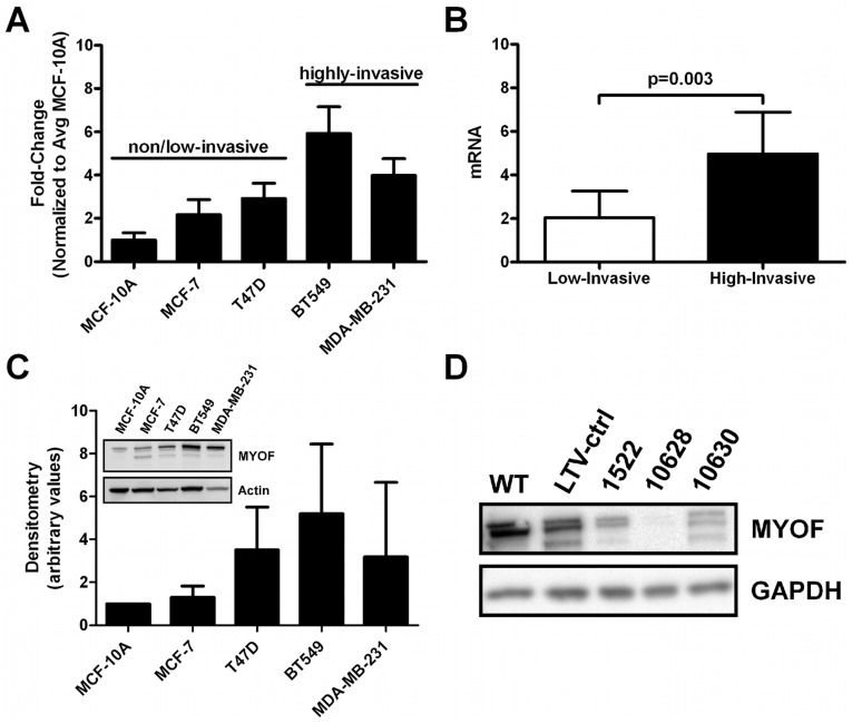
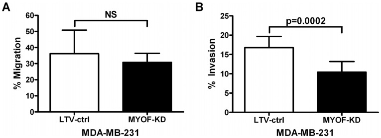
Myoferlin (MYOF) is a mammalian ferlin protein with homology to ancestral Fer-1, a nematode protein that regulates spermatic membrane fusion, which underlies the amoeboid-like movements of its sperm. Studies in muscle and endothelial cells have reported on the role of myoferlin in membrane repair, endocytosis, myoblast fusion, and the proper expression of various plasma membrane receptors. In this study, using an in vitro human breast cancer cell model, we demonstrate that myoferlin is abundantly expressed in invasive breast tumor cells. Depletion of MYOF using lentiviral-driven shRNA expression revealed that MDA-MB-231 cells reverted to an epithelial morphology, suggesting at least some features of mesenchymal to epithelial transition (MET). These observations were confirmed by the down-regulation of some mesenchymal cell markers (e.g., fibronectin and vimentin) and coordinate up-regulation of the E-cadherin epithelial marker. Cell invasion assays using Boyden chambers showed that loss of MYOF led to a significant diminution in invasion through Matrigel or type I collagen, while cell migration was unaffected. PCR array and screening of serum-free culture supernatants from shRNA(MYOF) transduced MDA-MB-231 cells indicated a significant reduction in the steady-state levels of several matrix metalloproteinases. These data when considered in toto suggest a novel role of MYOF in breast tumor cell invasion and a potential reversion to an epithelial phenotype upon loss of MYOF.


Conflict of interest statement

Competing Interests: The authors have declared that no competing interests exist.

Figures

Figure 1
Figure 1. Myoferlin expression in breast cancer.
(A) Quantitative RT-PCR results of MYOF mRNA levels in a panel of breast cancer cell lines, normalized to 18 S and compared to the nonmalignant breast epithelial cell line MCF-10A (mean ± s.d., n = 3 per cell type). No statistical significance was detected in comparing pairs of cell lines (Kruskal-Wallis with Dunn's multiple comparison test). (B) Representation of the data in subpanel A grouped by known invasive capacity of the cells (2 tailed, p = 0.003, Mann Whitney). (C) Immunoblotting for MYOF in breast cancer cell lines. Densitometry measurements was done by normalizing the density of MYOF to respective GAPDH or actin staining, and further normalized to the nonmalignant mammary epithelial cell line MCF-10A (n = 4, no statistical significance detected by Kruskal-Wallis with Dunn’s multiple comparison test). An immunoblot image is shown in the subpanel. (D) Myoferlin knockdown in breast cancer cells. Immunoblot showing the expression of MYOF in MDA-MB-231 wild type (WT), lentiviral transduction control (LTV-ctrl), and MYOF knockdown cell lysates using three Sigma-Aldrich MISSION® shRNA constructs (#1522, 10628, and 10630).
Figure 2
Figure 2. Morphology change following myoferlin depletion in MDA-MB-231 cells.
Immunofluorescence micrographs showing the morphology of MDA-MB-231 wild type (WT), lentiviral transduction control (LTV-ctrl), and myoferlin knockdown (MYOF-KD) stable cell lines in culture. Note the more epithelial morphology of MYOF-KD cells compared to the more mesenchymal appearance of the WT and LTV-ctrl cells. Bar = 50 µm.
Figure 3
Figure 3. Morphology change following myoferlin depletion in MDA-MB-231 cells.
Atomic force and scanning electron microscopy images showing the spindle, elongated shape of lentiviral control (LTV-ctrl) cells and the more flat and circular morphology of myoferlin depleted (MYOF-KD) cells. AFM imaging shows pronounced actin stress fibers (black arrows) oriented along the long axis being evident in the control but not in the MYOF depleted cells. Cytoplasmic poles, lamellipodia and filopodia are observable in the SEM images. White arrowheads indicate the leading edge of cells.
Figure 4
Figure 4. Expression of EMT markers following myoferlin depletion in MDA-MB-231 cells.
(A) Representative immunoblots (n = 3) of select EMT markers in lentiviral control (LTV-ctrl) and myoferlin depleted (MYOF-KD) cells serum-starved for 24 h. (B) Graphs illustrating the semi-quantitative evaluation of the expression of EMT markers by densitometry analysis of blots (n = 3). Density was presented in the graph as “relative density (%)” with density of lentiviral controls normalized to 100%, and statistical testing was done with the 1-sample t-test.
Figure 5
Figure 5. Myoferlin depletion reduces invasive but not migratory capacity of MDA-MB-231s control (LTV-ctrl) and MYOF depleted cells (MYOF-KD).
(A) Boyden chamber migration assay of MDA-MB-231 cells moving across 8 µm porous membranes towards a 10% serum gradient for 24 h (mean ± s.d., n = 3, unpaired 2-tailed t-test). (B) 24 h Boyden chamber invasion results of MDA-MB-231 cells across a 100% Matrigel coated 8 µm porous membrane towards a 10% serum chemoattractant (mean ± s.d., n = 3, unpaired 2-tailed t-test).
Figure 6
Figure 6. Myoferlin depletion attenuates MMP1 expression and collagen I invasion capacity of MDA-MB-231 cells.
(A) Representative (n = 3) immunoblotting results of secreted MMP1 in 24 h serum starved supernatant of MDA-MB-231 lentiviral control (LTV-ctrl) and myoferlin knockdown (MYOF-KD) cells. Recombinant human matrix metalloproteinase-1 was used as a standard. Verification of myoferlin knockdown was done in the corresponding cell lysates with GAPDH as a loading control. (B) Secreted pro-MMP1 was evaluated in 231LTV-ctrl and 231MYOF-KD cells (n = 2) using ELISA (mean ± s.d., Kruskal-Wallis test/Dunn's multiple comparison analysis). (C) Results from Boyden chamber invasion assays using a coating of 3 mg/ml of rat tail collagen I to evaluate the invasive capacity of 231LTV-ctrl and 231MYOF-KD cells (n = 3, mean ± s.d., Kruskal-Wallis test/Dunn's multiple comparison analysis).

References

    1. American Cancer Society. Breast Cancer Facts & Figures 2009–2010. 2009.
    1. Horner MRL, Krapcho M, Neyman N, Aminou R, Howlader N, et al. SEER Cancer Statistics Review, 1975–2006. National Cancer Institute. National Cancer Institute Surveillance Epidemiology and End Results website. 2009. Available: http://seer.cancer.gov/csr/1975_2006/. Accessed 2012 Jan 12.
    1. Hanahan D, Weinberg RA. The hallmarks of cancer. Cell. 2000;100:57–70. - PubMed
    1. Hanahan D, Weinberg RA. Hallmarks of cancer: the next generation. Cell. 2011;144:646–674. - PubMed
    1. Thiery JP, Acloque H, Huang RY, Nieto MA. Epithelial-mesenchymal transitions in development and disease. Cell. 2009;139:871–890. - PubMed

Publication types