. 2012;7(6):e39771.
doi: 10.1371/journal.pone.0039771. Epub 2012 Jun 25.

β-Catenin loss in hepatocytes promotes hepatocellular cancer after diethylnitrosamine and phenobarbital administration to mice

Affiliations

β-Catenin loss in hepatocytes promotes hepatocellular cancer after diethylnitrosamine and phenobarbital administration to mice

Prince Kwaku Awuah et al. PLoS One. 2012.

Abstract

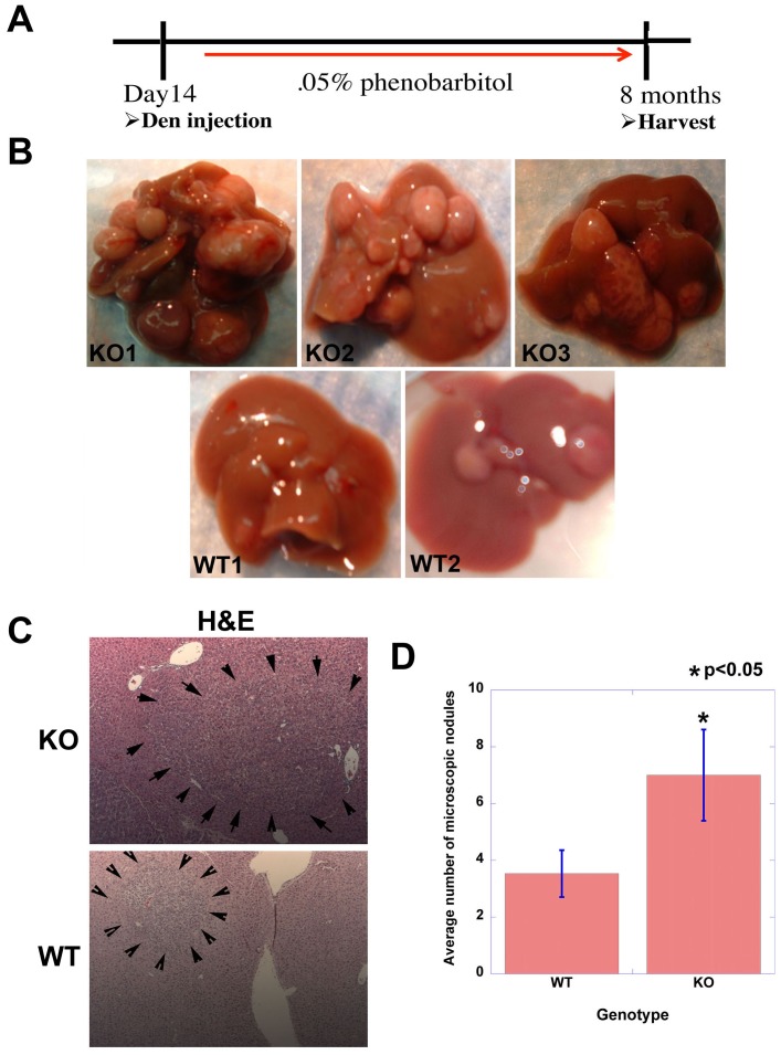
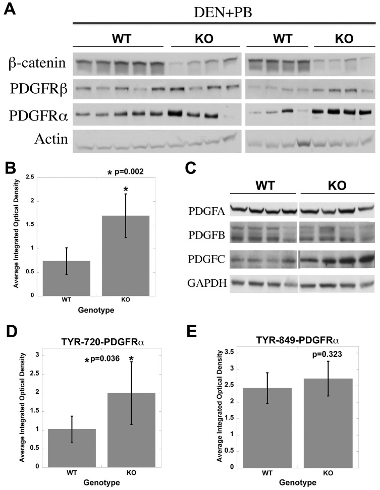
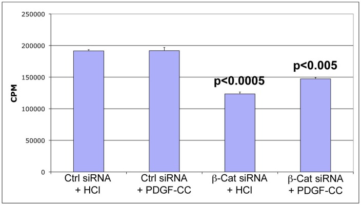
Hepatocellular Carcinoma (HCC) is the fifth most common cancer worldwide. β-Catenin, the central orchestrator of the canonical Wnt pathway and a known oncogene is paramount in HCC pathogenesis. Administration of phenobarbital (PB) containing water (0.05% w/v) as tumor promoter following initial injected intraperitoneal (IP) diethylnitrosamine (DEN) injection (5 µg/gm body weight) as a tumor inducer is commonly used model to study HCC in mice. Herein, nine fifteen-day male β-catenin knockout mice (KO) and fifteen wild-type littermate controls (WT) underwent DEN/PB treatment and were examined for hepatic tumorigenesis at eight months. Paradoxically, a significantly higher tumor burden was observed in KO (p<0.05). Tumors in KO were β-catenin and glutamine synthetase negative and HGF/Met, EGFR & IGFR signaling was unremarkable. A significant increase in PDGFRα and its ligand PDGF-CC leading to increased phosphotyrosine-720-PDGFRα was observed in tumor-bearing KO mice (p<0.05). Simultaneously, these livers displayed increased cell death, stellate cell activation, hepatic fibrosis and cell proliferation. Further, PDGF-CC significantly induced hepatoma cell proliferation especially following β-catenin suppression. Our studies also demonstrate that the utilized DEN/PB protocol in the WT C57BL/6 mice did not select for β-catenin gene mutations during hepatocarcinogenesis. Thus, DEN/PB enhanced HCC in mice lacking β-catenin in the liver may be due to their ineptness at regulating cell survival, leading to enhanced fibrosis and regeneration through PDGFRα activation. β-Catenin downregulation also made hepatoma cells more sensitive to receptor tyrosine kinases and thus may be exploited for therapeutics.


Conflict of interest statement

Competing Interests: The authors have declared that no competing interests exist.

Figures

Figure 1
Figure 1. Enhanced tumorigenesis in β-catenin KO mice exposed to DEN/PB regimen.
Experimental strategy summarizing DEN/PB treatment in KO and WT mice. A. Representative photographs of tumor-bearing livers in DEN/PB treated KO and WT mice at the time of harvest at 8 months of age. B. DEN/PB induced microscopic tumor foci (outlined by arrowheads) visualized by H&E in WT and KO livers at 8 months of age. C. A significant increase in microscopic tumor foci in KO as compared to WT (p<0.05). Tumors were counted from H&E stained sections representing 4 liver lobes from each KO and WT animals on DEN/PB protocol.
Figure 2
Figure 2. Tumorigenesis in KO mice following DEN/PB treatment is associated with greater injury, fibrosis and regeneration.
A. Representative tumor-bearing KO livers show increased parenchymal cell death (TUNEL), greater stellate cell activation (α-SMA) and increased parenchymal cell proliferation (PCNA) as compared to WT. B. Increased hepatic fibrosis (blue) in tumor-bearing KO livers is evident by Masson Trichrome staining as compared to control. C. PCNA- and TUNEL-positive cells were counted in 4 random sections of 5 representative KO and WT livers. A significant increase in both PCNA and TUNEL positive parenchymal cells was evident in KO livers as compared to WT after DEN/PB treatment.
Figure 3
Figure 3. Molecular signaling in tumor-bearing KO and WT mice.
A. Representative western blot analysis from 5 WT and 4 KO tumor-bearing livers shows low β-catenin, absent GS and dramatically lower cyclin-D1 in KO whereas c-Myc levels were increased. Actin verifies equal loading. B. Examination of RTK in the same sets of animals shows notably lower phospho-Met and phospho-IGFR and only marginally lower phospho-EGFR, in KO than WT livers by western blots. GAPDH verifies comparable loading.
Figure 4
Figure 4. β-Catenin and PDGFRα immunohistochemistry in tumors in WT and KO exposed to DEN/PB.
Tumors in WT were heterogeneous and were either positive for both β-catenin and PDGFRα, or for either one of them. In KO, almost all tumors lacked any β-catenin and showed intense PDGFRα-positivity.
Figure 5
Figure 5. Tumor-bearing KO mice display active PDGFRα signaling when compared to WT.
A. Representative western blots from 8 KO and 9 WT show a dramatic increase in total levels of PDGFRα and modest increase in PDGFRβ in the KO. Actin loading verifies equal loading. B. Average integrated optical density (IOD) obtained from scanned autoradiographs shown in Fig. 5A revealed significantly higher PDGFRα levels in KO (p = 0.002). C. Western blot from representative samples shows a dramatic increase in PDGF-CC, a ligand for PDGFRα in KO whereas PDGF-AA and BB remained unremarkable between the two groups. D. Bar graph depicts a significant increase in Tyr720-PDGFRα in KO as compared to WT (p<0.05). E. Insignificant differences were evident in Tyr-849-PDGFRα between the WT and KO.
Figure 6
Figure 6. β-Catenin suppression engenders mitogenicity to PDGF-CC in hepatoma cell culture.
PDGF-CC (10 ng/ml) treatment does not increase DNA synthesis as compared to HCl treatment of Hep3B cells. β-Catenin knockdown led to significant decrease in thymidine incorporation as compared to control siRNA (p<0.0005). However PDGF-CC treatment led to a significant increase in thymidine incorporation in β-catenin-suppressed as compared to control siRNA-transfected cells (p<0.005).

Comment in

References

    1. Parkin DM. Global cancer statistics in the year 2000. Lancet Oncol. 2001;2:533–543. - PubMed
    1. Lustig B, Behrens J. The Wnt signaling pathway and its role in tumor development. J Cancer Res Clin Oncol. 2003;129:199–221. - PubMed
    1. Clevers H. Wnt/beta-catenin signaling in development and disease. Cell. 2006;127:469–480. - PubMed
    1. Peifer M, Polakis P. Wnt signaling in oncogenesis and embryogenesis–a look outside the nucleus. Science. 2000;287:1606–1609. - PubMed
    1. Willert K, Brown JD, Danenberg E, Duncan AW, Weissman IL, et al. Wnt proteins are lipid-modified and can act as stem cell growth factors. Nature. 2003;423:448–452. - PubMed

Publication types

MeSH terms

LinkOut - more resources