. 2012;7(6):e39797.
doi: 10.1371/journal.pone.0039797. Epub 2012 Jun 25.

Genome-wide screening for genetic alterations in esophageal cancer by aCGH identifies 11q13 amplification oncogenes associated with nodal metastasis

Affiliations

Genome-wide screening for genetic alterations in esophageal cancer by aCGH identifies 11q13 amplification oncogenes associated with nodal metastasis

Jianming Ying et al. PLoS One. 2012.

Abstract

Background: Esophageal squamous cell carcinoma (ESCC) is highly prevalent in China and other Asian countries, as a major cause of cancer-related mortality. ESCC displays complex chromosomal abnormalities, including multiple structural and numerical aberrations. Chromosomal abnormalities, such as recurrent amplifications and homozygous deletions, directly contribute to tumorigenesis through altering the expression of key oncogenes and tumor suppressor genes.

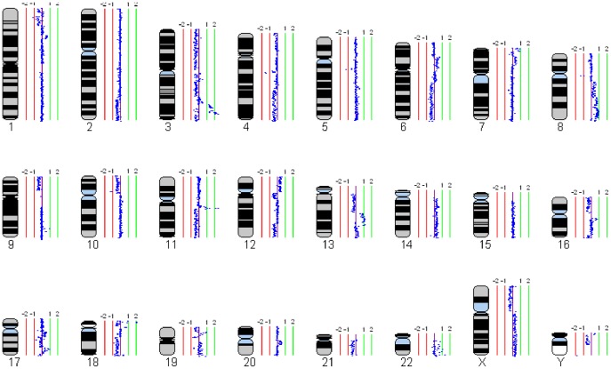
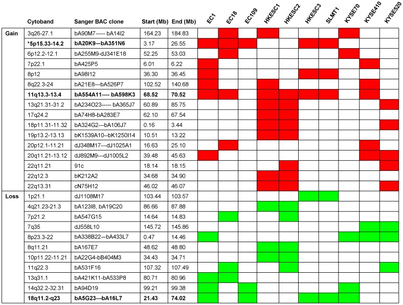
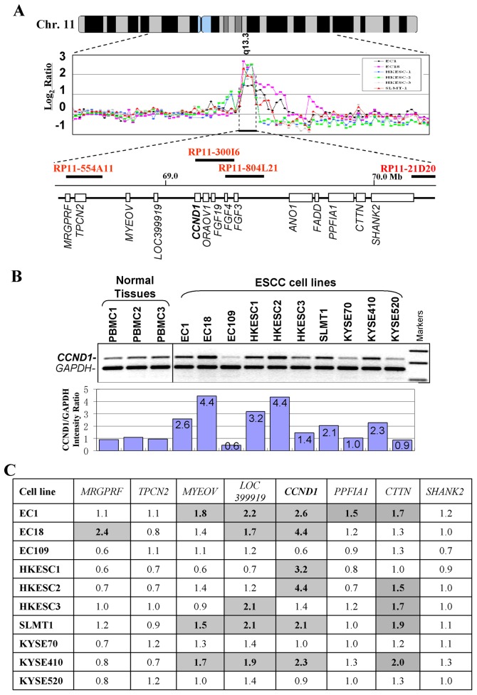
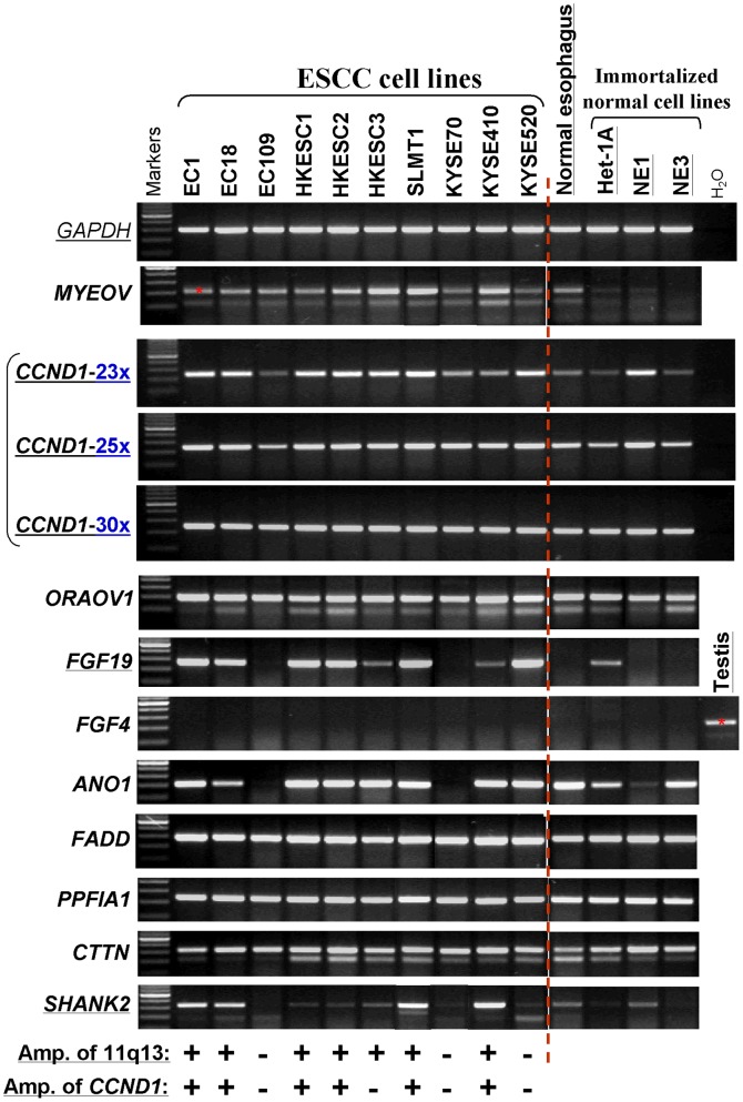
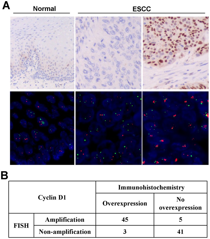
Methodology/principle findings: To understand the role of genetic alterations in ESCC pathogenesis and identify critical amplification/deletion targets, we performed genome-wide 1-Mb array comparative genomic hybridization (aCGH) analysis for 10 commonly used ESCC cell lines. Recurrent chromosomal gains were frequently detected on 3q26-27, 5p15-14, 8p12, 8p22-24, 11q13, 13q21-31, 18p11 and 20q11-13, with frequent losses also found on 8p23-22, 11q22, 14q32 and 18q11-23. Gain of 11q13.3-13.4 was the most frequent alteration in ESCC. Within this region, CCND1 oncogene was identified with high level of amplification and overexpression in ESCC, while FGF19 and SHANK2 was also remarkably over-expressed. Moreover, a high concordance (91.5%) of gene amplification and protein overexpression of CCND1 was observed in primary ESCC tumors. CCND1 amplification/overexpression was also significantly correlated with the lymph node metastasis of ESCC.

Conclusion: These findings suggest that genomic gain of 11q13 is the major mechanism contributing to the amplification. Novel oncogenes identified within the 11q13 amplicon including FGF19 and SHANK2 may play important roles in ESCC tumorigenesis.


Conflict of interest statement

Competing Interests: The authors have declared that no competing interests exist.

Figures

Figure 1
Figure 1. Whole genome profile of an ESCC cell line (EC18) by 1-Mb aCGH.
Normalized log2 signal intensity ratios were plotted using SeeGH software. A log2 signal ratio of 0 represents equivalent copy number between the sample and the reference DNA (details in Materials and Methods). Cytoband pattern for each chromosome is shown in the left. Vertical lines denote log2 signal ratios from −2 to +2 with copy number increasing in the right and decreasing in the left. Each dark blue dot represents a single BAC clone.
Figure 2
Figure 2. Minimal aberrant regions identified by aCGH.
Abnormalities with ≥20% frequency in 10 ESCC cell lines are shown. * Regions with abnormalities in ≥50% cell lines are shown in bold.
Figure 3
Figure 3. CCND1 is located at the center of the 11q13 amplicon in ESCC. A
, aCGH profiles of six ESCC cell lines at the CCND1 locus. Normalized log2 signal ratios were plotted. Amplifications were defined as log2 signal intensities ≥1. Horizontal lines denote log2 signal ratios from −1 to 3 with copy number increasing upwards. Each black/colorful dot represents a single BAC clone. Names of related BAC clones are also shown. Transcript map of the core 11q13 amplicon is shown in the bottom. B, Semi-quantitative duplex genomic DNA PCR analysis of CCND1 in 10 ESCC cell lines and 3 normal PBMC samples. Signal intensity ratios of CCND1/GAPDH are shown. C, Summary of gene copy number changes of several genes within the 11q13 amplicon. Numbers shown in the table are folds of copy numbers of ESCC cell lines relative to the mean values of three PBMC samples. PBMC, peripheral blood mononuclear cell.
Figure 4
Figure 4. Expression levels of several 11q13 genes around CCND1 in ESCC cell lines examined by semi-quantitative RT-PCR.
The amplification status of 11q13 region by aCGH and CCND1 by multiplex DNA PCR are listed at the bottom. +, amplified; -, not amplified. 23x, 25x, 30x: RT-PCR cycles. All other genes were examined by RT-PCR with 30 cycles, with GAPDH for only 23 cycles.
Figure 5
Figure 5. Representative immunohistochemical staining and FISH analysis
. A, Top panels, immunohistochemical staining for CCND1. Left, scattered positivity of CCND1, especially in the basal layer, is seen in normal esophageal epithelium (original magnification, ×200). Middle, a case of ESCC is negative for CCND1 expression (original magnification, ×200). Right, diffuse and strong nuclear staining for CCND1 in this case of ESCC (original magnification, ×200). Bottom panels, FISH analysis. Green signals refer to reference probe of chr 11 centromere while red signals are target probe for CCND1. Left, unamplified in normal esophageal epithelium, Middle, an unamplified ESCC case. Right, an amplified ESCC case. B. Results of CCND1 amplification and expression levels in 94 paraffin-embedded primary ESCC.

References

    1. Parkin DM, Pisani P, Ferlay J. Estimates of the worldwide incidence of eighteen major cancers in 1985. Int J Cancer. 1993;19:594–606. - PubMed
    1. Pera M, Manterola C, Vidal O, Grande L. Epidemiology of esophageal adenocarcinoma. J Surg Oncol. 2005;92:151–159. - PubMed
    1. Lambert R, Hainaut P. The multidisciplinary management of gastrointestinal cancer Epidemiology of oesophagogastric cancer. Best Pract Res Clin Gastroenterol. 2007;21:921–945. - PubMed
    1. Law FB, Chen YW, Wong KY, Ying J, Tao Q, et al. Identification of a novel tumor transforming gene GAEC1 at 7q22 which encodes a nuclear protein and is frequently amplified and overexpressed in esophageal squamous cell carcinoma. Oncogene. 2007;26:7490–7498. - PMC - PubMed
    1. Luo ML, Shen XM, Zhang Y, Wei F, Xu X, et al. Amplification and overexpression of CTTN (EMS1) contribute to the metastasis of esophageal squamous cell carcinoma by promoting cell migration and anoikis resistance. Cancer Res. 2006;66:11690–11699. - PubMed

Publication types