. 2012;7(6):e39878.
doi: 10.1371/journal.pone.0039878. Epub 2012 Jun 25.

Characterization of the autocrine/paracrine function of vitamin D in human gingival fibroblasts and periodontal ligament cells

Affiliations

Characterization of the autocrine/paracrine function of vitamin D in human gingival fibroblasts and periodontal ligament cells

Kaining Liu et al. PLoS One. 2012.

Abstract

Background: We previously demonstrated that 25-hydroxyvitamin D(3), the precursor of 1α,25-dihydroxyvitamin D(3), is abundant around periodontal soft tissues. Here we investigate whether 25-hydroxyvitamin D(3) is converted to 1α,25-dihydroxyvitamin D(3) in periodontal soft tissue cells and explore the possibility of an autocrine/paracrine function of 1α,25-dihydroxyvitamin D(3) in periodontal soft tissue cells.

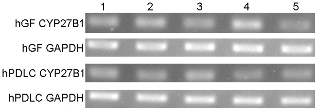
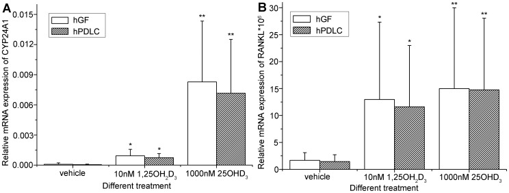
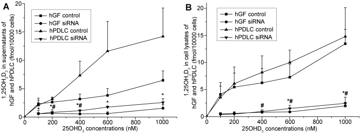
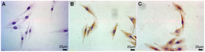
Methodology/principal findings: We established primary cultures of human gingival fibroblasts and human periodontal ligament cells from 5 individual donors. We demonstrated that 1α-hydroxylase was expressed in human gingival fibroblasts and periodontal ligament cells, as was cubilin. After incubation with the 1α-hydroxylase substrate 25-hydroxyvitamin D(3), human gingival fibroblasts and periodontal ligament cells generated detectable 1α,25-dihydroxyvitamin D(3) that resulted in an up-regulation of CYP24A1 and RANKL mRNA. A specific knockdown of 1α-hydroxylase in human gingival fibroblasts and periodontal ligament cells using siRNA resulted in a significant reduction in both 1α,25-dihydroxyvitamin D(3) production and mRNA expression of CYP24A1 and RANKL. The classical renal regulators of 1α-hydroxylase (parathyroid hormone, calcium and 1α,25-dihydroxyvitamin D(3)) and Porphyromonas gingivalis lipopolysaccharide did not influence 1α-hydroxylase expression significantly, however, interleukin-1β and sodium butyrate strongly induced 1α-hydroxylase expression in human gingival fibroblasts and periodontal ligament cells.

Conclusions/significance: In this study, the expression, activity and functionality of 1α-hydroxylase were detected in human gingival fibroblasts and periodontal ligament cells, raising the possibility that vitamin D acts in an autocrine/paracrine manner in these cells.


Conflict of interest statement

Competing Interests: The authors have declared that no competing interests exist.

Figures

Figure 1
Figure 1. mRNA expression of CYP27B1 in hGF and hPDLC.
mRNA expression of CYP27B1 was detected by RT-PCR in hGF and hPDLC from all five donors (each lane represents one donor). GAPDH was used as an internal control.
Figure 2
Figure 2. Protein expression of CYP27B1 in hGF and hPDLC.
Protein expression of CYP27B1 was detected by Western blot in hGF and hPDLC from all five donors (donors are numbered 1–5). β-actin was used as an internal control.
Figure 3
Figure 3. Activity of 1α-hydroxylase in hGF and hPDLC.
hGF and hPDLC from donors 2, 3 and 5 were incubated with 1000 nM 25OHD3 for the times indicated and the production of 1,25OH2D3 was determined in (A) supernatants and (B) cell lysates. After prolonged incubation, production of 1,25OH2D3 increased. hPDLC released more 1,25OH2D3 than hGF 12 h after incubation with 25OHD3. The data is presented as the mean±SE. ** hGF generated significantly less 1,25OH2D3 than hPDLC at the same time point (p<0.05). The time course of CYP27B1 (C) and CYP24A1 (D) mRNA expression is also presented. CYP27B1 mRNA expression peaked at 1 h and no difference was detected between 0 h and any other time point. CYP24A1 mRNA expression was significantly higher at 24 h and 48 h than 0 h. * CYP27B1 or CYP24A1 mRNA expression at the time point was significantly different from that at 0 h in hGF (p<0.05). # CYP27B1 or CYP24A1 mRNA expression at the time point was significantly different from that at 0 h in hPDLC (p<0.05).
Figure 4
Figure 4. Effect of 25OHD3 incubation on gene expression in hGF and hPDLC.
hGF and hPDLC from all five donors were treated with 1000 nM 25OHD3, 10 nM 1,25OH2D3 or vehicle for 48 h and mRNA expression of (A) CYP24A1 and (B) RANKL was examined by real-time PCR. The up-regulation observed after 1000 nM 25OHD3 treatment was significantly stronger than that observed after 10 nM 1,25OH2D3 treatment. The data are presented as the mean±SE. * denotes difference from vehicle (p<0.05). ** denotes difference from vehicle and 1,25OH2D3 (p<0.05).
Figure 5
Figure 5. Efficiency of RNA interference against CYP27B1.
All cells were transfected with either a siRNA oligonucleotide for CYP27B1 or a non-silencing control. Using real-time PCR as a measure, the efficiency of RNA interference against CYP27B1 was over 70% in hGF and hPDLC from all 5 donors. Donors are numbered 1–5. The data are presented as the mean±SD. * denotes difference from vehicle (p<0.05).
Figure 6
Figure 6. Effect of CYP27B1 silencing on 1,25OH2D3 generation.
hGF and hPDLC from donors 2, 3 and 5 were treated with 25OHD3 at various concentrations indicated in the figure for 48 h after transfection with the siRNA oligonucleotide for CYP27B1 or a non-silencing control and 1,25OH2D3 production was measured in (A) supernatants and (B) cell lysates. When CYP27B1 was not silenced, production of 1,25OH2D3 increased with increasing concentration of 25OHD3. When CYP27B1 was silenced, the generation of 1,25OH2D3 decreased significantly compared with when CYP27B1 was not silenced. The data are presented as the mean±SE. * hGF generated significantly less 1,25OH2D3 with the same amount of 25OHD3 when CYP27B1 was knocked down (p<0.05). # hPDLC generated significantly less 1,25OH2D3 (with the same amount of added 25OHD3) when CYP27B1 was knocked down (p<0.05).
Figure 7
Figure 7. Effect of CYP27B1 silencing on gene up-regulation by 25OHD3.
hGF and hPDLC from all five donors were treated with 1000 nM 25OHD3 for 48 h after transfection with the siRNA oligonucleotide for CYP27B1 or a non-silencing control and mRNA expression of (A) CYP24A1 and (B) RANKL was determined by real-time PCR. Compared with the transfection with a non-silencing control, transfection with the siRNA oligonucleotide for CYP27B1 resulted in a significantly weaker up-regulation of CYP24A1 and RANKL in both hGF and hPDLC. The data are presented as the mean±SE. * denotes difference from control (p<0.05).
Figure 8
Figure 8. Preliminary investigation of CYP27B1 regulation in hGF and hPDLC.
hGF and hPDLC from donors 2, 3, 4 and 5 were stimulated with different treatments indicated in the figure for 24 h and CYP27B1 expression was determined by real-time PCR. (A) Pg-LPS, parathyroid hormone, CaCl2 and 1,25OH2D3 did not significantly influence CYP27B1 mRNA expression. (B) IL-1β and sodium butyrate significantly up-regulated CYP27B1 mRNA expression independently of 1,25OH2D3. Additionally, the characteristics of CYP27B1 regulation in hGF and hPDLC were not significantly different. The data are presented as the mean±SE. * CYP27B1 mRNA expression was significantly different from that of the vehicle group in hGF (p<0.05). # CYP27B1 mRNA expression was significantly different from that of the vehicle group in hPDLC (p<0.05). IL-1β: interleukin-1β. Pg-LPS: Porphyromonas gingivalis lipopolysaccharide. PTH: parathyroid hormone.
Figure 9
Figure 9. mRNA expression of cubilin in hGF and hPDLC.
mRNA expression of cubilin was determined by RT-PCR in hGF and hPDLC from all five donors (each lane represents one donor). GAPDH was used as an internal control.
Figure 10
Figure 10. Protein expression of cubilin in hGF and hPDLC.
hGF and hPDLC from all five donors were used for immunocytochemical staining of cubilin; expression of cubilin was detected in all the cells examined. Panel A is the negative control for the immunocytochemical staining of cubilin (400×). Panel B and C contain images of the hGF and hPDLC, respectively, from donor 2 (400×). The primary antibody was replaced with PBS for the negative control.

Similar articles

Cited by

References

    1. Rachez C, Freedman LP. Mechanisms of gene regulation by vitamin D(3) receptor: a network of coactivator interactions. Gene. 2000;246:9–21. - PubMed
    1. von EM, Kongsbak M, Schjerling P, Olgaard K, Odum N, et al. Vitamin D controls T cell antigen receptor signaling and activation of human T cells. Nat Immunol. 2010;11:344–349. - PubMed
    1. Chen KS, DeLuca HF. Cloning of the human 1 alpha,25-dihydroxyvitamin D-3 24-hydroxylase gene promoter and identification of two vitamin D-responsive elements. Biochim Biophys Acta. 1995;1263:1–9. - PubMed
    1. Haussler MR, Whitfield GK, Haussler CA, Hsieh JC, Thompson PD, et al. The nuclear vitamin D receptor: biological and molecular regulatory properties revealed. J Bone Miner Res. 1998;13:325–349. - PubMed
    1. Christakos S, Dhawan P, Liu Y, Peng X, Porta A. New insights into the mechanisms of vitamin D action. J Cell Biochem. 2003;88:695–705. - PubMed

Publication types