. 2012;7(6):e39967.
doi: 10.1371/journal.pone.0039967. Epub 2012 Jun 27.

DOK3 negatively regulates LPS responses and endotoxin tolerance

Affiliations

DOK3 negatively regulates LPS responses and endotoxin tolerance

Qisheng Peng et al. PLoS One. 2012.

Abstract

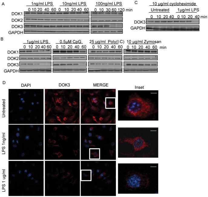
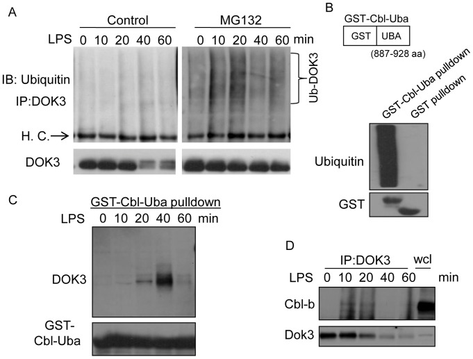
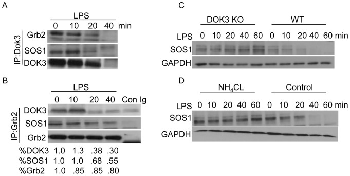
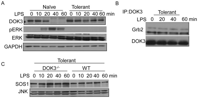
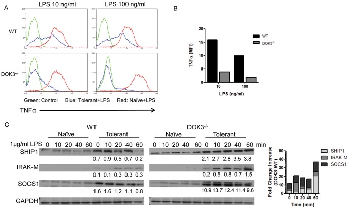
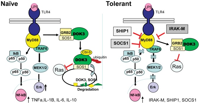
Innate immune activation via Toll-like receptors (TLRs), although critical for host defense against infection, must be regulated to prevent sustained cell activation that can lead to cell death. Cells repeatedly stimulated with lipopolysaccharide (LPS) develop endotoxin tolerance making the cells hypo-responsive to additional TLR stimulation. We show here that DOK3 is a negative regulator of TLR signaling by limiting LPS-induced ERK activation and cytokine responses in macrophages. LPS induces ubiquitin-mediated degradation of DOK3 leading to SOS1 degradation and inhibition of ERK activation. DOK3 mice are hypersensitive to sublethal doses of LPS and have altered cytokine responses in vivo. During endotoxin tolerance, DOK3 expression remains stable, and it negatively regulates the expression of SHIP1, IRAK-M, SOCS1, and SOS1. As such, DOK3-deficient macrophages are more sensitive to LPS-induced tolerance becoming tolerant at lower levels of LPS than wild type cells. Taken together, the absence of DOK3 increases LPS signaling, contributing to LPS-induced tolerance. Thus, DOK3 plays a role in TLR signaling during both naïve and endotoxin-induced tolerant conditions.


Conflict of interest statement

Competing Interests: The authors have declared that no competing interests exist.

Figures

Figure 1
Figure 1. LPS stimulation of macrophages induces DOK3 degradation.
BMM were stimulated with (A) 1, 10, or 100 ng/ml LPS; or (B) 1 µg/ml LPS (TLR4 ligand), 0.5 µM CpG (TLR9 ligand), 25 µg/ml Poly (I:C) (TLR3 ligand), or 10 µg/ml Zymosan (TLR2 ligand) for the indicated time and cell lysates were immunoblotted for DOK1, DOK2, DOK3, and GAPDH. (C) BMM were treated with 10 µg/ml of the translational inhibitor cycloheximide during LPS stimulation. Data are representative of three independent experiments. (D) BMM cells were stimulated with LPS (1 ng/ml and 1 µg/ml) for 1 h. The cells were fixed, permeabilized, and immunofluorescent staining for DOK3 (red) and DAPI (blue) was performed and visualized with a Leica TCS NT Confocal microscope. Bar is 10 µm.
Figure 2
Figure 2. LPS stimulation in macrophages induces phosphorylation-independent degradation of DOK3.
(A) BMM were stimulated with 1 µg/ml LPS and DMSO (control), proteasomal inhibitor (10 µM MG132), or lysosomal inhibitor (20 mM NH4Cl) or (B) BMM were stimulated with 1 µg/ml LPS in the presence of Src inhibitor (PP2), Syk inhibitor (Bay), PI3 kinase inhibitor (Ly294002), or Btk inhibitor (LFM) for the indicated time and cell lysates were immunoblotted for DOK3 and GAPDH. DOK3 phosphorylation was determined by immunoprecipitation (IP) with an anti-phosphotyrosine antibody and immunoblotting (IB) for DOK3. Data are representative of three independent experiments.
Figure 3
Figure 3. DOK3 is ubiquitinated through interaction with the E3 ligase Cbl-b in response to LPS stimulation.
(A) BMM were stimulated with 1 µg/ml LPS ±10 µM MG132 for the indicated time, and ubiquitinated DOK3 was determined by immunoprecipitating with an anti-DOK3 antibody and immunoblotting with an anti-ubiquitin antibody. DOK3 immunoblot confirms degradation of DOK3 in control but not MG32 treated cells. H.C., heavy chain of IgG (B) GST-Cbl-Uba, comprised of GST and 887–928 amino acids (aa) of Cbl-b, was used to immunoprecipitate ubiquitinated proteins (bottom panel shows input amount of GST or GST-fusion). (C) GST-Cbl-Uba pulldown of LPS stimulated BMM lysates were immunoblotted for DOK3. Input amount of GST-fusion protein is the bottom blot. (D) DOK3 immunoprecipitation of LPS treated BMM immunoblotted for Cbl-b and DOK3. wcl, whole cell lysate Data are representative of three independent experiments.
Figure 4
Figure 4. LPS-induced degradation of DOK3 mediates ERK activation.
(A) RAW264.7 macrophages, transduced with 23 nt plus hairpin shRNA designed to knock down DOK3 or a control luciferase knock down, were lysed and decreased DOK3 expression was confirmed by immunoblotting. Cells were and stimulated with 1 µg/ml LPS for the indicated time and cell lysates were immunoblotted for pERK, ERK, and GAPDH. (B) Wild type (WT) or DOK3-deficient BMM were stimulated with 1 µg/ml LPS and cell lysates were immunblotted for pERK, ERK, and the GAPDH. The amount of DOK3 and Grb2 was detected by immunoblot in cell lysates. (C) BMM cells were pretreated with DMSO or 10 µM MG132 and stimulated with 1 µg/ml LPS for the indicated times. Lysates were immunoblotted for phosphorylated ERK (pERK), ERK, and DOK3. (D) RAW264.7 macrophages transfected with FLAG-tagged mouse DOK3- were stimulated with 1 µg/ml LPS for the indicated time and cell lysates were immunoblotted for pERK, ERK, FLAG and GAPDH. Data are representative of three independent experiments.
Figure 5
Figure 5. DOK3 associates with Grb2 and SOS1 and promotes SOS1 degradation in response to LPS.
BMM were stimulated with 1 µg/ml LPS for indicated time and cell lysates were immunoprecipitated with (A) anti-DOK3 antibody or (B) anti-Grb2 antibody followed by immunoblotting for DOK3, Grb2, and SOS1. As a control, isotype matched antibody was used for immunoprecipitation (Con Ig). Protein levels were quantified using Image J. (C) Wild type and DOK3−/− BMM were stimulated with 1 µg/ml LPS for indicated time and immunoblotted for SOS1 and GAPDH. (D) BMM were treated with NH4CL or PBS (control) and were stimulated with 1 µg/ml LPS and immunoblotted for SOS1 and GAPDH. Data are representative of three independent experiments.
Figure 6
Figure 6. DOK3 alters LPS-induced inflammatory cytokine expression and is required for survival after LPS challenge.
(A) Wild type and DOK3-deficient BMM were stimulated with 1 µg/ml LPS and cell lysates were immunoblotted for IκBα and GAPDH. (B) Wild type and DOK3-deficient BMM were treated with 1 µg/ml LPS for 16 h and amounts of IL-10, IL-6, TNFα, and IL-1β in conditioned media were determined by ELISA. (C) Serum was collected from WT and DOK3−/− mice prior to and 60 min. following challenge with 5 mg/kg LPS and serum IL-6, TNFα, IL-10 and IL-1β levels were determined by ELISA. (D) DOK3−/− mice are hypersensitive (P<0.0027) compared to WT mice following i.p. challenge with a sublethal LPS dose (70 mg/kg). The * in C and D indicates a significant difference (P<0.05) between cell type/strain and results are expressed as means ± S.D.
Figure 7
Figure 7. DOK3 and SOS1 are stable during LPS-induced tolerance.
(A) BMM were pretreated with 1 µg/ml LPS for 18 hours, rested in fresh media for 2 hours and re-stimulated along with naïve cells with 1 µg/ml LPS for indicated time and cell lysates were immunoblotted for DOK3, pERK, ERK, and GAPDH. (B) Tolerant BMM were stimulated with 1 µg/ml LPS and cell lysates were immunoprecipitated with anti-DOK3 antibody and immunoblotted for Grb2 and DOK3. (C) Tolerant wild type and DOK3-deficient BMM were re-stimulated with 1 µg/ml LPS and lysates immunoblotted for SOS1 and JNK (loading control). Data are representative of three independent experiments.
Figure 8
Figure 8. DOK3-deficient macrophages are more sensitive to endotoxin tolerance.
Tolerance was induced in wild type and DOK3-deficient BMM with the indicated dose of LPS (10 or 100 ng/ml) overnight, followed by resting in fresh media for 2 h and re-challenged with the same dose of LPS for 12 h in the presence of Brefeldin A. Intracellular TNFα production was assessed with flow cytometry after the cells were fixed, permeabilized, and stained with anti-TNFα. Results are represented (A) by one-parameter histograms (green is control staining, red is naïve cells, and blue is tolerant cells) or (B) by MFI (mean fluorescence intensity) of TNFα-producing population and representative of two independent experiments. (C) Tolerant wild type and DOK3-deficient BMM or naïve cells were stimulated with 1 µg/ml LPS and immunoblotted for SHIP1, IRAK-M, SOCS1, and GAPDH. Densitometry of the bands was determined using image J normalizing to GAPDH, and the relative numerical value was written below the band. Data are representative of three independent experiments.
Figure 9
Figure 9. Proposed model of DOK3 regulation of TLR4 signaling and endotoxin tolerance.
In naïve cells, DOK3 associates with Grb2 and SOS1 constitutively. Upon LPS stimulation, DOK3 becomes ubiquitinated, possibly by Cbl-b, and degraded, thereby releasing Grb2. SOS1 is also degraded in a DOK3-dependent manner thus limiting ERK activation. In LPS-induced tolerant cells, DOK3 and SOS1 expression remains stable during repeated LPS challenge. DOK3 may limit ERK activation during tolerance by binding to and sequestering Grb2 and SOS1. Mediators of tolerance including IRAK-M, SHIP1, and SOCS1 are upregulated.

Similar articles

Cited by

References

    1. Janeway CA, Jr, Medzhitov R. Innate immune recognition. Annu Rev Immunol. 2002;20:197–216. - PubMed
    1. Nagai Y, Akashi S, Nagafuku M, Ogata M, Iwakura Y, et al. Essential role of MD-2 in LPS responsiveness and TLR4 distribution. Nat Immunol. 2002;3:667–672. - PubMed
    1. Takeda K, Kaisho T, Akira S. Toll-like receptors. Annu Rev Immunol. 2003;21:335–376. - PubMed
    1. Akashi S, Saitoh S, Wakabayashi Y, Kikuchi T, Takamura N, et al. Lipopolysaccharide interaction with cell surface Toll-like receptor 4-MD-2: higher affinity than that with MD-2 or CD14. J Exp Med. 2003;198:1035–1042. - PMC - PubMed
    1. Green NM, Marshak-Rothstein A. Toll-like receptor driven B cell activation in the induction of systemic autoimmunity. Semin Immunol. 2011;23:106–112. - PMC - PubMed

Publication types