. 2012 May 28;4(5):186-92.
doi: 10.4329/wjr.v4.i5.186.

Preoperative transarterial Embolisation in bone tumors

Affiliations

Preoperative transarterial Embolisation in bone tumors

Pankaj Gupta et al. World J Radiol. .

Abstract

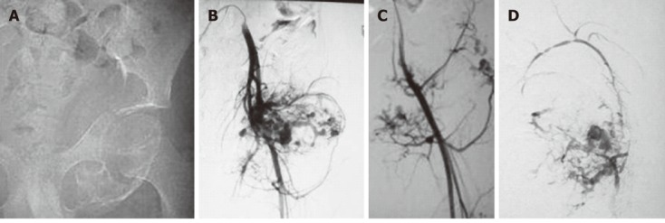
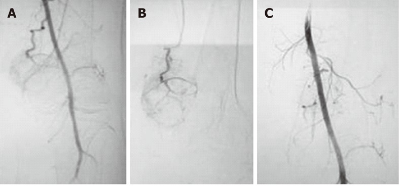
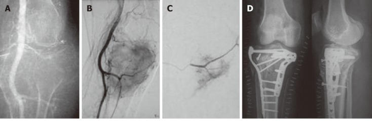
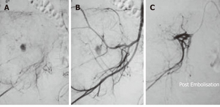
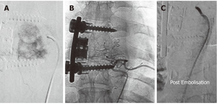
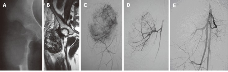
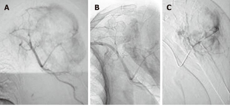
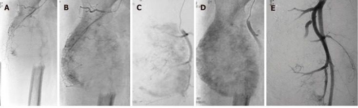
Bone tumors include a variety of lesions, both primary and metastatic. The treatment modalities for bone tumors vary with the individual lesion, but in general surgical excision is the treatment of choice with other adjunctive therapies. However, surgery for many bone tumors is complex due to several factors including tumor bulk, vascularity, vicinity to vital structures and potentially inaccessible location of the lesion. Transarterial Embolisation (TAE) is one of the important adjuvant treatment modalities and in some cases it may be the primary and curative treatment. Preoperative TAE has proved to be effective in both primary and metastatic bone tumors. It reduces tumor vascularity and intraoperative blood loss, the need for blood transfusion and associated complications, allows better definition of tissue planes at surgery affording more complete excision, and hence reduced recurrence. Preoperative chemoEmbolisation has also been shown to increase the sensitivity of some tumors to subsequent chemotherapy and radiotherapy. There are several techniques and embolic agents available for this purpose, but the ultimate aim is to achieve tumor devascularization. In this review, we discuss the techniques including the choice of embolic agent, application to individual lesions and potential complications.

Keywords: Blood loss; Bone tumors; Embolisation; Interventional; Radiology; Surgical.


Figures

Figure 1
Figure 1
Pre-operative Embolisation of aneurysmal bone cyst of left pelvic bones in a 13-year-old male. A: An image from a digital subtraction angiography examination of a 13-year-old male showed an expansile lesion involving the left inferior pubic ramus and ischium with no cortical breech or extraosseous soft tissue component. The lesion proved to be an aneurysmal bone cyst on histopathology; B: Angiogram obtained after cannulation of left common iliac artery revealed marked vascularity of the lesion with multiple tumor feeders arising from both the internal and external iliac artery; C: Selective left common femoral angiogram showed the tumour supply from this artery; D: Post-Embolisation angiogram after injection of 95% alcohol selectively into the major feeder arising from both the internal and external iliac arteries showed a marked reduction in tumor blush.
Figure 2
Figure 2
Embolisation of left distal femoral aneurysmal bone cyst in a 42-year-old male. A: Forty-two-year-old male with an expansile lytic lesion involving the left medial femoral condyle with histopathologic confirmation of aneurysmal bone cyst. Left common femoral angiogram revealed a prominent arterial feeder arising from the superficial femoral artery at the level of the adductor canal; B: Angiogram obtained after selective cannulation of the arterial feeder revealed the marked vascularity of the lesion; C: Embolisation with polyvinyl alcohol particles 300-500 μm was performed following selective cannulation of the feeder. Post-Embolisation angiogram demonstrated marked reduction in tumor blush.
Figure 3
Figure 3
Embolisation of left proximal tibial giant cell tumor in a 29-year-old male. A: Twenty-nine-year-old male with an eccentric expansile lytic lesion of the left proximal tibial epiphysis extending to the subarticular surface which proved to be a giant cell tumor on histopathology; B: Left common femoral angiogram revealed prominent arterial feeders from the superficial femoral artery with marked tumor blush; C: Angiogram obtained after Embolisation with polyvinyl alcohol particles 300-500 μm shows considerable reduction in tumor blush; D: Postoperative radiographs (AP and lateral) reveal bone cements placed in the defect with multiple screws and fixation plates.
Figure 4
Figure 4
Right iliac giant cell tumor Embolisation in a 25-year-old female. A: Twenty-five-year old female with a large lytic expansile lesion involving the right iliac wing confirmed to be a giant cell tumor on histopathology. Internal iliac artery angiogram showed marked tumor vascularity from the posterior division; B: A delayed phase image showed marked tumor blush; C: Embolisation was performed with gelfoam. Post-Embolisation angiogram showed the occlusion of a large tumor feeder with reduction in tumor blush.
Figure 5
Figure 5
Pre-operative trans-arterial Embolisation of a thoracic vertebral hemangioma in a young male. A: Twenty-three-year-old male with pain in the upper back for 3 mo. Radiographs revealed an expansile lytic lesion of the left transverse process of the sixth thoracic vertebra. Computed tomographic-guided biopsy of the lesion confirmed it to be a hemangioma. A flush aortogram with an angiographic pigtail placed at the level of the aortic arch identified the tumor feeding artery arising from the sixth left posterior intercostal artery which was selectively cannulated; B: Selective angiogram obtained from the feeder revealed marked tumor blush; C: Angiogram obtained after Embolisation with gelfoam showed almost complete loss of tumor blush.
Figure 6
Figure 6
Telengectiatic osteosarcoma in a young male managed preoperatively by trans-arterial embolisation. A: Twenty-year-old male with a large eccentric expansile lytic lesion of the right femoral metaphysis which was confirmed to be a telangiectatic osteosarcoma on histopathology; B: Magnetic resonance imaging was performed for tumor staging. The lesion was well circumscribed with no involvement of neurovascular bundle, no satellite lesions and no extension to the hip joint; C: Right common femoral angiogram revealed tumor supply from profunda femoris artery; D: Selective angiogram revealed marked tumor blush; E: Post-Embolisation angiogram after injection of polyvinyl alcohol particles 300-500 μm showed marked reduction in tumor blush.
Figure 7
Figure 7
Preoperative embolisation in a metastatic lesion from thyroid carcinoma in an elderly male. A: Sixty-two-year-old male with papillary carcinoma of thyroid and an expansile lytic lesion involving the left proximal humerus; B: Left axillary arteriogram revealed marked tumour vascularity; C: Post-Embolisation angiogram after injection of polyvinyl alcohol particles 300-500 μm showing 90% reduction in tumour vascularity.
Figure 8
Figure 8
Preoperative embolisation in a metastatic lesion from renal cell carcinoma in an elderly male. A: Fifty-two-year-old male with renal cell carcinoma (RCC) and a large destructive lesion of the right femoral metaphysis extending to involve the proximal diaphysis. Preoperative Embolisation was planned to reduce tumour vascularity. Metastases from thyroid carcinoma and RCC are highly vascular; B and C: Right common femoral angiogram revealed marked tumor vascularity; D: Delayed phase image shows profound tumor vascularity; E: Embolisation was performed with 300-500 μm polyvinyl alcohol particles and marked reduction (90%) in tumour vascularity was achieved.

Similar articles

Cited by

References

    1. Börüban S, Sancak T, Yildiz Y, Sağlik Y. Embolisation of benign and malignant bone and soft tissue tumors of the extremities. Diagn Interv Radiol. 2007;13:164–171. - PubMed
    1. Kauffmann G, Wimmer B, Bischoff W, Adler C, Strecker EP. [Fundamental experiments for therapeutic artery occlusion by angiography catheters (author’s transl)] Radiologe. 1977;17:489–491. - PubMed
    1. Radeleff B, Eiers M, Lopez-Benitez R, Noeldge G, Hallscheidt P, Grenacher L, Libicher M, Zeifang F, Meeder PJ, Kauffmann GW, et al. Transarterial Embolisation of primary and secondary tumors of the skeletal system. Eur J Radiol. 2006;58:68–75. - PubMed
    1. Brado M, Hansmann HJ, Richter GM, Kauffmann GW. [Interventional therapy of primary and secondary tumors of the spine] Orthopade. 1998;27:269–273. - PubMed
    1. Munk PL, Legiehn GM. Musculoskeletal interventional radiology: applications to oncology. Semin Roentgenol. 2007;42:164–174. - PubMed