

Spinal fusion-hardware construct: Basic concepts and imaging review
- PMID: 22761979
- PMCID: PMC3386531
- DOI: 10.4329/wjr.v4.i5.193
Spinal fusion-hardware construct: Basic concepts and imaging review
Abstract
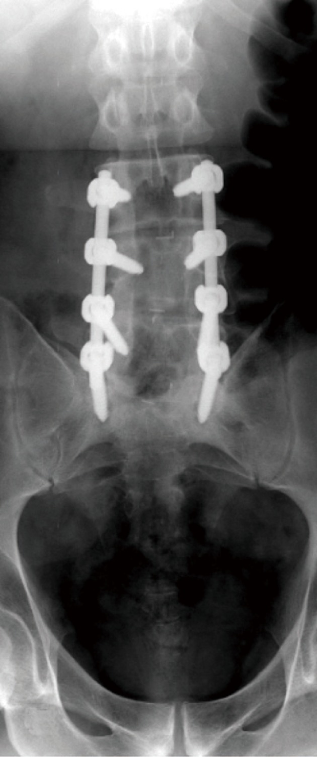
The interpretation of spinal images fixed with metallic hardware forms an increasing bulk of daily practice in a busy imaging department. Radiologists are required to be familiar with the instrumentation and operative options used in spinal fixation and fusion procedures, especially in his or her institute. This is critical in evaluating the position of implants and potential complications associated with the operative approaches and spinal fixation devices used. Thus, the radiologist can play an important role in patient care and outcome. This review outlines the advantages and disadvantages of commonly used imaging methods and reports on the best yield for each modality and how to overcome the problematic issues associated with the presence of metallic hardware during imaging. Baseline radiographs are essential as they are the baseline point for evaluation of future studies should patients develop symptoms suggesting possible complications. They may justify further imaging workup with computed tomography, magnetic resonance and/or nuclear medicine studies as the evaluation of a patient with a spinal implant involves a multi-modality approach. This review describes imaging features of potential complications associated with spinal fusion surgery as well as the instrumentation used. This basic knowledge aims to help radiologists approach everyday practice in clinical imaging.
Keywords: Hardware; Imaging; Instrumentation; Spinal fusion; Spine.
Figures
Similar articles
-
Imaging features of the postoperative spine: a guide to basic understanding of spine surgical procedures.Insights Imaging. 2023 Jun 6;14(1):103. doi: 10.1186/s13244-023-01447-0. Insights Imaging. 2023. PMID: 37278946 Free PMC article. Review.
-
New techniques in lumbar spinal instrumentation: what the radiologist needs to know.Radiology. 2011 Aug;260(2):317-30. doi: 10.1148/radiol.11101104. Radiology. 2011. PMID: 21778450 Review.
-
Lumbar spine fusion and stabilization: hardware, techniques, and imaging appearances.Radiographics. 2007 Nov-Dec;27(6):1737-49. doi: 10.1148/rg.276065205. Radiographics. 2007. PMID: 18025515 Review.
-
Decision making in surgical treatment of chronic low back pain: the performance of prognostic tests to select patients for lumbar spinal fusion.Acta Orthop Suppl. 2013 Feb;84(349):1-35. doi: 10.3109/17453674.2012.753565. Acta Orthop Suppl. 2013. PMID: 23427903
-
Imaging of the postoperative spine.Radiol Clin North Am. 2006 May;44(3):407-18. doi: 10.1016/j.rcl.2006.01.002. Radiol Clin North Am. 2006. PMID: 16644358 Review.
Cited by
-
Pedicle subtraction metallectomy with complex posterior reconstruction for fixed cervicothoracic kyphosis: illustrative case.J Neurosurg Case Lessons. 2023 Jul 17;6(3):CASE23180. doi: 10.3171/CASE23180. Print 2023 Jul 17. J Neurosurg Case Lessons. 2023. PMID: 37486908 Free PMC article.
-
Uncommon Iatrogenic Devices Seen on Chest Radiographs.Indian J Radiol Imaging. 2021 Jan;31(1):172-184. doi: 10.1055/s-0041-1729487. Epub 2021 May 31. Indian J Radiol Imaging. 2021. PMID: 34316125 Free PMC article.
-
Nanohydroxyapatite Effect on the Degradation, Osteoconduction and Mechanical Properties of Polymeric Bone Tissue Engineered Scaffolds.Open Orthop J. 2016 Dec 30;10:900-919. doi: 10.2174/1874325001610010900. eCollection 2016. Open Orthop J. 2016. PMID: 28217213 Free PMC article.
-
Complications exclusive to long strut grafts used following multilevel cervical corpectomy: Utilization of advanced imaging techniques.Indian J Radiol Imaging. 2017 Jul-Sep;27(3):263-267. doi: 10.4103/ijri.IJRI_211_16. Indian J Radiol Imaging. 2017. PMID: 29089670 Free PMC article.
-
Human Mesenchymal Stem Cell Morphology and Migration on Microtextured Titanium.Front Bioeng Biotechnol. 2016 May 10;4:41. doi: 10.3389/fbioe.2016.00041. eCollection 2016. Front Bioeng Biotechnol. 2016. PMID: 27243001 Free PMC article.
References
-
- Hibbs RA. An operation for progressive spinal deformities. NY Med J. 1911;93:1013–1016.
-
- Albee FH. Transplantation of a portion of the tibia into the spine for Pott’s disease. JAMA. 1911;57:855. - PubMed
-
- Young PM, Berquist TH, Bancroft LW, Peterson JJ. Complications of spinal instrumentation. Radiographics. 2007;27:775–789. - PubMed
-
- Rutherford EE, Tarplett LJ, Davies EM, Harley JM, King LJ. Lumbar spine fusion and stabilization: hardware, techniques, and imaging appearances. Radiographics. 2007;27:1737–1749. - PubMed
-
- Slone RM, McEnery KW, Bridwell KH, Montgomery WJ. Fixation techniques and instrumentation used in the thoracic, lumbar, and lumbosacral spine. Radiol Clin North Am. 1995;33:233–265. - PubMed
LinkOut - more resources
Full Text Sources
