

Lung parenchymal changes in patients with ankylosing spondylitis
- PMID: 22761981
- PMCID: PMC3386533
- DOI: 10.4329/wjr.v4.i5.215
Lung parenchymal changes in patients with ankylosing spondylitis
Abstract
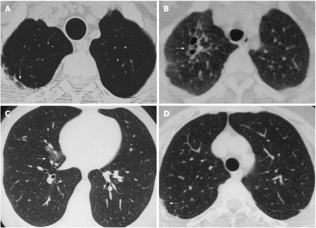
Aim: To assess lung parenchymal changes in ankylosing spondylitis (AS) using high resolution computed tomography (HRCT).
Methods: We included 78 AS patients whose average age was 33.87 (18-56) years with a ratio of 53 males to 25 females who were followed up for 3.88 (1-22) years on average. Pneumonia and tuberculosis were excluded. In a detailed examination of lung HRCT findings, we investigated the presence of parenchymal micronodules, parenchymal bands, subpleural bands, interlobular and intralobular septal thickening, irregularity of interfaces, ground-glass opacity, consolidation, mosaic pattern, bronchial wall thickening, bronchial dilatation, tracheal dilatation, pleural thickening, emphysema, thoracic cage asymmetry, honeycomb appearance, structural distortion, apical fibrosis and other additional findings.
Results: In detailed HRCT evaluations, lung parenchymal changes were found in 46 (59%) of all patients. We found parenchymal bands in 21 (27%) cases, interlobular septal thickening in 9 (12%), emphysema in 9 (12%), apical fibrosis in 8 (10%), ground-glass opacities in 7 (9%), parenchymal micronodules in 5 (6%), irregularity in interfaces in 3 (4%), bronchial dilatation in 3 (4%), mosaic pattern in 2 (3%), pleural thickening in 2 (3%), consolidation in 1 (1%), bronchial wall thickening in 1 (1%) and a subpleural band in 1 (1%) case. Furthermore, we detected subsegmental atelectasis in 2 patients and a cavitary lesion in 1 patient.
Conclusion: Our study had the highest number of AS cases of all previous studies in evaluating lung parenchymal changes. The rate of lung parenchymal changes was slightly lower than that reported in recent literature.
Keywords: Ankylosing spondylitis; High-resolution computed tomography; Lung.
Figures
Similar articles
-
Asbestosis: high-resolution CT-pathologic correlation.Radiology. 1990 Aug;176(2):389-94. doi: 10.1148/radiology.176.2.2367652. Radiology. 1990. PMID: 2367652
-
Comparison of early and late pleuropulmonary findings of ankylosing spondylitis by high-resolution computed tomography and effects on patients' daily life.Clin Rheumatol. 2005 Feb;24(1):22-8. doi: 10.1007/s10067-004-0960-1. Epub 2004 Jul 20. Clin Rheumatol. 2005. PMID: 15674655
-
Pulmonary findings in Churg-Strauss syndrome in chest X-rays and high resolution computed tomography at the time of initial diagnosis.Clin Rheumatol. 2010 Oct;29(10):1127-34. doi: 10.1007/s10067-010-1530-3. Epub 2010 Jul 12. Clin Rheumatol. 2010. PMID: 20623310
-
The idiopathic interstitial pneumonias.Curr Probl Diagn Radiol. 2004 Sep-Oct;33(5):189-99. doi: 10.1067/j.cpradiol.2004.04.003. Curr Probl Diagn Radiol. 2004. PMID: 15459629 Review.
-
CT signs and patterns of lung disease.Radiol Clin North Am. 2001 Nov;39(6):1115-35. doi: 10.1016/s0033-8389(05)70334-1. Radiol Clin North Am. 2001. PMID: 11699664 Review.
Cited by
-
Rapidly Progressive Pulmonary Apical Fibrosis and Parenchymal Destruction in a Patient with Ankylosing Spondylitis.Case Rep Rheumatol. 2020 Sep 14;2020:8852515. doi: 10.1155/2020/8852515. eCollection 2020. Case Rep Rheumatol. 2020. PMID: 33014502 Free PMC article.
-
Pulmonary high-resolution computed tomography findings in patients with synovitis, acne, pustulosis, hyperostosis and osteitis syndrome.PLoS One. 2018 Dec 5;13(12):e0206858. doi: 10.1371/journal.pone.0206858. eCollection 2018. PLoS One. 2018. PMID: 30517110 Free PMC article. Clinical Trial.
-
Apical fibrobullous lung disease in ankylosing spondylitis: case report and literature review.Eur Clin Respir J. 2022 Jun 10;9(1):2086359. doi: 10.1080/20018525.2022.2086359. eCollection 2022. Eur Clin Respir J. 2022. PMID: 35712130 Free PMC article.
-
Progressive Pulmonary Fibrocystic Changes of Both Upper Lungs in a Patient with Ankylosing Spondylitis.Tuberc Respir Dis (Seoul). 2015 Oct;78(4):459-62. doi: 10.4046/trd.2015.78.4.459. Epub 2015 Oct 1. Tuberc Respir Dis (Seoul). 2015. PMID: 26508946 Free PMC article.
-
Evaluation of the Swallowing and Voice Functions in Ankylosing Spondylitis Patients.Dysphagia. 2022 Apr;37(2):455-462. doi: 10.1007/s00455-021-10340-1. Epub 2021 Jul 14. Dysphagia. 2022. PMID: 34259915
References
-
- Kiris A, Ozgocmen S, Kocakoc E, Ardicoglu O, Ogur E. Lung findings on high resolution CT in early ankylosing spondylitis. Eur J Radiol. 2003;47:71–76. - PubMed
-
- Senocak O, Manisali M, Ozaksoy D, Sevinç C, Akalin E. Lung parenchyma changes in ankylosing spondylitis: demonstration with high resolution CT and correlation with disease duration. Eur J Radiol. 2003;45:117–122. - PubMed
-
- Rosenow E, Strimlan CV, Muhm JR, Ferguson RH. Pleuropulmonary manifestations of ankylosing spondylitis. Mayo Clin Proc. 1977;52:641–649. - PubMed
-
- Chakera TM, Howarth FH, Kendall MJ, Lawrence DS, Whitfield AG. The chest radiograph in ankylosing spondylitis. Clin Radiol. 1975;26:455–459. - PubMed
-
- Casserly IP, Fenlon HM, Breatnach E, Sant SM. Lung findings on high-resolution computed tomography in idiopathic ankylosing spondylitis--correlation with clinical findings, pulmonary function testing and plain radiography. Br J Rheumatol. 1997;36:677–682. - PubMed
LinkOut - more resources
Full Text Sources
Research Materials
