Review
. 2012 Jul;2(7):a006247.
doi: 10.1101/cshperspect.a006247.

Biochemistry and cell biology of tau protein in neurofibrillary degeneration

Affiliations
Review

Biochemistry and cell biology of tau protein in neurofibrillary degeneration

Eva-Maria Mandelkow et al. Cold Spring Harb Perspect Med. 2012 Jul.

Abstract

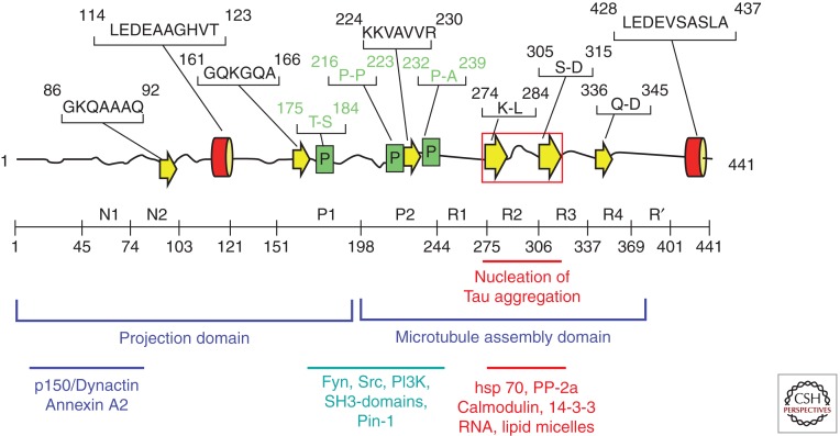
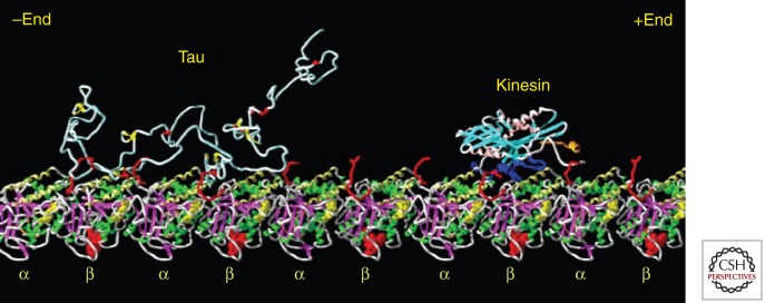
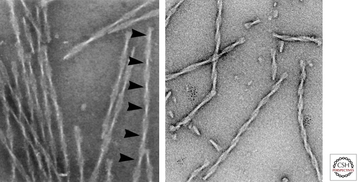
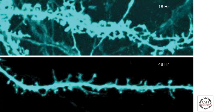
Tau represents the subunit protein of one of the major hallmarks of Alzheimer disease (AD), the neurofibrillary tangles, and is therefore of major interest as an indicator of disease mechanisms. Many of the unusual properties of Tau can be explained by its nature as a natively unfolded protein. Examples are the large number of structural conformations and biochemical modifications (phosphorylation, proteolysis, glycosylation, and others), the multitude of interaction partners (mainly microtubules, but also other cytoskeletal proteins, kinases, and phosphatases, motor proteins, chaperones, and membrane proteins). The pathological aggregation of Tau is counterintuitive, given its high solubility, but can be rationalized by short hydrophobic motifs forming β structures. The aggregation of Tau is toxic in cell and animal models, but can be reversed by suppressing expression or by aggregation inhibitors. This review summarizes some of the structural, biochemical, and cell biological properties of Tau and Tau fibers. Further aspects of Tau as a diagnostic marker and therapeutic target, its involvement in other Tau-based diseases, and its histopathology are covered by other chapters in this volume.


Figures

Figure 1.
Figure 1.
Domains and structural elements in Tau. Top: Representation of Tau deduced from NMR (Mukrasch et al. 2009). Most of the chain is unfolded (black lines), with a few short and transient elements of secondary structure (α-helix red, β-strand yellow, poly-proline helix green). The red box indicates the region of the two hexapeptide motifs responsible for Tau aggregation. Middle: Domain subdivision (following Gustke et al. 1994). The carboxy-terminal half promotes microtubule assembly, and the amino-terminal half projects out from the microtubule surface. N1, N2 and R2 may be absent owing to alternative splicing. R1–R4 represent the repeat domain; together with the flanking domains, this represents the microtubule interaction domain. Bottom: Approximate location of interaction sites with other proteins.
Figure 2.
Figure 2.
Visualization of Tau and kinesin bound to microtubules. The diagram shows a Tau molecule and a kinesin motor domain bound to a microtubule protofilament (row of αβ-tubulin heterodimers). All molecules are in the same size range (∼350–450 residues), but tubulin and kinesin are compactly folded. Tau is not and therefore occupies a much larger volume, loosely filled with polypeptide chain and highly mobile. Structures modeled after Nogales et al. (1999) (tubulin), Sack et al. (1997) (kinesin), and Hoenger et al. (1998) (docking of kinesin on microtubule). Tau is shown as a random coil; its microtubule-bound conformation is not known. (Figure composed by A. Marx.)
Figure 3.
Figure 3.
Tau fibers. Left: Twisted fibers appearing as “paired helical filaments” isolated from Alzheimer brain tissue, with ∼80 nm periodicity (arrowheads). Right: Fibers assembled in vitro from the proaggregant Tau repeat domain (K18ΔK280). Note the similarity of the twisted structures, even though the repeat domain contains only ∼27% of the full-length protein. (Micrographs by E.M. Mandelkow and S. Barghorn.)
Figure 4.
Figure 4.
Dendritic missorting of Tau and synaptic decay. Top: Mature primary rat hippocampal neuron at 21 DIV with numerous dendritic spines, 18 h after transfection with full-length Tau (2N4R, tagged with CFP, blue). Note that Tau has invaded dendritic shafts and spines. Bottom: At 48 h after transfection most spines have shrunk or disappeared. (Adapted from Thies and Mandelkow 2007; reprinted, with permission, from the author.)
Figure 5.
Figure 5.
Loss and recovery of memory in regulatable Tau-transgenic mice. The mice express the proaggregant Tau repeat domain (K18ΔK280). Expression is switched OFF in the presence of doxycyclin, and ON without doxycyclin in the drinking water (tet-off system; Gossen and Bujard 2002), and tested in the Morris Water Maze. Left: Control mouse learns position of hidden platform quickly, short length of swimming path. Center: Mouse after switching the Tau repeat domain ON for 12 months has severe learning deficits, and requires a long time and long path length to find the platform. Right: Same mouse as before, after switching Tau expression OFF again for 4 weeks. The memory has returned to control levels (details in Sydow et al. 2011).

Similar articles

Cited by

References

    1. Aamodt EJ, Williams RC Jr 1984. Microtubule-associated proteins connect microtubules and neurofilaments in vitro. Biochemistry 23: 6023–6031 - PubMed
    1. Ackmann M, Wiech H, Mandelkow E 2000. Nonsaturable binding indicates clustering of tau on the microtubule surface in a paired helical filament-like conformation. J Biol Chem 275: 30335–30343 - PubMed
    1. Aguzzi A, Rajendran L 2009. The transcellular spread of cytosolic amyloids, prions, and prionoids. Neuron 64: 783–790 - PubMed
    1. Ahn JS, Radhakrishnan ML, Mapelli M, Choi S, Tidor B, Cuny GD, Musacchio A, Yeh LA, Kosik KS 2005. Defining Cdk5 ligand chemical space with small molecule inhibitors of tau phosphorylation. Chem Biol 12: 811–823 - PubMed
    1. Al-Bassam J, Ozer RS, Safer D, Halpain S, Milligan RA 2002. MAP2 and tau bind longitudinally along the outer ridges of microtubule protofilaments. J Cell Biol 157: 1187–1196 - PMC - PubMed