. 2012 Oct;123(1):147-58.
doi: 10.1111/j.1471-4159.2012.07859.x. Epub 2012 Aug 14.

Calpain-cleaved type 1 inositol 1,4,5-trisphosphate receptor impairs ER Ca(2+) buffering and causes neurodegeneration in primary cortical neurons

Affiliations

Calpain-cleaved type 1 inositol 1,4,5-trisphosphate receptor impairs ER Ca(2+) buffering and causes neurodegeneration in primary cortical neurons

Catherine M Kopil et al. J Neurochem. 2012 Oct.

Abstract

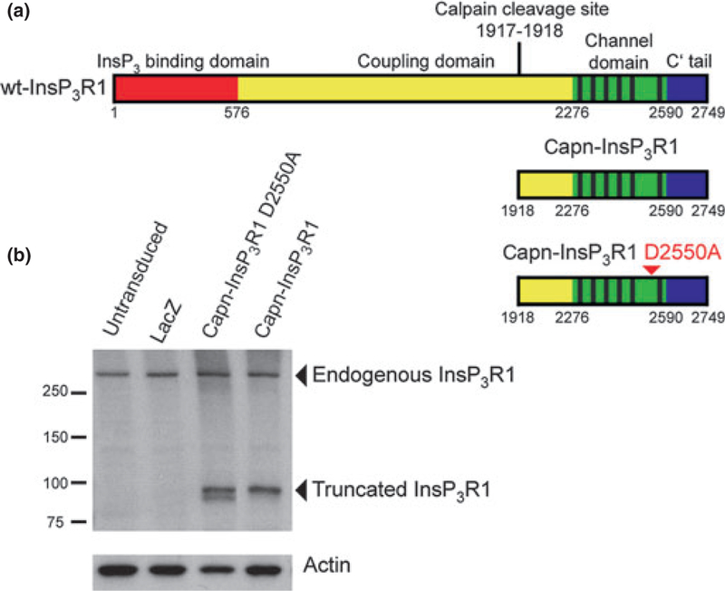
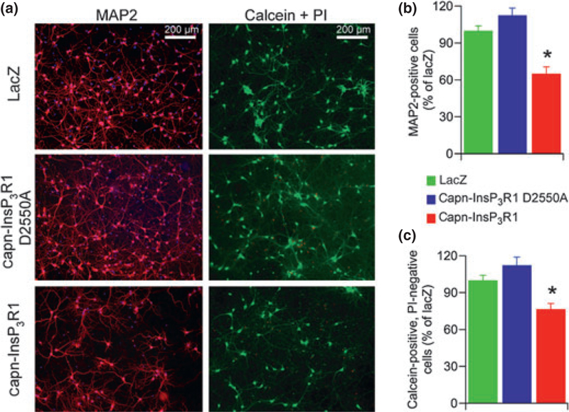
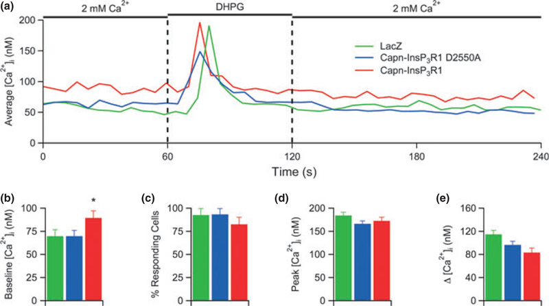
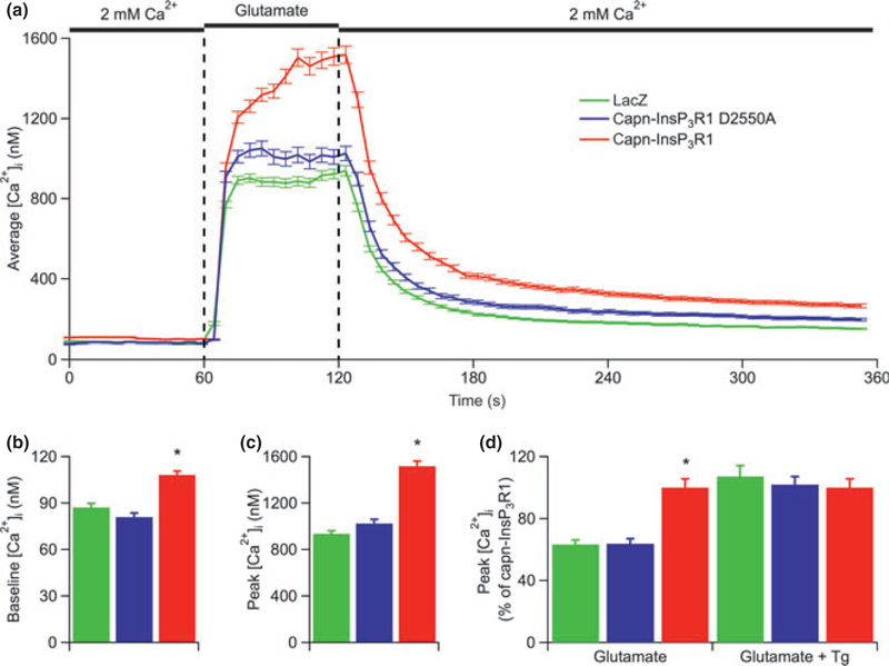
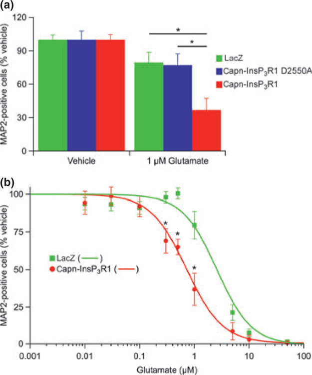
Disruption of neuronal Ca(2+) homeostasis plays a well-established role in cell death in a number of neurodegenerative disorders. Recent evidence suggests that proteolysis of the type 1 inositol 1,4,5-trisphosphate receptor (InsP(3)R1), a Ca(2+) release channel on the endoplasmic reticulum, generates a dysregulated channel, which may contribute to aberrant Ca(2+) signaling and neurodegeneration in disease states. However, the specific effects of InsP(3)R1 proteolysis on neuronal Ca(2+) homeostasis are unknown, as are the functional contributions of this pathway to neuronal death. This study evaluates the consequences of calpain-mediated InsP(3)R1 proteolysis on neuronal Ca(2+) signaling and survival using adeno-associated viruses to express a recombinant cleaved form of the channel (capn-InsP(3)R1) in rat primary cortical neurons. Here, we demonstrate that expression of capn-InsP(3)R1 in cortical cultures reduced cellular viability. This effect was associated with increased resting cytoplasmic Ca(2+) concentration ([Ca(2+)](i)), increased [Ca(2+)](i) response to glutamate, and enhanced sensitivity to excitotoxic stimuli. Together, our results demonstrate that InsP(3)R1 proteolysis disrupts neuronal Ca(2+) homeostasis, and potentially acts as a feed-forward pathway to initiate or execute neuronal death.


Conflict of interest statement

Conflict of interest

The authors declare no conflict of interest.

Figures

Fig. 1
Fig. 1
Schematic representation and expression of recombinant InsP3R1 constructs in primary neurons. (a) Protein domain structure of wt-InsP3R1 (top), calpain-cleaved InsP3R1 recombinant (middle), and calpain-cleaved InsP3R1 with the D2550A point mutation (bottom; arrowhead). Residues numbered according to rat type 1 SI+, SII+, SIII-sequence (protein accession NP_001007236.1). (b) Western blot analysis of whole-cell lysates from untransduced rat primary cortical cultures and primary cortical cultures transduced with AAV 2/1 expressing lacZ, capn-InsP3R1 D2550A, or capn-InsP3R1 at 1 week post-transduction (14 DIV). Carboxyl-terminal InsP3R1 antibody was used to detect endogenous and recombinant truncated rat InsP3R1. Antibody against actin was used as a loading control.
Fig. 2
Fig. 2
Primary cortical neuron viability is reduced by expression of capn-InsP3R1. Rat primary cortical cultures were transduced with adeno-associated virus 2/1 expressing lacZ, capn-InsP3R1 D2550A, or capn-InsP3R1 (7 DIV). (a) One week following transduction (14 DIV), cultures were stained for microtubule-associated protein 2 (MAP2) (red) and nuclei (blue; left panel), or incubated with calcein-AM (green) and propidium iodide (PI) (red; right panel). Representative 100· epifluorescence images captured for counting are shown. (b) Percent neuronal survival as determined by counts of MAP2-reactive cells (one-way ANOVA; *p < 0.001). (c) Percent cell survival as determined by counts of calcein-AM positive, PI-negative cells (unpaired t-tests with unequal variance; *p < 0.001). Expression of capn-InsP3R1 resulted in a significant decrease in percent viable neurons compared with lacZ and capn-InsP3R1 D2550A as determined by both assays.
Fig. 3
Fig. 3
Capn-InsP3R1 does not deplete InsP3-sensitive stores. (a) Representative single cell [Ca2+]i responses to (S)-3,5-dihydroxyphenylglycine (DHPG) in Fura-2-loaded primary cortical neurons (14 DIV) transduced with lacZ, capn-InsP3R1 D2550A, or capn-InsP3R1. Total number of single-cell Ca2+ responses analyzed in these experiments was 219, 246, and 172 for lacZ-, capn-InsP3R1 D2550A-, and capn-InsP3R1-transduced cultures, respectively. (b) Summary of average resting [Ca2+]i in neurons from cultures used for DHPG Ca2+ imaging experiments. Cultures expressing capn-InsP3R1 demonstrate increased baseline [Ca2+]i (unpaired t-tests with unequal variance; *p < 0.001). (c) Percentage of cells in transduced cortical cultures that exhibited > 10% increase in [Ca2+]i in response to DHPG. (d) Summary of average peak [Ca2+]i responses elicited by DHPG. (e) Summary of average change in [Ca2+]i from resting to peak [Ca2+]i. Despite significant differences in resting [Ca2+]i in these experiments, no statistical differences were observed in average change in [Ca2+]i.
Fig. 4
Fig. 4
Neurons expressing capn-InsP3R1 have increased glutamate-induced rises in [Ca2+]i. (a) Averaged single-cell [Ca2+]i responses to glutamate in Fura-2-loaded primary cortical neurons (14 DIV) transduced with lacZ, capn-InsP3R1 D2550A, or capn-InsP3R1. Total number of single-cell Ca2+ responses analyzed in these experiments was 491, 271, and 396 for lacZ-, capn-InsP3R1 D2550A-, and capn-InsP3R1-transduced cultures, respectively. (b) Summary of average resting [Ca2+]i in neurons from cultures used for glutamate Ca2+ imaging experiments. Expression of capn-InsP3R1 significantly increased resting [Ca2+]i in neurons (108.2 ± 2.6 nM) compared with capn-InsP3R1 D2550A (81.0 ± 2.5 nM) and lacZ controls(87.3 ± 2.6 nM; unpaired t-tests with unequal variance; *p < 0.001).(c) Summary of average peak [Ca2+]i responses elicited by glutamate shows an increased maximum [Ca2+]i achieved in capn-InsP3R1-expressing cells (unpaired t-tests with unequal variance; *p < 0.001).(d) Summary of average peak [Ca2+]i elicited by glutamate in the absence or presence of Tg (normalized data for glutamate alone (left) are the same as absolute data shown Fig. 4c). Total number of single-cell Ca2+ responses analyzed in experiments with Tg was 182, 209, and 146 for lacZ-, capn-InsP3R1 D2550A-, and capn-InsP3R1-transduced cultures, respectively. Tg eliminated the capn-InsP3R1-associated glutamate-induced enhanced peak [Ca2+]i (lacZ, 1,357 ± 66 nM; capn-InsP3R1 D2550a, 1,276 ± 44 nM; capn-InsP3R1, 1,288 ± 54 nM).
Fig. 5
Fig. 5
Expression of capn-InsP3R1 increases neuronal susceptibility to glutamate-mediated injury. (a) Transduced primary cortical cultures (14 DIV) were exposed to 1 μM glutamate or HEPES-buffered saline vehicle. Twenty-four hours later, cultures were stained for microtubule-associated protein 2 (MAP2) and percent neuronal survival quantified relative to vehicle-treated cultures. Expression of capn-InsP3R1 resulted in a significant decrease in percentage of MAP2-positive cells following glutamate injury compared with lacZ and capn-InsP3R1 D2550A (one-way ANOVA; *p < 0.01). (b) Glutamate dose-response curves for lacZ and capn-InsP3R1-transduced cultures (data at 1 μM glutamate are the same as shown in Fig. 5a). Expression of capn-InsP3R1 significantly decreased percent MAP2-positive cells following treatment with 0.3, 0.5, and 1 lM glutamate compared with lacZ (unpaired t tests; *p < 0.01) and resulted in a shift in the glutamate dose-response curve. LD50 for glutamate in neurons expressing capn-InsP3R1 (0.75 ± 0.19 μM) is reduced compared with neurons expressing lacZ (2.61 ± 0.78 μM).

Similar articles

Cited by

References

    1. Alzayady KJ and Wojcikiewicz RJ (2005) The role of Ca2+ in triggering inositol 1,4,5-trisphosphate receptor ubiquitination. Biochem. J 392, 601–606. - PMC - PubMed
    1. Assefa Z, Bultynck G, Szlufcik K, Nadif Kasri N, Vermassen E, Goris J, Missiaen L, Callewaert G, Parys JB and De Smedt H (2004) Caspase-3-induced truncation of type 1 inositol trisphosphate receptor accelerates apoptotic cell death and induces inositol trisphosphate-independent calcium release during apoptosis.J. Biol. Chem 279, 43227–43236. - PubMed
    1. Bardo S, Cavazzini MG and Emptage N (2006) The role of the endoplasmic reticulum Ca2+ store in the plasticity of central neurons. Trends Pharmacol. Sci 27, 78–84. - PubMed
    1. Berridge MJ, Lipp P and Bootman MD (2000) The versatility and universality of calcium signalling. Nat. Rev. Mol. Cell Biol 1, 11–21. - PubMed
    1. Berridge MJ, Bootman MD and Roderick HL (2003) Calcium signalling: dynamics, homeostasis and remodelling. Nat. Rev. Mol. Cell Biol 4, 517–529. - PubMed

Publication types

MeSH terms