. 2013 Aug;23(8):1874-83.
doi: 10.1093/cercor/bhs175. Epub 2012 Jul 4.

Dissecting the anticipation of aversion reveals dissociable neural networks

Affiliations

Dissecting the anticipation of aversion reveals dissociable neural networks

Daniel W Grupe et al. Cereb Cortex. 2013 Aug.

Abstract

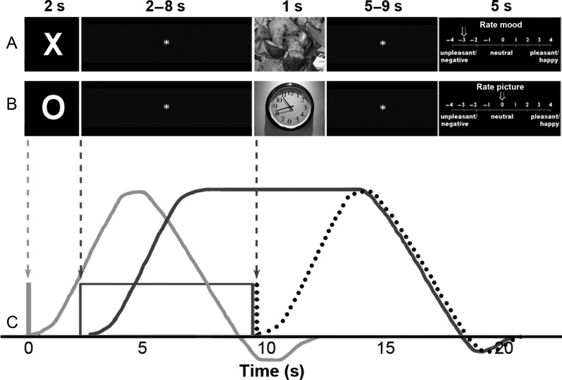
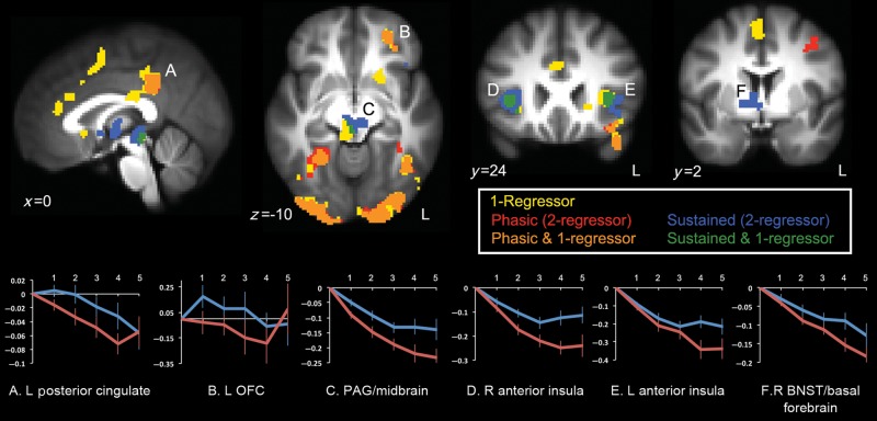
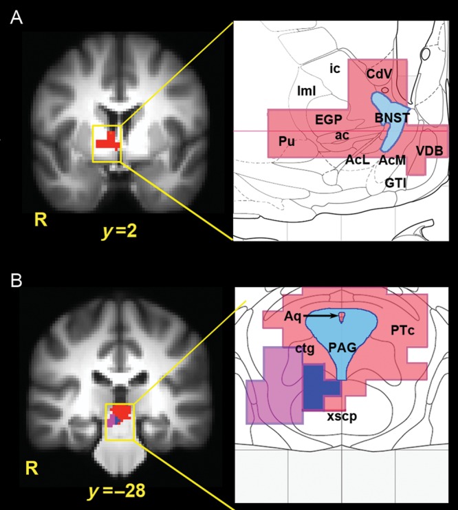
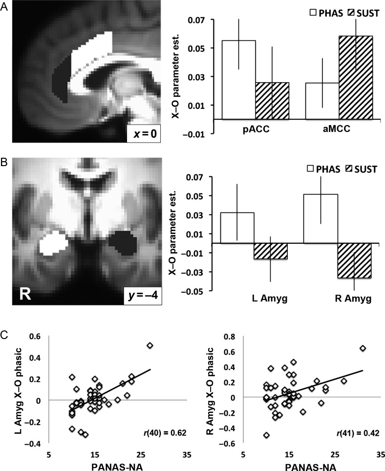
The anticipation of future adversity confers adaptive benefits by engaging a suite of preparatory mechanisms, but this process can also be deleterious when carried out in excess. Neuroscientific investigations have largely treated anticipation as a unitary process, but we show here using functional magnetic resonance imaging that distinct stages of aversive anticipation are supported by dissociable neural mechanisms. Immediate anticipatory responses were observed in regions associated with threat detection and early processing of predictive cues, including the orbitofrontal cortex and pregenual anterior cingulate cortex, as well as the amygdala for individuals with elevated anxiety symptoms. Sustained anticipatory activity was observed in the forebrain/bed nucleus of the stria terminalis, anterior insula, anterior mid-cingulate cortex (aMCC), and midbrain/periaqueductal gray, regions associated with anxiety, interoception, and defensive behavior. The aMCC showed increased functional coupling with the midbrain during sustained anticipation of aversion, highlighting a circuit critical for the expression of preparatory fear responses. These data implicate distinct sets of regions that are active during different temporal stages of anticipation, and provide insight into how the human brain faces the future both adaptively and maladaptively.

Keywords: BNST; amygdala; anterior insula; fMRI; rostral cingulate.


Figures

Figure 1.
Figure 1.
Schematic of the paradigm presented to subjects during fMRI scanning. (A) On aversive trials, subjects viewed an X cue for 2 s, followed by a 2- to 8-s ISI and an aversive picture for 1 s. Following a 5- to 9-s ISI, subjects rated either their mood or the picture valence. (B) Neutral trials had an identical structure to aversive trials, with an O cue preceding a neutral picture. (C) Modeling of the BOLD signal during anticipation included 2 distinct regressors, each of which was convolved with a standard hemodynamic response function: An event regressor at the onset of the anticipatory cue (light gray line and curve), and a boxcar regressor lasting the duration of the anticipatory ISI (dark gray box and curve). Responses during the picture period were modeled using an event regressor (dotted line and curve). The x-axis shows time (in seconds) from the trial onset.
Figure 2.
Figure 2.
(Top panel) Anticipatory activation for the 1-regressor model (yellow), and phasic (red) and sustained anticipatory activation (blue) for the 2-regressor model at P < 0.05 (corrected using Monte Carlo simulation). Overlap between 1-regressor and phasic clusters is shown in orange; overlap between 1-regressor and sustained clusters is shown in green. Activation of the posterior cingulate (A) and the left OFC (B) in the 1-regressor model was attributed to the phasic regressor in the 2-regressor model. Activation of the midbrain/PAG (C) and bilateral anterior insula (D/E) in the 1-regressor model was attributed to the sustained regressor in the 2-regressor model. Sustained activity was observed in the right basal forebrain including the BNST (F), whereas no activity was observed in this region for the 1-regressor model. At the corrected threshold, neither regressor in the 2-regressor model independently accounted for the rostral cingulate activation in the 1-regressor model (Fig. 4A and Supplementary Fig. S1). (Bottom panel) Time course data extracted from clusters showing significant phasic (A,B) and sustained (C–F) activity. The x-axis reflects 2-s volumes from the cue onset and the y-axis reflects percent signal change from the baseline. The error bars (standard errors of the mean) increase in magnitude for later time points, which contained fewer averages.
Figure 3.
Figure 3.
(A) Activation of the right basal forebrain during sustained anticipation of aversion, as overlaid on an anatomical atlas (Mai et al. 1998). At P < 0.05 (corrected using Monte Carlo simulation), this activation included the dorsal and central BNST (shown in blue), as well as the external globus pallidus (EGP), ventral caudate (CdV), putamen (Pu), vertical limb of the diagonal band (VDB), great terminal island (GTI), and dorsal/posterior portions of the nucleus accumbens (AcL = lateral accumbens core; AcM = medial accumbens shell). ac, anterior commissure; ic, internal capsule, lml, external medullary lamina of the globus pallidus. (B) Sustained anticipation of aversion was accompanied by increased midbrain activity (red) overlapping with the anatomical location of the PAG (blue) and pretectal area (PTc) when overlaid on an anatomical atlas (Mai et al. 1998). PPI analysis revealed an overlapping midbrain cluster (purple) showing increased functional coupling with the anterior mid-cingulate cortex during sustained anticipation. All activations shown at P < 0.05 (corrected using Monte Carlo simulation). Aq, cerebral aqueduct; ctg, central tegmental tract; xscp, decussation of the cerebellar peduncle. Adapted from Mai et al. 1998.
Figure 4.
Figure 4.
ROI analyses for the anatomically defined rostral cingulate cortex and amygdala. (A) The pregenual anterior cingulate cortex (pACC) showed phasic (PHAS) anticipatory activation, while the anterior mid-cingulate cortex (aMCC) showed sustained (SUST) anticipatory activation. (B) The amygdala (AMYG) showed marginally greater phasic relative to sustained anticipatory activation. For (A) and (B), error bars show standard error of the mean. (C) Scatter plots reflecting significant correlations between phasic amygdala activation and negative affect (NA) scores on the Positive and Negative Affect Schedule (PANAS). One regression outlier (i.e., studentized residual with Bonferonni corrected P < 0.05) was removed from the left amygdala correlation, which thus includes only 42 data points.

References

    1. Alvarez RP, Chen G, Bodurka J, Kaplan R, Grillon C. Phasic and sustained fear in humans elicits distinct patterns of brain activity. Neuroimage. 2011;55:389–400. - PMC - PubMed
    1. An X, Bandler R, Ongur D, Price JL. Prefrontal cortical projections to longitudinal columns in the midbrain periaqueductal gray in macaque monkeys. J Comp Neurol. 1998;401:455–479. - PubMed
    1. Bandler R, Keay KA, Floyd N, Price J. Central circuits mediating patterned autonomic activity during active vs. passive emotional coping. Brain Res Bull. 2000;53:95–104. - PubMed
    1. Barlow DH. Unraveling the mysteries of anxiety and its disorders from the perspective of emotion theory. Am Psychol. 2000;55:1247–1263. - PubMed
    1. Birbaumer N, Elbert T, Canavan AGM, Rockstroh B. Slow potentials of the cerebral cortex and behavior. Physiol Rev. 1990;70:1–41. - PubMed

Publication types