

Delivery of PDGF-B and BMP-7 by mesoporous bioglass/silk fibrin scaffolds for the repair of osteoporotic defects
- PMID: 22763224
- PMCID: PMC5995476
- DOI: 10.1016/j.biomaterials.2012.06.021
Delivery of PDGF-B and BMP-7 by mesoporous bioglass/silk fibrin scaffolds for the repair of osteoporotic defects
Abstract
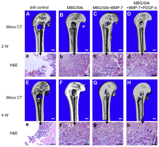
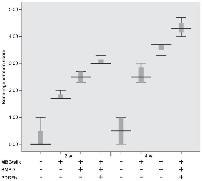
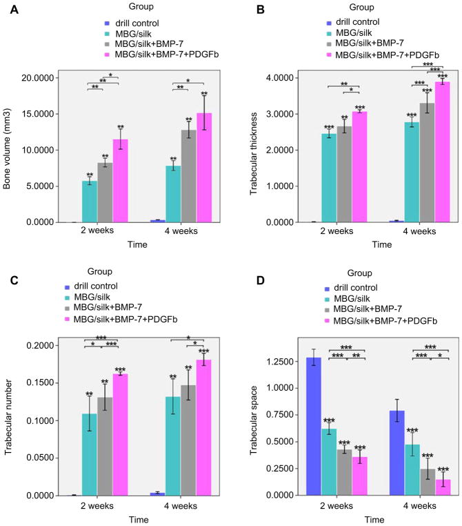
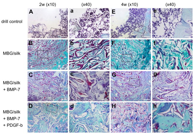
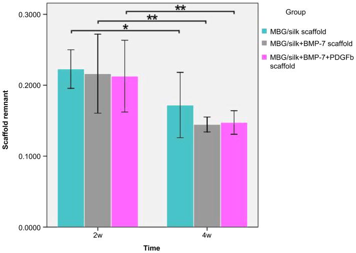
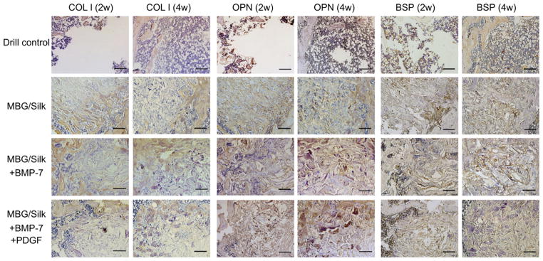
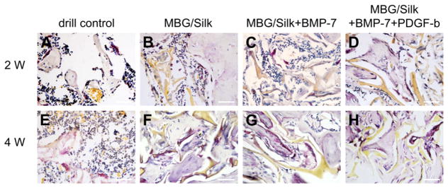
Osteoporosis is a chronic disease affecting millions of people worldwide caused by an imbalance between bone-forming osteoblasts and bone-resorbing osteoclasts. Despite recent developments in pharmacological agents to prevent osteoporotic-related fractures, much less attention has been placed on the repair of bone defects following fracture. Critical to this process is the recruitment of mesenchymal stem cells (MSCs) to defect sites by growth factors. One method which has been effective for the sustained release of growth factors is that of gene therapy. The aim of the present study was to investigate newly developed mesoporous bioglass/silk fibrin scaffolds containing adPDGF-b and adBMP-7 into osteoporotic critical-sized femur defects in ovariectomised rats following treatment periods of 2 and 4 weeks. In vivo osteogenetic efficiency evaluated by μ-CT analysis, hematoxylin and eosin staining, and immunohistochemical (type I collagen, osteopontin and BSP) revealed significantly new bone formation in defects containing adenovirus for both PDGF-b and BMP-7 when compared to scaffolds alone and scaffolds containing BMP-7. TRAP-positive staining also demonstrated the ability for these scaffolds to be degraded over time and initiate bone turnover/remodeling. Although the use of gene therapy for clinical applications is still in its infancy, results from the present study demonstrate their potent ability to recruit mesenchymal progenitor cells through sustained release of PDGF-b and BMP-7 which may be beneficial for patients suffering from osteoporotic-related fractures.
Copyright © 2012 Elsevier Ltd. All rights reserved.
Figures
Similar articles
-
Novel MesoPorous BioGlass/silk scaffold containing adPDGF-B and adBMP7 for the repair of periodontal defects in beagle dogs.J Clin Periodontol. 2015 Mar;42(3):262-71. doi: 10.1111/jcpe.12364. Epub 2015 Feb 16. J Clin Periodontol. 2015. PMID: 25580515
-
In vitro and in vivo evaluation of adenovirus combined silk fibroin scaffolds for bone morphogenetic protein-7 gene delivery.Tissue Eng Part C Methods. 2011 Aug;17(8):789-97. doi: 10.1089/ten.tec.2010.0453. Epub 2011 Apr 20. Tissue Eng Part C Methods. 2011. PMID: 21506685
-
Porous CaP/silk composite scaffolds to repair femur defects in an osteoporotic model.J Mater Sci Mater Med. 2013 Aug;24(8):1963-75. doi: 10.1007/s10856-013-4945-y. Epub 2013 May 15. J Mater Sci Mater Med. 2013. PMID: 23674058 Free PMC article.
-
Gene therapy approaches for bone regeneration.Cells Tissues Organs. 2004;176(1-3):95-108. doi: 10.1159/000075031. Cells Tissues Organs. 2004. PMID: 14745239 Free PMC article. Review.
-
Clinical considerations of regenerative medicine in osteoporosis.Curr Osteoporos Rep. 2014 Jun;12(2):230-4. doi: 10.1007/s11914-014-0201-8. Curr Osteoporos Rep. 2014. PMID: 24619559 Review.
Cited by
-
[Mesenchymal stem cells modified with Runt-related transcription factor 2 promote bone regeneration in rabbit mandibular distraction osteogenesis].Hua Xi Kou Qiang Yi Xue Za Zhi. 2016 Apr;34(2):125-9. doi: 10.7518/hxkq.2016.02.004. Hua Xi Kou Qiang Yi Xue Za Zhi. 2016. PMID: 27337919 Free PMC article. Chinese.
-
Bone union formation in the rat mandibular symphysis using hydroxyapatite with or without simvastatin: effects on healthy, diabetic, and osteoporotic rats.Clin Oral Investig. 2020 Apr;24(4):1479-1491. doi: 10.1007/s00784-019-03180-9. Epub 2020 Jan 11. Clin Oral Investig. 2020. PMID: 31925587
-
The osteogenic potential of mesoporous bioglasses/silk and non-mesoporous bioglasses/silk scaffolds in ovariectomized rats: in vitro and in vivo evaluation.PLoS One. 2013 Nov 12;8(11):e81014. doi: 10.1371/journal.pone.0081014. eCollection 2013. PLoS One. 2013. PMID: 24265840 Free PMC article.
-
HnRNPL inhibits the osteogenic differentiation of PDLCs stimulated by SrCl2 through repressing Setd2.J Cell Mol Med. 2019 Apr;23(4):2667-2677. doi: 10.1111/jcmm.14166. Epub 2019 Feb 12. J Cell Mol Med. 2019. PMID: 30746871 Free PMC article.
-
Osteoporosis: the current status of mesenchymal stem cell-based therapy.Cell Mol Biol Lett. 2016 Aug 12;21:12. doi: 10.1186/s11658-016-0013-1. eCollection 2016. Cell Mol Biol Lett. 2016. PMID: 28536615 Free PMC article. Review.
References
-
- Genant HK, Cooper C, Poor G, Reid I, Ehrlich G, Kanis J, et al. Interim report and recommendations of the World Health Organization Task-Force for osteoporosis. Osteoporos Int. 1999;10(4):259–64. - PubMed
-
- Rodan GA, Martin TJ. Therapeutic approaches to bone diseases. Science. 2000;289:1508–14. - PubMed
-
- Roush K. Prevention and treatment of osteoporosis in postmenopausal women: a review. Am J Nurs. 2011;111:26–35. - PubMed
Publication types
MeSH terms
Substances
Grants and funding
LinkOut - more resources
Full Text Sources
Medical
Research Materials
