

Estradiol selectively reduces central neural activation induced by hypertonic NaCl infusion in ovariectomized rats
- PMID: 22763321
- PMCID: PMC3590917
- DOI: 10.1016/j.physbeh.2012.06.015
Estradiol selectively reduces central neural activation induced by hypertonic NaCl infusion in ovariectomized rats
Abstract
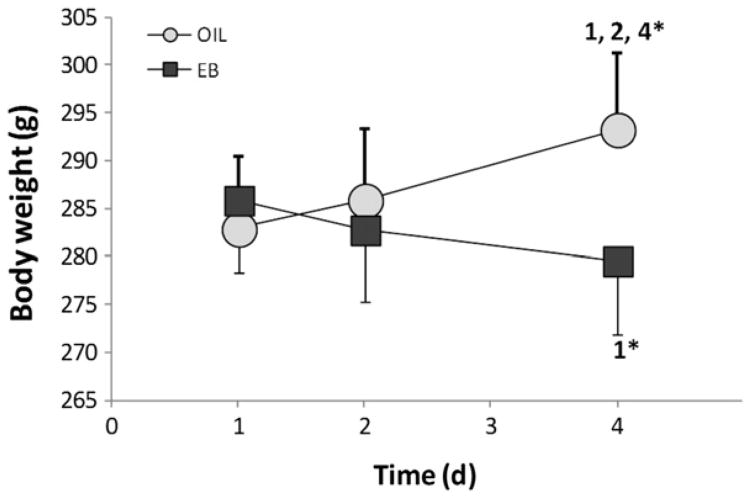
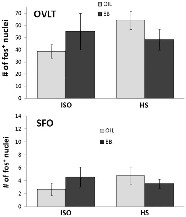
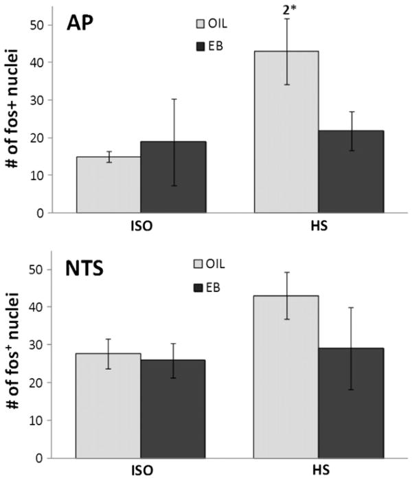
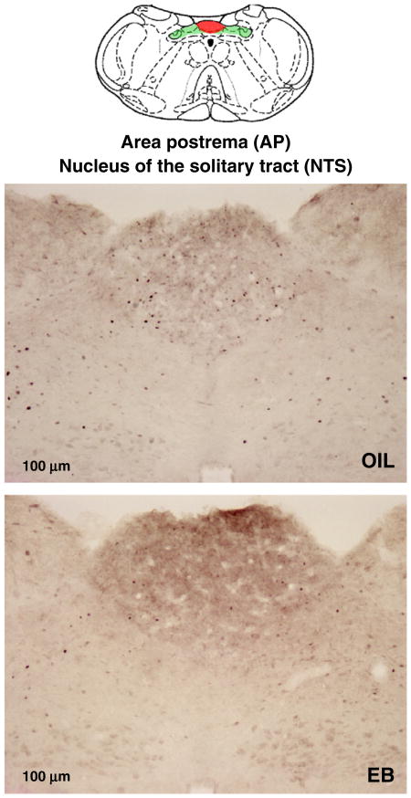
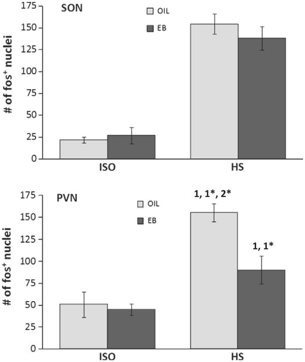
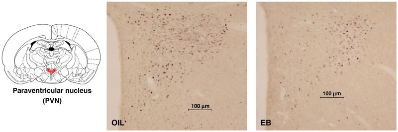
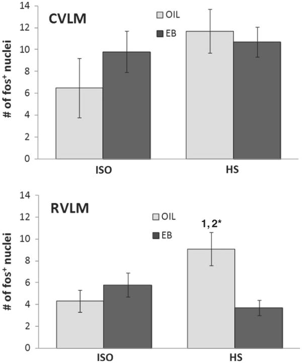
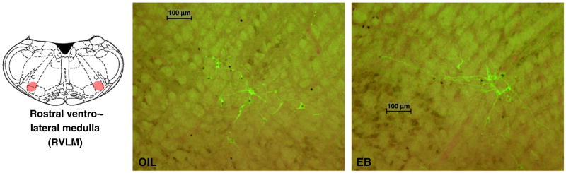
We recently reported that the latency to begin drinking water during slow, intravenous infusion of a concentrated NaCl solution was shorter in estradiol-treated ovariectomized rats compared to oil vehicle-treated rats, despite comparably elevated plasma osmolality. To test the hypothesis that the decreased latency to begin drinking is attributable to enhanced detection of increased plasma osmolality by osmoreceptors located in the CNS, the present study used immunocytochemical methods to label fos, a marker of neural activation. Increased plasma osmolality did not activate the subfornical organ (SFO), organum vasculosum of the lamina terminalis (OVLT), or the nucleus of the solitary tract (NTS) in either oil vehicle-treated rats or estradiol-treated rats. In contrast, hyperosmolality increased fos labeling in the area postrema (AP), the paraventricular nucleus of the hypothalamus (PVN) and the rostral ventrolateral medulla (RVLM) in both groups; however, the increase was blunted in estradiol-treated rats. These results suggest that estradiol has selective effects on the sensitivity of a population of osmo-/Na(+)-receptors located in the AP, which, in turn, alters activity in other central areas associated with responses to increased osmolality. In conjunction with previous reports that hyperosmolality increases blood pressure and that elevated blood pressure inhibits drinking, the current findings of reduced activation in AP, PVN, and RVLM-areas involved in sympathetic nerve activity-raise the possibility that estradiol blunts HS-induced blood pressure changes. Thus, estradiol may eliminate or reduce the initial inhibition of water intake that occurs during increased osmolality, and facilitate a more rapid behavioral response, as we observed in our recent study.
Copyright © 2012 Elsevier Inc. All rights reserved.
Figures
Similar articles
-
Subfornical organ disconnection alters Fos expression in the lamina terminalis, supraoptic nucleus, and area postrema after intragastric hypertonic NaCl.Am J Physiol Regul Integr Comp Physiol. 2005 Apr;288(4):R947-55. doi: 10.1152/ajpregu.00570.2004. Epub 2004 Dec 2. Am J Physiol Regul Integr Comp Physiol. 2005. PMID: 15576664
-
Regional differences in the expression of Fos-like immunoreactivity after central salt loading in conscious rats: modulation by endogenous vasopressin and role of the area postrema.Brain Res. 2004 Oct 1;1022(1-2):182-94. doi: 10.1016/j.brainres.2004.02.082. Brain Res. 2004. PMID: 15353228
-
Intragastric hypertonic saline increases vasopressin and central Fos immunoreactivity in conscious rats.Am J Physiol. 1997 Mar;272(3 Pt 2):R750-8. doi: 10.1152/ajpregu.1997.272.3.R750. Am J Physiol. 1997. PMID: 9087636
-
Neurochemical Circuits Subserving Fluid Balance and Baroreflex: A Role for Serotonin, Oxytocin, and Gonadal Steroids.In: De Luca LA Jr, Menani JV, Johnson AK, editors. Neurobiology of Body Fluid Homeostasis: Transduction and Integration. Boca Raton (FL): CRC Press/Taylor & Francis; 2014. Chapter 9. In: De Luca LA Jr, Menani JV, Johnson AK, editors. Neurobiology of Body Fluid Homeostasis: Transduction and Integration. Boca Raton (FL): CRC Press/Taylor & Francis; 2014. Chapter 9. PMID: 24829993 Free Books & Documents. Review.
-
Physiological actions of angiotensin II mediated by AT1 and AT2 receptors in the brain.Clin Exp Pharmacol Physiol Suppl. 1996;3:S99-104. Clin Exp Pharmacol Physiol Suppl. 1996. PMID: 8993847 Review.
Cited by
-
Bidirectional effects of estradiol on the control of water intake in female rats.Horm Behav. 2021 Jul;133:104996. doi: 10.1016/j.yhbeh.2021.104996. Epub 2021 May 18. Horm Behav. 2021. PMID: 34020111 Free PMC article.
-
Control of fluid intake by estrogens in the female rat: role of the hypothalamus.Front Syst Neurosci. 2015 Mar 4;9:25. doi: 10.3389/fnsys.2015.00025. eCollection 2015. Front Syst Neurosci. 2015. PMID: 25788879 Free PMC article. Review.
-
Estradiol and osmolality: Behavioral responses and central pathways.Physiol Behav. 2015 Dec 1;152(Pt B):422-30. doi: 10.1016/j.physbeh.2015.06.017. Epub 2015 Jun 12. Physiol Behav. 2015. PMID: 26074202 Free PMC article. Review.
-
Aging affects isoproterenol-induced water drinking, astrocyte density, and central neuronal activation in female Brown Norway rats.Physiol Behav. 2018 Aug 1;192:90-97. doi: 10.1016/j.physbeh.2018.03.005. Epub 2018 Mar 5. Physiol Behav. 2018. PMID: 29518407 Free PMC article.
-
Estrogen Replacement Reduces Oxidative Stress in the Rostral Ventrolateral Medulla of Ovariectomized Rats.Oxid Med Cell Longev. 2016;2016:2158971. doi: 10.1155/2016/2158971. Epub 2015 Nov 10. Oxid Med Cell Longev. 2016. PMID: 26640612 Free PMC article.
References
-
- Andersen LJ, Jensen TU, Bestle MH, Bie P. Gastrointestinal osmoreceptors and renal sodium excretion in humans. Am J Physiol Regul Integr Comp Physiol. 2000;278:R287–94. - PubMed
-
- Barron WM, Schreiber J, Lindheimer MD. Effect of ovarian sex steroids on osmoregulation and vasopressin secretion in the rat. Am J Physiol. 1986;250:E352–61. - PubMed
-
- Bealer SL. Central control of cardiac baroreflex responses during peripheral hyperosmolality. Am J Physiol Regul Integr Comp Physiol. 2000;278:R1157–63. - PubMed
-
- Bisley JW, Rees SM, McKinley MJ, Hards DK, Oldfield BJ. Identification of osmoresponsive neurons in the forebrain of the rat: a Fos study at the ultrastructural level. Brain Res. 1996;720:25–34. - PubMed
-
- Bossmar T, Forsling M, Akerlund M. Circulating oxytocin and vasopressin is influenced by ovarian steroid replacement in women. Acta Obstet Gynecol Scand. 1995;74:544–8. - PubMed
Publication types
MeSH terms
Substances
Grants and funding
LinkOut - more resources
Full Text Sources
