. 2013 Feb;27(2):430-40.
doi: 10.1038/leu.2012.183. Epub 2012 Jul 5.

The epoxyketone-based proteasome inhibitors carfilzomib and orally bioavailable oprozomib have anti-resorptive and bone-anabolic activity in addition to anti-myeloma effects

Affiliations

The epoxyketone-based proteasome inhibitors carfilzomib and orally bioavailable oprozomib have anti-resorptive and bone-anabolic activity in addition to anti-myeloma effects

M A Hurchla et al. Leukemia. 2013 Feb.

Abstract

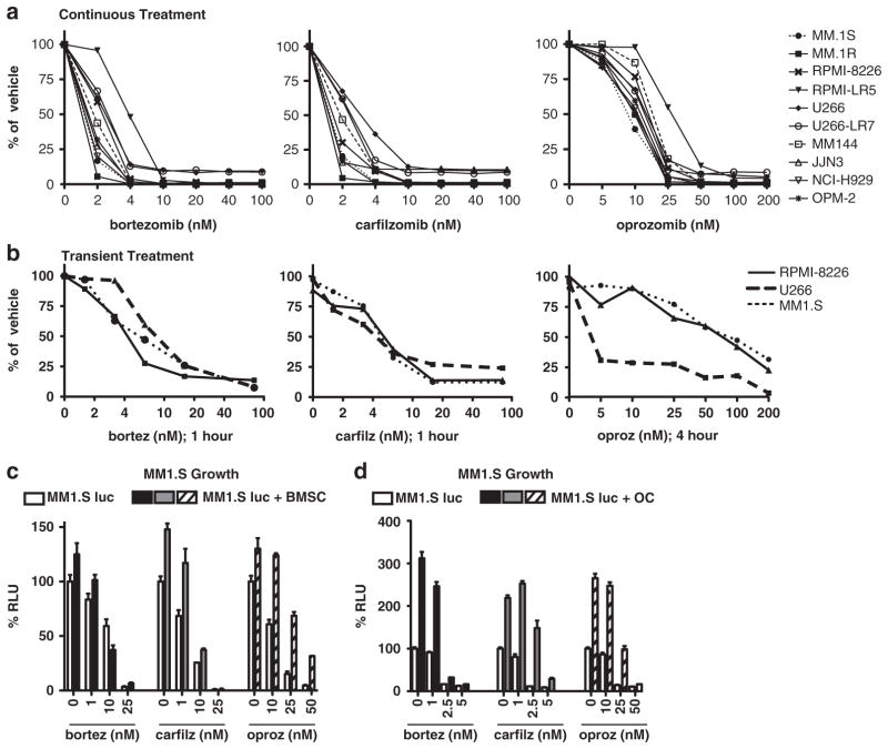
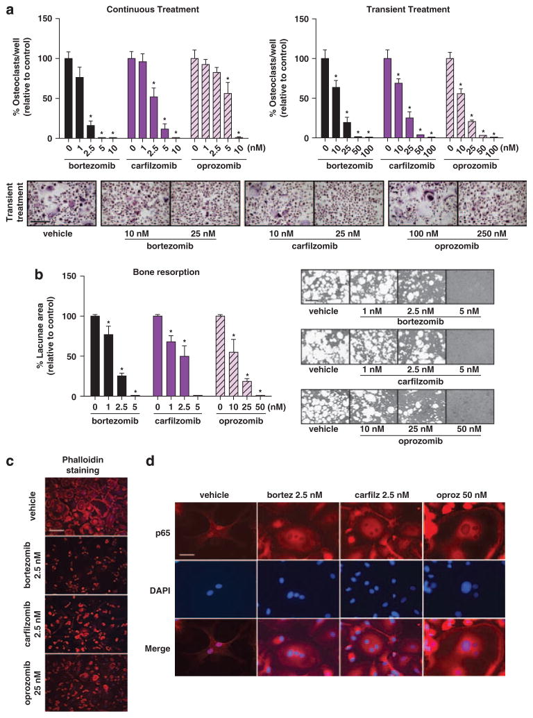
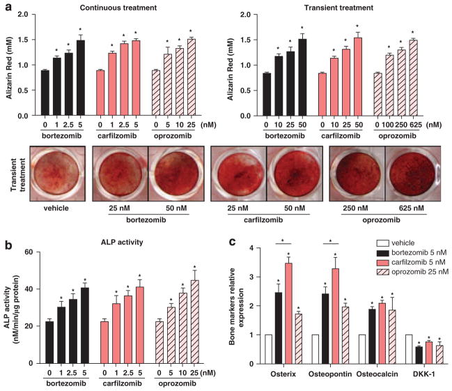
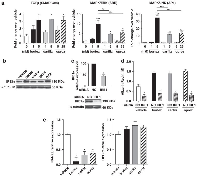
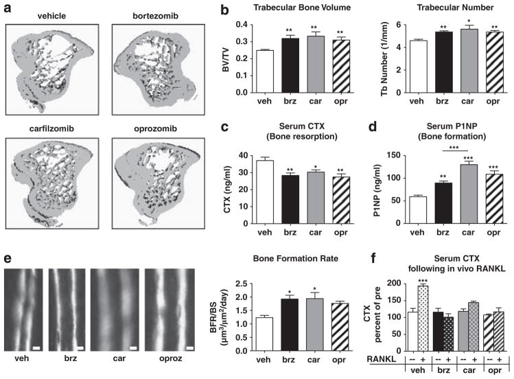
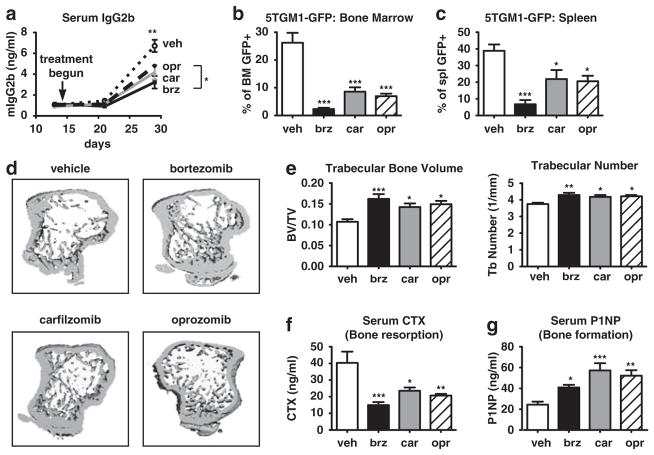
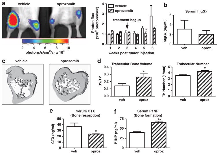
Proteasome inhibitors (PIs), namely bortezomib, have become a cornerstone therapy for multiple myeloma (MM), potently reducing tumor burden and inhibiting pathologic bone destruction. In clinical trials, carfilzomib, a next generation epoxyketone-based irreversible PI, has exhibited potent anti-myeloma efficacy and decreased side effects compared with bortezomib. Carfilzomib and its orally bioavailable analog oprozomib, effectively decreased MM cell viability following continual or transient treatment mimicking in vivo pharmacokinetics. Interactions between myeloma cells and the bone marrow (BM) microenvironment augment the number and activity of bone-resorbing osteoclasts (OCs) while inhibiting bone-forming osteoblasts (OBs), resulting in increased tumor growth and osteolytic lesions. At clinically relevant concentrations, carfilzomib and oprozomib directly inhibited OC formation and bone resorption in vitro, while enhancing osteogenic differentiation and matrix mineralization. Accordingly, carfilzomib and oprozomib increased trabecular bone volume, decreased bone resorption and enhanced bone formation in non-tumor bearing mice. Finally, in mouse models of disseminated MM, the epoxyketone-based PIs decreased murine 5TGM1 and human RPMI-8226 tumor burden and prevented bone loss. These data demonstrate that, in addition to anti-myeloma properties, carfilzomib and oprozomib effectively shift the bone microenvironment from a catabolic to an anabolic state and, similar to bortezomib, may decrease skeletal complications of MM.


Conflict of interest statement

CONFLICT OF INTEREST

CJK is an employee of Onyx Pharmaceuticals. All other authors declare no conflict of interest.

Figures

Figure 1
Figure 1
Continuous or physiologic transient administration of carfilzomib or oprozomib is cytotoxic to human MM cells in vitro. (a) A panel of 10/human MM cell lines were treated with the indicated doses of bortezomib, carfilzomib or oprozomib continuously for 48 h and subjected to MTT assay for viability. (b) MM cell lines were treated with indicated drug doses on day 0 for 1 h (bortezomib, carfilzomib) or 4 h (oprozomib). Cells were then washed and cultured in drug-free media for 48 additional h and viability assessed by MTT assay. (c, d) PIs overcome the proliferative and protective effects of bone microenvironment cells. MM.1S cells labeled with firefly luciferase (MM.1S-luc) were cultured in the presence (filled bars) or absence (open bars) of human (c) CD138 BMSCs or (d) OCs. Cultures were treated with indicated doses of drugs for 48 h (BMSC) or 5 days (OC) and MM cell viability readout by luciferase activity. Results are expressed as mean±s.d. RLU, relative luminescence units.
Figure 2
Figure 2
Oprozomib and carfilzomib inhibit OC differentiation and function in vitro. (a) Human OCs were generated from PBMCs cultured in osteoclastogenic medium for 21 days in the presence or absence of indicated concentrations of the PIs (continuous treatment, left panel); alternatively, PIs were present only for the initial 4 h of differentiation (transient treatment, right panel). OCs were identified as multinucleated (≥3 nuclei) TRAP + cells. Representative micrographs of differentiated OCs after transient treatment are shown. (b) To assess inhibition of mineralized matrix resorption, PBMCs were seeded on calcium-coated slides and maintained in osteoclastogenic medium for 17 days with or without indicated concentrations of the drugs. Graphs represent mean values of samples from OCs derived from three healthy donors±s.d. *P<0.05 between treated cultures and vehicle control. Bar =50 μm. (c) PIs disrupted the integrity of the actin ring in multinucleated pre-OCs (14 days in osteoclastogenic medium under continuous PI-treatment). Actin =phalloidin–rhodamine, DAPI =nuclei; representative micrographs are reported. Bar =50 μm. (d) Pre-OCs were treated with indicated concentrations of PIs for 3 h prior to stimulation with RANKL for 30 min. In absence of PIs (vehicle), RANKL induces p65 translocation to the nucleus, whereas in PI-treated cells the p65 subunit of NF-κB is retained in the cytoplasm. p65 subunit =visualized in red, DAPI =nuclei; representative micrographs for each PI are shown (Bar =12.5 μm).
Figure 3
Figure 3
PIs promote osteogenic differentiation and mineralization in vitro. (a) Primary MSCs from MM patients (6/9 with osteolytic lesions) were cultured in osteogenic medium either in the continuous presence of PIs (left panel) or transiently treated on day 0 for 4 h (right panel). Mineralization was analyzed by alizarin red staining and subsequent dye quantification in OBs differentiated for 21 days; representative images of alizarin red staining following transient treatment are shown. Results are expressed as the mean±s.d. (b) In the presence of PIs and at day 11 of osteogenic differentiation, ALP activity was measured in OBs derived from MSCs from five MM patients (3/5 with osteolytic lesions). Graphs illustrate mean values±s.e.m. (c) Total RNA from the hMSC-TERT cell line was isolated at day 14 of differentiation in the presence of PIs at indicated doses, and expression of osteogenic-related markers (Osterix, osteopontin, osteocalcin) and DKK-1 were evaluated using real-time reverse transcription-PCR. Expression levels for each gene were normalized with respect to GAPDH expression and referred to vehicle control. Data are represented as the mean±s.d. from three different experiments. In all panels: *P<0.05, versus vehicle control or between indicated groups.
Figure 4
Figure 4
PI treatment diminishes RANKL expression in OBs and promotes osteogenic differentiation and function through activation of the transforming growth factor β (TGFβ), MAPK and UPR pathways. (a) Reporter assays demonstrated that the activity of the Smad2/3/4, MAPK/ERK (serum response element; SRE) and MAPK/JNK (AP1) pathways were increased in MC3T3-E1 OB-progenitor cells following 24 h of continuous treatment with PIs; results are expressed as mean±s.e.m. (b) Western blot for the IRE1α component of the UPR in hMSC-TERT cells treated with PIs for 24 h (25 nM bortezomib and carfilzomib, 250 nM oprozomib). Brefeldin A (600 ng/ml) was used as a positive control. (c) Expression of IRE1α was reduced at the mRNA and protein levels 48 h after transfection with IRE1 targeting siRNAs. (d) The hMSC-TERT cell line was maintained for 14 days in osteogenic medium with PIs (5 nM bortezomib/carfilzomib or 25 nM oprozomib) and transfected 3 times/week with IRE1 targeting or non-targeting (NC) siRNAs. Mineralization was greatly reduced when IRE1α was silenced even in the presence of PIs. (e) The hMSC-TERT cell line was maintained in osteogenic medium for 21 days in the presence of PIs and expression of RANKL and osteoprotegerin was assessed by real-time reverse transcription-PCR. Maximal effect on the relative expression of RANKL (day 14) or osteoprotegerin (day 7) is shown; results are expressed as mean±s.d. In all panels: *P<0.05, **P<0.01, ***P<0.001 versus vehicle or between indicated groups.
Figure 5
Figure 5
Epoxyketone-based PIs exert bone anabolic effects on non-tumor bearing mice. C57Bl/6 mice (n =10/group) were treated for 2 weeks with vehicle, bortezomib, carfilzomib or oprozomib on dosing schedules outlined in Materials and Methods. (a, b) MicroCT analyses show that all PIs induced an equivalent increase in trabecular bone volume and number. (c) Bone resorption (serum carboxy-terminal telopeptide collagen crosslinks (serum CTX)) was significantly and equivalently decreased with each PI. (d) OB function (serum N-terminal propeptide of type I procollagen (serum P1NP)) was significantly increased in all PI-treated animals compared with vehicle-treated. Notably, P1NP levels in carfilzomib-treated mice were significantly greater than those of bortezomib-treated mice. (e) Trabecular bone formation rate was measured by double calcein labeling; calcein incorporates into actively mineralizing bone with the distance between labels being proportional to the amount of newly formed bone within the 5-day interlabel period. PI treatment increased the bone formation rate per bone surface (BFR/BS) as assessed by dynamic histomorphometry (Bar =10 μm). (f) PIs inhibited RANKL-induced pathological bone resorption in the absence of tumor. After 2 weeks of PI treatment as above, mice were given three doses of purified RANKL (n =5/drug) or PBS vehicle (n =5) to stimulate OC activity. Serum CTX measured 90 min following the final RANKL dose is expressed as a percent of the same mouse prior to RANKL stimulation. All results are expressed as mean±s.e.m. *P<0.05, **P<0.01, ***P<0.001 versus vehicle or between indicated groups.
Figure 6
Figure 6
PIs decrease myeloma burden and associated bone destruction in an immunocompetent murine model. 5TGM1-GFP murine myeloma cells were injected into syngeneic C57Bl/KaLwRij mice. After 14 days, mice were randomized into PI treatment groups (n≥7/group) and dosed for an additional 2 weeks. All PIs decreased tumor burden as measured by (a) levels of serum IgG2b (clonotype of 5TGM1 cells) and (b, c) percent of GFP + tumor cells within the BM or spleen upon sacrifice at day 28. (d, e) MicroCT analysis demonstrated that treatment with PIs protected animals from myeloma-induced loss of trabecular bone volume and number. (f) Serum carboxy-terminal telopeptide collagen crosslinks (serum CTX) was significantly decreased and (g) serum N-terminal propeptide of type I procollagen (serum P1NP) was significantly increased in all PI-treated animals compared with vehicle-treated animals. All results are expressed as mean±s.e.m. *P<0.05, **P<0.01, ***P<0.001 versus vehicle or between indicated groups.
Figure 7
Figure 7
Oprozomib decreases human MM tumor burden and protects mice from bone destruction. Immunocompromised NOD-SCID-IL2Rγ−/− mice (n =5/group) were intravenously injected with RMPI-8226 human MM cells stably labeled with firefly luciferase. Tumors were allowed to establish for 3 weeks after which mice were randomized into treatment groups. During weeks 3–6 animals were treated with oprozomib (n =5) or vehicle (n =5). (a) Tumor burden was monitored weekly by in vivo bioluminescence imaging. Mice treated with oprozomib had decreased tumor burden compared with those in the vehicle treated group. Representative image of hind limb tumor burden as visualized on week 6. (b) Serum human Igλ (secreted by RPMI-8226 cells) was decreased in oprozomib-treated mice at the time of killing, indicating decreased tumor burden. (c, d) Although tumor-associated bone loss was evident in vehicle-treated mice, trabecular bone was preserved with oprozomib treatment as measured by micro CT. (c) Representative 3D reconstructions. (d) Oprozomib significantly increased trabecular bone volume and number. Oprozomib treatment decreased serum carboxy-terminal telopeptide collagen crosslinks (serum CTX) (e) and increased serum N-terminal propeptide of type I procollagen (serum P1NP) (f). Results are expressed as mean±s.e.m. *P<0.05, **P<0.01.

Similar articles

Cited by

References

    1. Basak GW, Srivastava AS, Malhotra R, Carrier E. Multiple myeloma bone marrow niche. Curr Pharm Biotechnol. 2009;10:345–346. - PubMed
    1. Esteve FR, Roodman GD. Pathophysiology of myeloma bone disease. Best Pract Res Clin Haematol. 2007;20:613–624. - PubMed
    1. Yaccoby S. Osteoblastogenesis and tumor growth in myeloma. Leuk Lymphoma. 2010;51:213–220. - PMC - PubMed
    1. Kumar SK, Rajkumar SV, Dispenzieri A, Lacy MQ, Hayman SR, Buadi FK, et al. Improved survival in multiple myeloma and the impact of novel therapies. Blood. 2008;111:2516–2520. - PMC - PubMed
    1. Kyle RA, Rajkumar SV. Multiple myeloma. Blood. 2008;111:2962–2972. - PMC - PubMed

Publication types

MeSH terms