. 2012 Jul 5;487(7405):64-9.
doi: 10.1038/nature11220.

Biophysical mechanism of T-cell receptor triggering in a reconstituted system

Affiliations

Biophysical mechanism of T-cell receptor triggering in a reconstituted system

John R James et al. Nature. .

Abstract

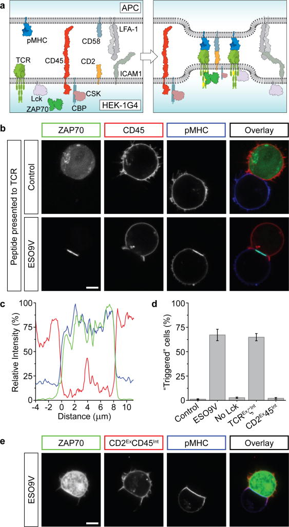
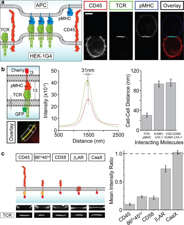
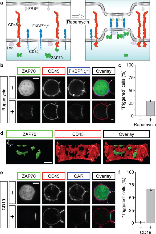
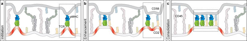
A T-cell-mediated immune response is initiated by the T-cell receptor (TCR) interacting with peptide-bound major histocompatibility complex (pMHC) on an infected cell. The mechanism by which this interaction triggers intracellular phosphorylation of the TCR, which lacks a kinase domain, remains poorly understood. Here, we have introduced the TCR and associated signalling molecules into a non-immune cell and reconstituted ligand-specific signalling when these cells are conjugated with antigen-presenting cells. We show that signalling requires the differential segregation of a phosphatase and kinase in the plasma membrane. An artificial, chemically controlled receptor system generates the same effect as TCR–pMHC, demonstrating that the binding energy of an extracellular protein–protein interaction can drive the spatial segregation of membrane proteins without a transmembrane conformational change. This general mechanism may extend to other receptors that rely on extrinsic kinases, including, as we demonstrate, chimaeric antigen receptors being developed for cancer immunotherapy.


Figures

Figure 1
Figure 1. Regulatable TCR triggering in an engineered HEK cell line
a, Schematic representation of molecules transfected into HEK cells. b, Western blot of phosphorylated proteins after transfection of HEK cells with selected molecules (green circles). c, Cells transfected with the TCR, Lck and ZAP70, were transfected with additional molecules (green circles), showing the synergistic action of modulatory proteins CSK/CBP and CD45 to restrain Lck. The decreased phosphorylation between lanes 5 and 7 is due to endogenous CSK recruitment. d, Confocal images of HEK-1G4 showing ZAP70-GFP recruitment to the plasma membrane in the presence of Lck. e, Quantitation of ZAP70 relocalisation with indicated molecules transfected in HEK-1G4 cells. Data presented as mean ± sem of 3 independent experiments (~300 cells per experiment). f, Addition of 100 μM pervanadate to HEK-1G4 cells (expressing components in lane 8 of c) caused the accumulation of membrane-localised ZAP70. Scale bars, 5 μm.
Figure 2
Figure 2. The exclusion of CD45 phosphatase is necessary and sufficient for TCR triggering
a, Proteins expressed in HEK-1G4 for cell conjugation. b, HEK-1G4, expressing all components shown in a, were conjugated with APC (Raji cells) expressing cognate pMHC (ESO9V), or a control pMHC. Coloured boxes denote protein representation in the overlay image. c, A representative line profile of membrane fluorescence from CD45 (red), pMHC (blue) and ZAP70 (green) at the conjugate interface. d, Quantitation of triggering (defined as unambiguous recruitment of ZAP70 to conjugate region) for all conjugates described in text. Data is presented as the mean ± sem of independent experiments (n=4 or 5, 30-150 conjugates per experiment). e, Forcing CD45 into conjugate region by fusing to CD2 (CD2ExCD45Int) blocks TCR triggering (ZAP70 remains cytosolic). Quantitation is shown in d. Scale bars, 5 μm.
Figure 3
Figure 3. The TCR-pMHC interaction drives protein exclusion at conjugate regions
a, A schematic and representative image dataset showing the TCR-pMHC interaction is sufficient to drive CD45 exclusion and its own clustering (scale bar, 5 μm). b, The intermembrane distance between the conjugates (see Supplementary Methods) was measured, shown schematically as the separation between the two fluorophores over a normal line (white line) averaged across the conjugate region (dotted box). This procedure was performed for the cognate TCR/pMHC interaction (n=20 cells), LFA-1/ICAM1 (n=23 cells) and CD2/CD58+LFA-1/ICAM1; n=20 cells) interactions in the presence of control pMHC, with mean ± sem shown. c, HEK cells were transfected with TCR-GFP and indicated molecule (fused to mCherry) and conjugated with APCs (CD45 phosphatase domains shown in orange). Representative images of conjugate region are shown, with quantitation of the ratio of fluorescence inside and outside of the interface. Data presented as mean ± sem (n=20) for each construct.
Figure 4
Figure 4. Artificial receptor systems can cause CD45 exclusion and triggering
a, Schematic of the chemically-inducible receptor system. FRB fused to a transmembrane segment (FRBEx) replaces pMHC on the APC and FKBPExζInt (fusion of FKBP, CD86 and intracellular CD3ζsignalling motifs) replaces the TCR. Rapamycin induces FKBPExζInt-FRBEx interaction. Additional molecules have been omitted for clarity. b. Rapamycin addition causes ZAP70 accumulation, denoting receptor triggering (gamma correction applied to lower images). FRBEx-expressing cells shown by dotted lines (scale bar 5 μm). c, Quantitation of rapamycin-induced triggering (mean ± sem over four experiments). d, A deconvolved 3D-rendering of the reconstituted cell interface shown in b (scale bar 2 μm). e, Reconstituted HEK cells with the TCR replaced by a chimaeric antigen receptor (CAR) specific for CD19 (see text) were conjugated with either CD19- (Jurkat) or CD19+ (Raji) cells, marked by dotted lines (scale bar 5 μm). f, Quantitation of CAR-mediated triggering (mean ± sem over three experiments).
Figure 5
Figure 5. A model for steps in TCR-mediated segregation based on membrane bending and energy minimisation
The schematic uses equivalent molecule representations as previous figures (only extracellular domains are shown for simplicity), and boxed regions highlight features of each panel. a, After initial adhesion driven by large receptors such as LFA-1, transient fluctuations in the intermembrane distance permit encounters between TCR and pMHC whose binding interaction overcomes energetically unfavourable membrane bending (red regions). b, Additional molecules (such as CD2/CD58) provide additional binding energy that stabilise regions of local membrane bending and may enhance the local exclusion of larger molecules by TCR-pMHC interactions. c, Consolidation of discrete contact regions serves to minimise unfavourable membrane bending and leads to the exclusion of smaller proteins that do not provide any countering ligand-binding free energy. See text for details.

References

    1. Smith-Garvin JE, Koretzky GA, Jordan MS. T cell activation. Annu Rev Immunol. 2009;27:591–619. - PMC - PubMed
    1. Lemmon MA, Schlessinger J. Cell signaling by receptor tyrosine kinases. Cell. 2010;141:1117–1134. - PMC - PubMed
    1. Palacios EH, Weiss A. Function of the Src-family kinases, Lck and Fyn, in T-cell development and activation. Oncogene. 2004;23:7990–8000. - PubMed
    1. Love PE, Hayes SM. ITAM-mediated signaling by the T-cell antigen receptor. Cold Spring Harb Perspect Biol. 2010;2:a002485. - PMC - PubMed
    1. Au-Yeung BB, et al. The structure, regulation, and function of ZAP-70. Immunol Rev. 2009;228:41–57. - PubMed

Publication types

MeSH terms

Substances