. 2012 Jul 26;487(7408):514-8.
doi: 10.1038/nature11182.

Structural insights into electron transfer in caa3-type cytochrome oxidase

Affiliations

Structural insights into electron transfer in caa3-type cytochrome oxidase

Joseph A Lyons et al. Nature. .

Abstract

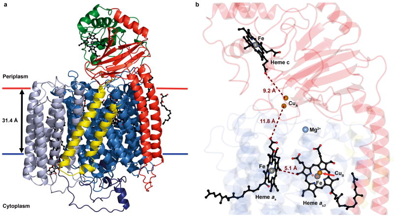
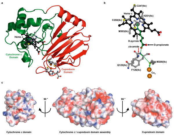
Cytochrome c oxidase is a member of the haem copper oxidase superfamily (HCO). HCOs function as the terminal enzymes in the respiratory chain of mitochondria and aerobic prokaryotes, coupling molecular oxygen reduction to transmembrane proton pumping. Integral to the enzyme's function is the transfer of electrons from cytochrome c to the oxidase via a transient association of the two proteins. Electron entry and exit are proposed to occur from the same site on cytochrome c. Here we report the crystal structure of the caa3-type cytochrome oxidase from Thermus thermophilus, which has a covalently tethered cytochrome c domain. Crystals were grown in a bicontinuous mesophase using a synthetic short-chain monoacylglycerol as the hosting lipid. From the electron density map, at 2.36 Å resolution, a novel integral membrane subunit and a native glycoglycerophospholipid embedded in the complex were identified. Contrary to previous electron transfer mechanisms observed for soluble cytochrome c, the structure reveals the architecture of the electron transfer complex for the fused cupredoxin/cytochrome c domain, which implicates different sites on cytochrome c for electron entry and exit. Support for an alternative to the classical proton gate characteristic of this HCO class is presented.


Figures

Figure 1
Figure 1. Structure of and cofactor arrangement in caa3-oxidase
a, Structure (ribbon model). SU I/III is colored to highlight its canonical subunits: SU I, blue; SU III, blue-grey and the fusion linker, dark blue. SU IIc is colored to highlight the classical SU II (red) and the fused cytochrome c domain (green). SU IV is in yellow. The hemes are in ball and stick with the iron and copper metal centers as grey and copper spheres, respectively. Membrane boundaries are based on hydrophobic thickness calculations from the OPM server. b, Cofactor arrangement. Hemes c, as and as3, and the iron and copper ions are shown as in a. The magnesium ion is represented as a light blue sphere. Distances are shown in brown. SU I/III, IIc and IV are color coded as faded blue, red and yellow ribbons, respectively.
Figure 2
Figure 2. Active site, water pool and oxygen channel in caa3-oxidase
a, b, The active site of heme as3 viewed from (i) the oxygen entry side and (ii) the periplasm. In a is shown a 2mFo-DFc electron density map contoured to 1σ (blue) and an anomalous difference map contoured to 5σ (red). In b is shown an mFo-DFc electron density difference map calculated without the bridging ligands contoured to 4σ (green). For clarity, the anomalous map is not shown in a(ii). c, Water pool. A water filled cavity centered on a magnesium ion is situated above heme as3. Coordinate bonds are shown as dashed black lines. d, The calculated oxygen channel is Y-shaped and hydrophobic. Two extremities of the channel (red) contact the apolar surface of the protein (black arrows); a third contacts the binuclear centre.
Figure 3
Figure 3. Proton pathways in caa3-oxidase
a, The D-pathway begins at Asp 103 and leads through a solvent filled cavity to Tyr 248. Alternative conformations for the Asp 103 side chain were modeled into electron density and are shown. The conformer pointing towards Asn 110 represents the conformation observed in other known oxidases. b, Detailed view of the YS gate, the structural equivalent in caa3-oxidase of the classical Type A1 gating glutamate. c, The K-pathway originates at Glu 84 of SU IIc and continues up to the binuclear centre via the cross-linked His 250 - Tyr 254, by way of Lys 328. Hydrogen bonds are shown as dashed black lines in a–c. The known covalent linkage between His 250 and Tyr 254 was modeled based on separate biochemical characterization.
Figure 4
Figure 4. Cytochrome c/cupredoxin complex and electron transfer pathway in caa3-oxidase
a, Structure of the cytochrome c (green) and cupredoxin (red) domains of SU IIc. b, The optimum electron transfer pathway between the heme c iron and the CuA center was calculated with HARLEM. Through bond and through space tunneling are shown in solid and dashed green lines, respectively. c, “Open book” view of surface complementarities between the cytochrome c and cupredoxin domains in caa3-oxidase. Electrostatic potentials are viewed at ±10 kbT. The linker (residues 217-235) between the two domains has been removed for clarity. Surfaces are at 25 % transparency to reveal the positions of heme c and the CuA centre shown in black ball and stick and copper spheres, respectively.

References

    1. Ferguson-Miller S, Babcock GT. Heme/copper terminal oxidases. Chem Rev. 1996;96:2889–2908. - PubMed
    1. Muresanu L, et al. The electron transfer complex between cytochrome c552 and the CuA domain of the Thermus thermophilus ba3 oxidase. A combined NMR and computational approach. J Biol Chem. 2006;281:14503–14513. - PubMed
    1. Maneg O, Ludwig B, Malatesta F. Different interaction modes of two cytochrome-c oxidase soluble CuA fragments with their substrates. J Biol Chem. 2003;278:46734–46740. - PubMed
    1. Maneg O, Malatesta F, Ludwig B, Drosou V. Interaction of cytochrome c with cytochrome oxidase: two different docking scenarios. Biochim Biophys Acta. 2004;1655:274–281. - PubMed
    1. Pereira MM, Santana M, Teixeira M. A novel scenario for the evolution of haem-copper oxygen reductases. Biochim Biophys Acta. 2001;1505:185–208. - PubMed

Publication types

MeSH terms

Associated data