. 2012 Jul 4;487(7405):109-13.
doi: 10.1038/nature11130.

Reduced airway surface pH impairs bacterial killing in the porcine cystic fibrosis lung

Affiliations

Reduced airway surface pH impairs bacterial killing in the porcine cystic fibrosis lung

Alejandro A Pezzulo et al. Nature. .

Abstract

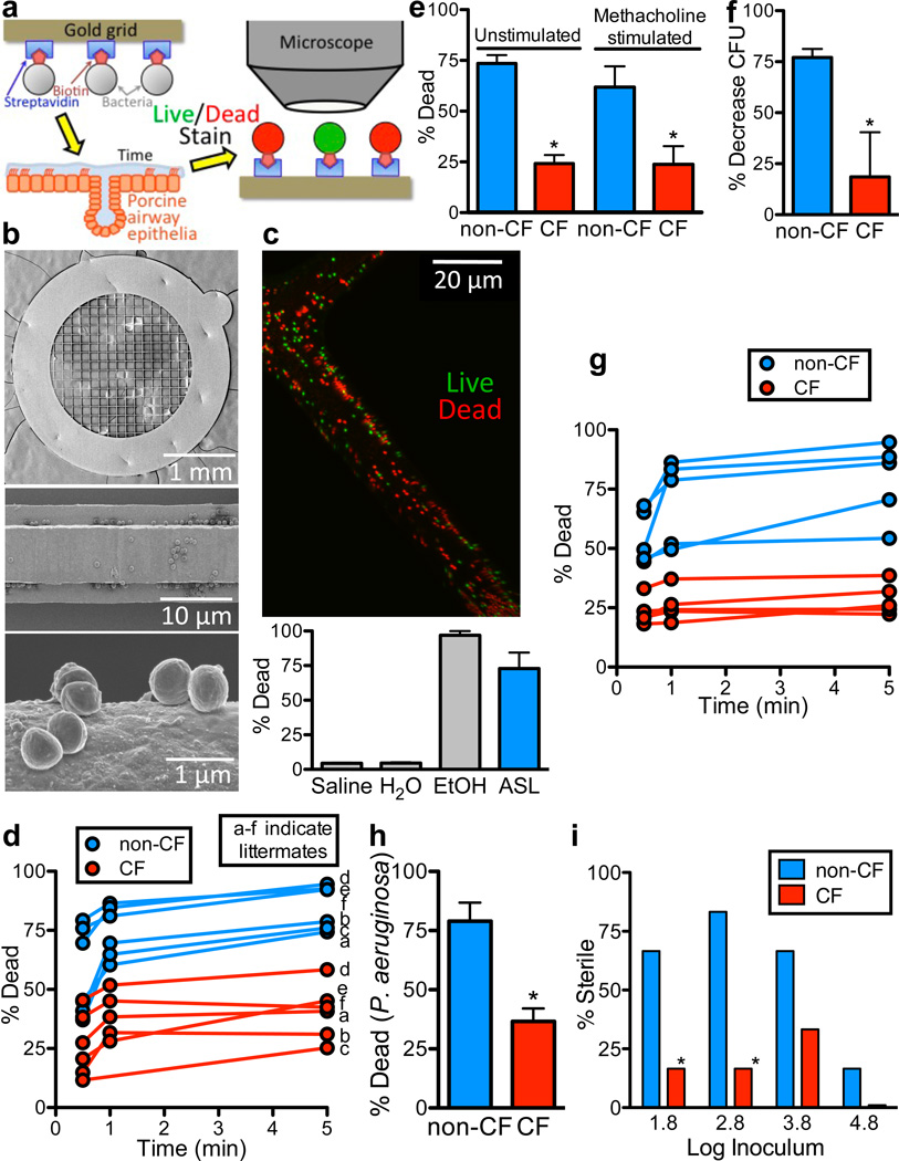
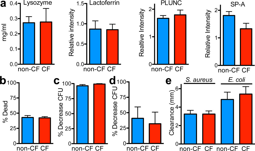
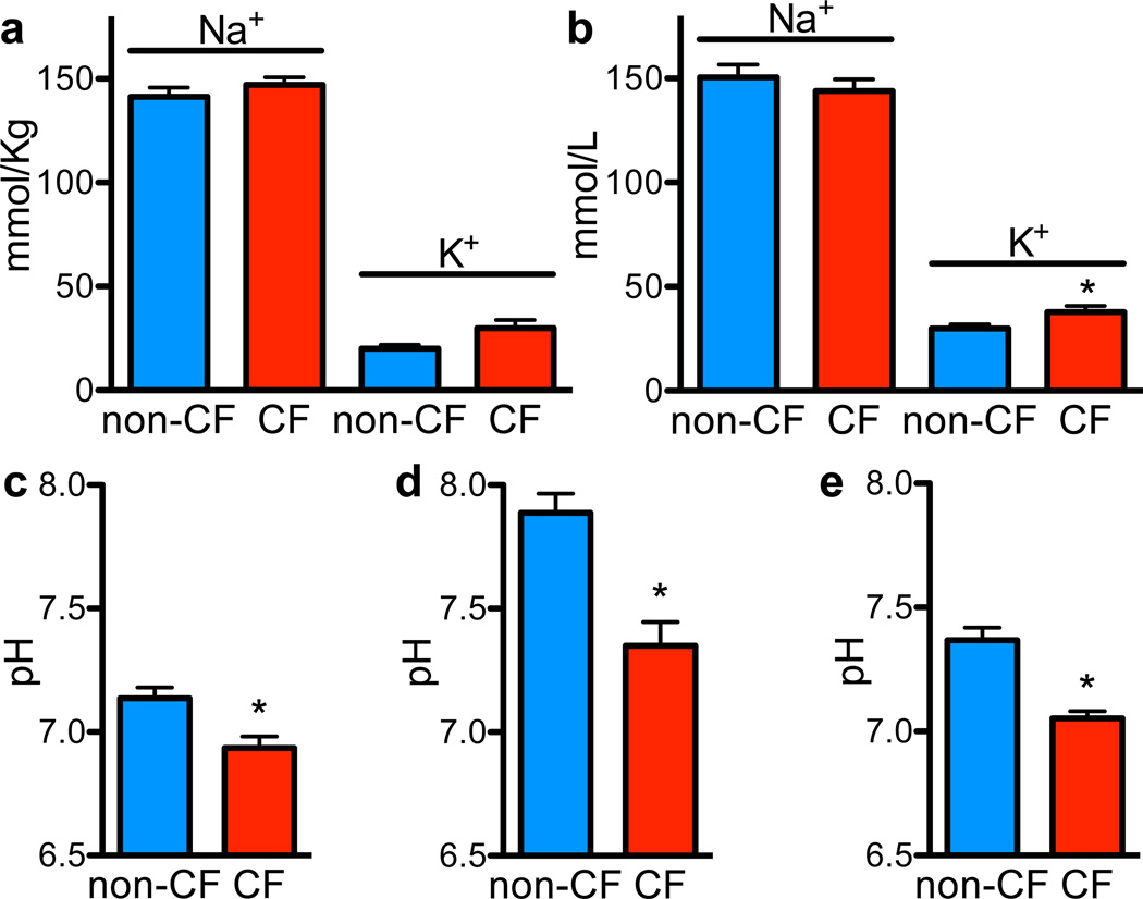
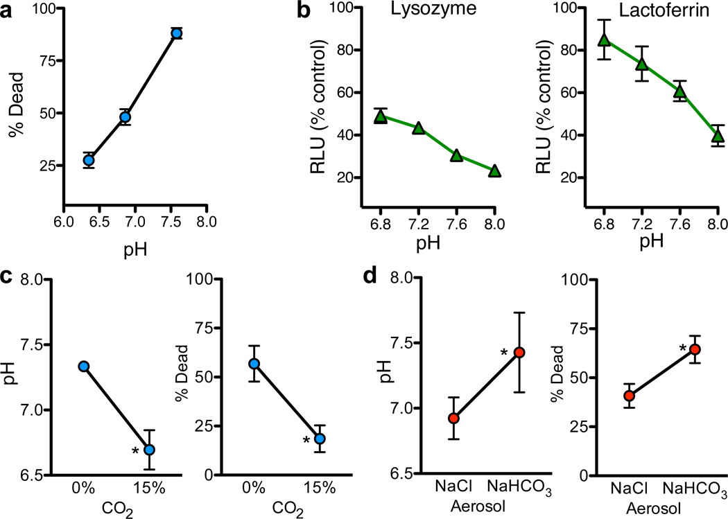
Cystic fibrosis (CF) is a life-shortening disease caused by mutations in the cystic fibrosis transmembrane conductance regulator (CFTR) gene. Although bacterial lung infection and the resulting inflammation cause most of the morbidity and mortality, how the loss of CFTR function first disrupts airway host defence has remained uncertain. To investigate the abnormalities that impair elimination when a bacterium lands on the pristine surface of a newborn CF airway, we interrogated the viability of individual bacteria immobilized on solid grids and placed onto the airway surface. As a model, we studied CF pigs, which spontaneously develop hallmark features of CF lung disease. At birth, their lungs lack infection and inflammation, but have a reduced ability to eradicate bacteria. Here we show that in newborn wild-type pigs, the thin layer of airway surface liquid (ASL) rapidly kills bacteria in vivo, when removed from the lung and in primary epithelial cultures. Lack of CFTR reduces bacterial killing. We found that the ASL pH was more acidic in CF pigs, and reducing pH inhibited the antimicrobial activity of ASL. Reducing ASL pH diminished bacterial killing in wild-type pigs, and, conversely, increasing ASL pH rescued killing in CF pigs. These results directly link the initial host defence defect to the loss of CFTR, an anion channel that facilitates HCO(3)(-) transport. Without CFTR, airway epithelial HCO(3)(-) secretion is defective, the ASL pH falls and inhibits antimicrobial function, and thereby impairs the killing of bacteria that enter the newborn lung. These findings suggest that increasing ASL pH might prevent the initial infection in patients with CF, and that assaying bacterial killing could report on the benefit of therapeutic interventions.


Figures

Figure 1
Figure 1. Bacterial killing is impaired in CF ASL
a. Schematic showing biotin/streptavidin linking S. aureus to gold grids that were placed on the airway surface. After removal, bacteria were exposed to fluorescent live/dead stain (SYTO 9/propidium iodide), imaged, and counted. b. Scanning electron photomicrographs of bacteria-coated grid (top), grid bar (middle), and individual bacteria (bottom). c. Image of bacteria-coated grid (green=live, red=dead) after placement for 5min on tracheal surface of 1-month-old, wild-type pig. Bottom shows percentage of bacteria that were dead after immersion in saline, water, 70% ethanol, or placement on tracheal surface. n=3 each. Here and elsewhere, bars are SEM. d. S. aureus-coated grids were placed on tracheal surface of newborn CF and non-CF pigs for indicated times. Data are percentage dead bacteria. Each set of 3 time-points is from a single animal. Letters (a–f) indicate littermates; there is no 1min time point for CF pig in litter c due to experimental error. For each pig, 2–3 grids were used at each time point, 5–16 fields were counted per grid, each field contained ~100–1000 bacteria (Fig. S6), and data from each field were averaged. Operators were blinded to genotype. CF was different from non-CF at all time points, P<0.01. e. S. aureus-coated grids were placed for 30sec on tracheal surface before and ~5min after methacholine stimulated secretion. n=6/genotype. *P<0.02. For each genotype, data with and without methacholine do not differ significantly. f. ASL was removed from methacholine-stimulated pigs, bacteria (1×10⁶ CFU/ml) were incubated in 1µl ASL for 10min. in a micro CFU assay, and bacteria were counted by dilution plating. n=6/genotype. *P<0.05. g. S. aureus-coated grids were placed on surface of primary epithelial cultures for indicated times, and percentage dead bacteria was determined. Each set of data points represents mean from epithelia from a different animal. CF was different from non-CF at all time points, P<0.001. h. P. aeruginosa-coated grids were placed on surface of cultured epithelia for 5min. n=5 cultures from different pigs per genotype. *P<0.001. i. S. aureus in 100nl H2O were applied to apical surface of cultured epithelia. Apical surface was washed 24hr later and bacteria counted by dilution plating. Data are % of epithelia that showed no bacterial growth. n=18/genotype. *P<0.005, Fisher's exact test.
Figure 2
Figure 2. CF and non-CF ASL have similar antimicrobial concentrations and aggregate antimicrobial activity under optimal conditions
a. Lysozyme concentration was measured with lysoplates; n=8/genotype. Quantitative western blots assayed lactoferrin, PLUNC, and SP-A; data are relative intensity of blots, n=12/genotype. b. 60µl isotonic xylitol was added to apical surface of airway cultures (diluting ASL ~100:1). 3min later, two S. aureus-coated grids were placed on epithelial surface for 1min, then removed and counted. n=6 epithelia/genotype, each from different pigs. c. ASL was removed from cultured epithelia by washing with 100µl H2O. ASL was incubated with S. aureus (3.3×10³ CFU/ml) for 60min, and micro CFU assays were used to measure antimicrobial activity. n=12/genotype. See also Fig. S2. d. Methacholine-stimulated ASL was diluted 1:100 in H2O, incubated with S. aureus (1×10⁶ CFU/ml) for 60min, and CFU assays were used to measure antimicrobial activity. n=5/genotype. e. Radial diffusion assays were used to measure antimicrobial activity of ASL collected from epithelial cultures. n=6/genotype.
Figure 3
Figure 3. ASL pH is more acidic in CF than non-CF
a. ASL was collected under basal conditions from tracheal surface using Parafilm-coated paper; Na+ and K+ concentrations were measured as described in Methods (Table S3). n=8 non-CF and 6 CF. b. ASL Na+ and K+ concentrations in methacholine-stimulated ASL. n=16 non-CF and 14 CF pigs. *P<0.05. c. ASL pH measured in vivo using pH-sensitive planar optical probe placed on tracheal surface. n=6 non-CF and 7 CF; littermates were used with one extra CF. *P<0.05. Studies were done with animals in 5% CO2; therefore, ASL CO2 concentration was likely >5% due to CO2 production by the pigs. d. Methacholine-stimulated ASL was removed, and pH was measured with a micro-optical pH probe. pH was measured 10min after removal in ambient CO2, which likely increased pH values compared to in vivo. n=10 pigs/genotype; littermates were used. *P<0.0005. e. ASL pH measured in cultured airway epithelia using fluorescent pH indicator. N=5epithelia/genotype, each from a different pig. *P<0.01. Calculated HCO3 concentrations using measured pH, the 5% CO2 concentration, and Henderson-Hasselbalch equation were non-CF 28.1±4.2mM (n=8) and CF 13.1±2.4mM (n=8), p=0.007.
Figure 4
Figure 4. Increasing ASL pH enhances antimicrobial activity
a. Methacholine-stimulated ASL was removed from non-CF pigs, pH was adjusted with HCl, and antimicrobial activity was measured with S. aureus-coated grids. n=6. See Fig. S3, Table S3. b. Effect of pH on antimicrobial activity of 1.25mg/ml lysozyme and 4mg/ml lactoferrin in S. aureus luminescence assay. Data are relative luminescence compared to control at 30min. for lysozyme and 60min. for lactoferrin (Fig. S5). Similar results were obtained with E. coli (Fig. S4). c. Tracheas of non-CF pigs were exposed to 0% or 15% CO2 in vivo. pH was measured with pH-sensitive planar electrode. n=6/genotype. Bacterial killing was measured with S. aureus-coated grids placed on surface for 30sec. n=4/genotype. *P<0.05. d. NaHCO3 or NaCl (50µl, 100mM) was aerosolized onto airway surface of CF pigs. pH (n=6/genotype) and bacterial killing (n=5/genotype) were measured as in 4c. *P<0.05.

References

    1. Welsh MJ, Ramsey BW, Accurso F, Cutting GR. In: The Metabolic and Molecular Basis of Inherited Disease. Scriver CR, et al., editors. McGraw-Hill; 2001. pp. 5121–5189.
    1. Quinton PM. Role of epithelial HCO3− transport in mucin secretion: lessons from cystic fibrosis. Am. J. Physiol. Cell Physiol. 2010;299:C1222–C1233. - PMC - PubMed
    1. Gibson RL, Burns JL, Ramsey BW. Pathophysiology and management of pulmonary infections in cystic fibrosis. Am. J. Respir. Crit. Care Med. 2003;168:918–951. - PubMed
    1. Wine JJ. The genesis of cystic fibrosis lung disease. J. Clin. Invest. 1999;103:309–312. - PMC - PubMed
    1. Guggino WB. Cystic fibrosis and the salt controversy. Cell. 1999;96:607–610. - PubMed

METHODS REFERENCES

    1. Rogers CS, et al. Production of CFTR null and ΔF508 heterozygous pigs by AAV-mediated gene targeting and somatic cell nuclear transfer. J. Clin. Invest. 2008;118:1571–1577. - PMC - PubMed
    1. Karp PH, et al. Wise C. Epithelial Cell Culture Protocols. Vol. 188. Humana Press, Inc.; 2002. pp. 115–137.
    1. Smith TC, et al. Methicillin-resistant Staphylococcus aureus MRSA strain ST398 is present in midwestern U.S. swine and swine workers. PLoS ONE. 2009;4:e4258. - PMC - PubMed
    1. Fantner GE, Barbero RJ, Gray DS, Belcher AM. Kinetics of antimicrobial peptide activity measured on individual bacterial cells using high-speed atomic force microscopy. Nat. Nanotechnol. 2010;5:280–285. - PMC - PubMed
    1. Cole AM, Wu M, Kim YH, Ganz T. Microanalysis of antimicrobial properties of human fluids. J. Microbiol. Methods. 2000;41:135–143. - PubMed

Publication types

MeSH terms

Substances

Associated data