Review
. 2012 Jul;25(3):471-506.
doi: 10.1128/CMR.05031-11.

Molecular biology, epidemiology, and pathogenesis of progressive multifocal leukoencephalopathy, the JC virus-induced demyelinating disease of the human brain

Affiliations
Review

Molecular biology, epidemiology, and pathogenesis of progressive multifocal leukoencephalopathy, the JC virus-induced demyelinating disease of the human brain

Michael W Ferenczy et al. Clin Microbiol Rev. 2012 Jul.

Abstract

Progressive multifocal leukoencephalopathy (PML) is a debilitating and frequently fatal central nervous system (CNS) demyelinating disease caused by JC virus (JCV), for which there is currently no effective treatment. Lytic infection of oligodendrocytes in the brain leads to their eventual destruction and progressive demyelination, resulting in multiple foci of lesions in the white matter of the brain. Before the mid-1980s, PML was a relatively rare disease, reported to occur primarily in those with underlying neoplastic conditions affecting immune function and, more rarely, in allograft recipients receiving immunosuppressive drugs. However, with the onset of the AIDS pandemic, the incidence of PML has increased dramatically. Approximately 3 to 5% of HIV-infected individuals will develop PML, which is classified as an AIDS-defining illness. In addition, the recent advent of humanized monoclonal antibody therapy for the treatment of autoimmune inflammatory diseases such as multiple sclerosis (MS) and Crohn's disease has also led to an increased risk of PML as a side effect of immunotherapy. Thus, the study of JCV and the elucidation of the underlying causes of PML are important and active areas of research that may lead to new insights into immune function and host antiviral defense, as well as to potential new therapies.


Figures
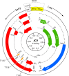
Fig 1
Fig 1
Schematic diagram of the JCV genome. The circular Mad-1 genome is 5,130 bp and codes for 9 proteins: large T antigen (T), small t antigen (t), T′135, T′136, T′165, VP1, VP2, VP3, and agnoprotein. At the top is the NCCR, composed of the origin of replication (ORI) and two 98-bp repeats. Map units are denoted in the center circle. The early open reading frame (ORF) proceeds from the NCCR in the counterclockwise orientation and the late ORF in the clockwise direction. Proteins in the same reading frame are depicted in the same color. Start and stop sites are indicated. All transcripts are polyadenylated [(A)n].
Fig 2
Fig 2
Representation of the early events involved in JCV infection. JCV (indicated by green viral capsids with supercoiled circular DNA) initially binds to carbohydrate receptors (likely α2,6-linked sialic acid) on the cell surface. The sialic acid could be attached to the G-protein-coupled 7-transmembrane receptor for serotonin (5HT2AR) or to another cell surface glycoprotein or glycolipid. The virus is then internalized into clathrin-coated pits and sorted into the early endosome. JCV colocalizes with cholera toxin, most likely in maturing or late endosomes. The virus then traffics to the to the ER by analogy with other polyomaviruses. It is likely that JCV interacts with PDI and ERAD proteins similar to those that interact with SV40 and mPyV in the ER. This is expected to cause conformational changes to the JCV virion, denoted by red shading of the capsid, and retrotranslocation of the virion into the cytoplasm. From there, it is likely that JCV enters the nucleus through the nuclear pore.
Fig 3
Fig 3
DNA sequence block representation of the noncoding control regions (NCCRs) of selected viral variants, showing DNA sequence block arrangements of the NCCRs of the prototype variant Mad-1 (A), Mad-8, which has similarity to and is illustrative of the majority of NCCR arrangements found in PML tissue (B), and the archetype variant CY, which is commonly found in urine of healthy individuals (C). Changes in NCCR sequence result in changes in transcription factor binding sites, which affect tissue specificity and activity of viral transcription and DNA replication. Changes in transcription factor binding are complex and differ markedly between variants of JCV found in PML patients. Depicted are the DNA sequence blocks known to make up JCV NCCR rearrangements, with the direction of transcription indicated by arrows. The numbering scheme is that of Frisque et al. (154). The origin of replication is denoted “ORI.” The lowercase letters “a,” “b,” “c,” “d,” “e,” and “f” indicate sequence blocks. The italicized “b” in the Mad-8 sequence represents a deletion of part of the “b” sequence. Red lines in Mad-8 represent an insertion of a single extra adenine between sequence blocks. The locations of TATA boxes, as well as binding sites for JCV large T antigen (black box with “T”) and sequences similar to the HIV tat-responsive element, known as the transactivation response element (TAR), are above the DNA sequence representation. Known binding sites for cellular proteins are underneath the DNA sequence blocks, and Most sites have been determined in Mad-1, with sites in the “b” and “d” sequence blocks determined in archetype. Sites in Mad-8 and “a,” “c,” and “e” sites of archetype have not been experimentally shown and have been determined through sequence similarity to known, experimentally determined binding sites in Mad-1, Mad-4, or archetype variants. Sites found only in Mad-1 are shown in yellow. Binding sites in both Mad-1 and Mad-8 are orange, those found in both Mad-8 and archetype are green, and those found only in archetype are red. Abbreviations are as follows: 1, Sp1; 2, SF2/ASF; 4, NFAT4; A, AP-1 (c-jun); B, Bag-1; C, C/EBPβ; D, DDX-1; G, GF-1/SμBP-2; H, HIF-1α; i, GBP-i; K, NF-κB; L, LCP-1; N, NFI; O, Tst-1/Oct-6/SCIP; P, Purα and YB-1; S, Spi-B. References can be found in the text.
Fig 4
Fig 4
Magnetic resonance imaging of PML. (A and B) T2-weighted images show a progressive and exponential increase in high-signal-intensity lesions over a period of 1 month in a patient with HIV infection. Lesions are seen in the frontal lobe, the internal capsule, and the splenium of the corpus callosum with spread to the opposite hemisphere. (C) A section from the frontal lobe of the same patient shows effacement of the cortical sulci and some midline shift suggestive of inflammation due IRIS.
None
None
None
None
None
None
None

References

    1. Agostini HT, et al. 1995. BK virus and a new type of JC virus excreted by HIV-1 positive patients in rural Tanzania. Arch. Virol. 140:1919–1934 - PubMed
    1. Agostini HT, et al. 2000. Influence of JC virus coding region genotype on risk of multiple sclerosis and progressive multifocal leukoencephalopathy. J. Neurovirol. 6 (Suppl. 2): S101–S108 - PubMed
    1. Agostini HT, Ryschkewitsch CF, Brubaker GR, Shao J, Stoner GI. 1997. Five complete genomes of JC virus type 3 from Africans and African Americans. Arch. Virol. 142:637–655 - PubMed
    1. Agostini HT, Yanagihara R, Davis V, Ryschkewitsch CF, Stoner GL. 1997. Asian genotypes of JC virus in Native Americans and in a Pacific Island population: markers of viral evolution and human migration. Proc. Natl. Acad. Sci. U. S. A. 94:14542–14546 - PMC - PubMed
    1. Ahmed W, Wan C, Goonetilleke A, Gardner T. 2010. Evaluating sewage-associated JCV and BKV polyomaviruses for sourcing human fecal pollution in a coastal river in Southeast Queensland, Australia. J. Environ. Qual. 39:1743–1750 - PubMed

Publication types

MeSH terms

Substances

LinkOut - more resources