Review
. 2012 Jul;25(3):507-44.
doi: 10.1128/CMR.00012-12.

Human and animal dirofilariasis: the emergence of a zoonotic mosaic

Affiliations
Review

Human and animal dirofilariasis: the emergence of a zoonotic mosaic

Fernando Simón et al. Clin Microbiol Rev. 2012 Jul.

Abstract

Dirofilariasis represents a zoonotic mosaic, which includes two main filarial species (Dirofilaria immitis and D. repens) that have adapted to canine, feline, and human hosts with distinct biological and clinical implications. At the same time, both D. immitis and D. repens are themselves hosts to symbiotic bacteria of the genus Wolbachia, the study of which has resulted in a profound shift in the understanding of filarial biology, the mechanisms of the pathologies that they produce in their hosts, and issues related to dirofilariasis treatment. Moreover, because dirofilariasis is a vector-borne transmitted disease, their distribution and infection rates have undergone significant modifications influenced by global climate change. Despite advances in our knowledge of D. immitis and D. repens and the pathologies that they inflict on different hosts, there are still many unknown aspects of dirofilariasis. This review is focused on human and animal dirofilariasis, including the basic morphology, biology, protein composition, and metabolism of Dirofilaria species; the climate and human behavioral factors that influence distribution dynamics; the disease pathology; the host-parasite relationship; the mechanisms involved in parasite survival; the immune response and pathogenesis; and the clinical management of human and animal infections.


Figures

Fig 1
Fig 1
Biological life cycles of D. immitis and D. repens. mf, microfilaremia.
Fig 2
Fig 2
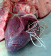
Male and female adult worms of D. immitis in the heart of a dog.
Fig 3
Fig 3
Immunohistochemical positive reaction (red) against the Wolbachia surface protein (WSP) reveals the presence of symbiont bacteria in the hypodermal cords of a D. repens adult worm. (Courtesy of L. H. Kramer, University of Parma, Parma, Italy.)
Fig 4
Fig 4
Nutrient uptake and energy metabolism of D. immitis. There is a selective uptake of nutrients through both the digestive tract and the cuticle in microfilariae and adult worms. The main route for energy generation is anaerobic glycolysis with lactate as a main end product. Enzymes involved in glycolysis identified in D. immitis are indicated. PGI, phosphoglucose isomerase; PFK, phosphofructokinase; TPI, triosephosphate isomerase; GAPDH, glyceraldehyde-3-phosphate dehydrogenase; PGK, phosphoglycerate kinase; PGM, phosphoglycerate mutase; PK, pyruvate kinase; LDH, lactate dehydrogenase; bisP, bisphosphate; CoA, coenzyme A.
Fig 5
Fig 5
Current geographical distribution of canine dirofilariasis. Blue, D. immitis infections; green, D. repens infections; orange, presence of both species.
Fig 6
Fig 6
Current distribution of human dirofilariasis. Purple, countries in which D. immitis cases predominate; gray, countries in which D. repens cases predominate; ■, sporadic pulmonary cases; ▲, sporadic subcutaneous/ocular cases. The asterisk indicates that data from European countries except for the former Soviet Union were included.
Fig 7
Fig 7
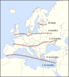
Approximate length of the transmission periods of Dirofilaria spp. per year in Europe (calculated with data from references and 149).
Fig 8
Fig 8
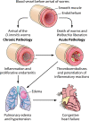
Progress of heartworm disease in dogs. The disease usually has a chronic progression. Initially, the damages affect the arteries, spreading later to the lung parenchyma and the right heart chambers. The simultaneous death of many worms contributes to the acute presentation.
Fig 9
Fig 9
Pathological alterations in pulmonary arteries associated with canine heartworm disease. (A) Surface of the vascular endothelium of a pulmonary artery from a dog with heartworm disease showing well-developed intravascular villi (yellow arrow). The black arrow indicates the presence of an adult worm. (B) Large thromboembolism (yellow arrow) in a pulmonary artery of a dog that died of cardiopulmonary dirofilariasis. (Courtesy of L. Venco, Clinica Veterinaria Città di Pavia, Pavia, Italy.)
Fig 10
Fig 10
X-ray images of canine and feline heartworm disease. (A) Lateral thoracic radiograph of an 8-year-old dog heavily parasitized by D. immitis. Cardiomegaly on the right heart (white arrow), a mixed pulmonary pattern (red arrow), enlarged pulmonary vessels (blue arrow), perihilar edema (yellow arrow), and large areas of pulmonary densification are noted. (B) Lateral thoracic radiograph of a cat with heartworm-associated respiratory disease. Note the lung air trapping as evidenced by the flattened and caudally displaced diaphragm (white arrow) and the gas-filled stomach, caused by aerophagia (red arrow).
Fig 11
Fig 11
Anti-Wolbachia surface protein (WSP)-positive immunohistochemical reaction in a microfilaria from the kidney of a dog with heartworm disease (arrow).
Fig 12
Fig 12
Canine subcutaneous dirofilariasis caused by D. repens. (A) Subcutaneous nodule in the scrotum of a male dog. (B) Adult worm in an open subcutaneous nodule. (Courtesy of Sergey Kartashov, Rostov, Russia.)
Fig 13
Fig 13
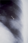
Human pulmonary dirofilariasis. Shown is a thoracic radiograph showing a pulmonary nodule attributed to D. immitis (arrow).
Fig 14
Fig 14
Human subcutaneous and ocular dirofilariasis. (A) External appearance of a subcutaneous nodule in the ocular region. (B) Histological section of a nodule showing sections of adult D. repens worms. (C) Intravitreal location of an adult D. repens worm in a human patient. (Panels A and B courtesy of Vladimir Kartashev, University of Rostov Na Donu, Rostov, Russia; panel C reprinted from reference with permission.)
Fig 15
Fig 15
Management of human pulmonary and subcutaneous dirofilariasis.
Fig 16
Fig 16
Predicted immune events occurring during D. immitis infections. A dual (Th2/Th1) host immune response is shown. (1) The Th2-type anti-inflammatory response is stimulated by D. immitis antigens and the presence of microfilariae. The expression levels of IL-4 and IL-10 are increased, and antibodies related to the Th2-type response arise: IgG1 (dogs) or IgE (humans). (2) The Th1-type proinflammatory response is stimulated by Wolbachia bacteria released from dying worms. The Wolbachia surface protein (WSP) stimulates the expression of IFN-γ interacting with monocytes (probably through Toll-like receptors [TLR]) and inhibits their apoptosis. Wolbachia also stimulates the production of the Th1-type antibody response and the expression of proinflammatory mediators by vascular endothelial cells. Endothelial cells also increase the expression levels of adhesion/transmigration molecules (intercellular adhesion molecule [ICAM], platelet endothelial cell adhesion molecule [PECAM], and migration vascular cell adhesion molecule [VCAM]) and cellular proliferation molecules (E-cadherin and vascular endothelial grow factor [VEGF]). Some of these stimuli are also triggered by D. immitis somatic antigen (DiSA).
Fig 17
Fig 17
Mechanisms of survival and immune evasion in D. immitis. (1) Short-term immune evasion by L3 larvae. Infective larvae avoid the host immune response, releasing large amounts of two surface antigens, of 6 and 35 kDa. (2) Long-term immune evasion. Preadult/adult worms mask their surface, adsorbing different host molecules and cells. They have surface nonimmunogenic glycolipids and many isoforms of heat shock proteins (HSPs) and detoxificant enzymes that eliminate toxic products synthesized by macrophages and neutrophils. Microfilariae have surface proteases that digest host antibodies. (3) Modulation of the vascular environment. Live adult worms release metabolic products (E/S antigens) that stimulate anti-inflammatory prostaglandin E2 (PGE2) eicosanoid and decrease monocyte transmigration. Additionally, E/S antigens bind plasminogen and activate plasmin to eliminate thromboembolisms in the presence of the tissue plasminogen activator (t-PA), whose expression is also stimulated by E/S antigens in vascular endothelial cells. DiES, D. immitis excretory/secretory products; PLG, plasminogen.
None
None
None
None
None
None
None

References

    1. Abraham D, Grieve RD, Oaks JA. 1990. Dirofilaria immitis: moulting process of third-stage larvae. Exp. Parasitol. 70:314–322 - PubMed
    1. Abramowsky CR, Powers KG, Aikawa M, Swinehart G. 1981. Dirofilaria immitis. 5. Immunopathology of filarial nephropathy in dogs. Am. J. Pathol. 104:1–12 - PMC - PubMed
    1. Adhami J, Reiter P. 1998. Introduction and establishment of Aedes (Stegomyia) albopictus skuse (Diptera: Culicidae) in Albania. J. Am. Mosq. Control Assoc. 14:340–343 - PubMed
    1. Ahid SM, Lourenço-De-Oliveira R. 1999. Mosquitoes potential vectors of canine heartworm in the Northeast Region from Brazil. Rev. Saude Publica 33:560–565 - PubMed
    1. Aiello A, Aiello P, Aiello F. 2005. A case of palpebral dirofilariasis. Eur. J. Ophthalmol. 15:407–408 - PubMed

Publication types

MeSH terms

Substances