Review
. 2012 Sep 1;4(9):a005645.
doi: 10.1101/cshperspect.a005645.

Synaptic vesicle endocytosis

Affiliations
Review

Synaptic vesicle endocytosis

Yasunori Saheki et al. Cold Spring Harb Perspect Biol. .

Abstract

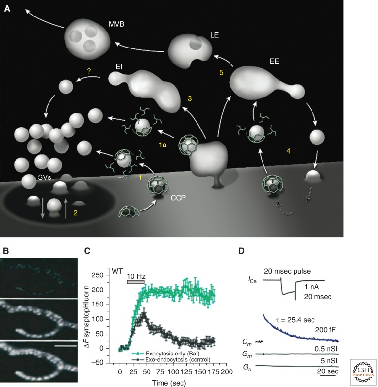
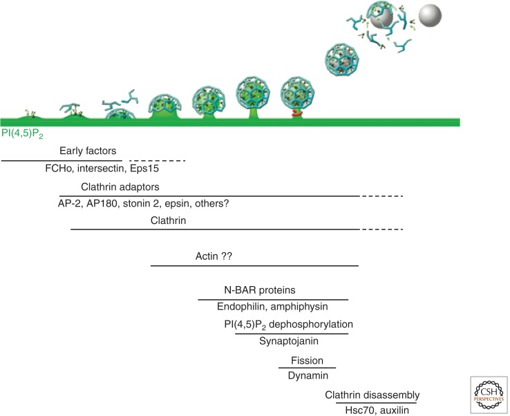
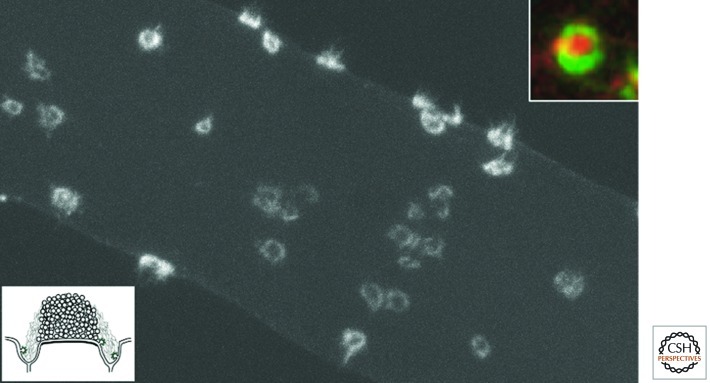
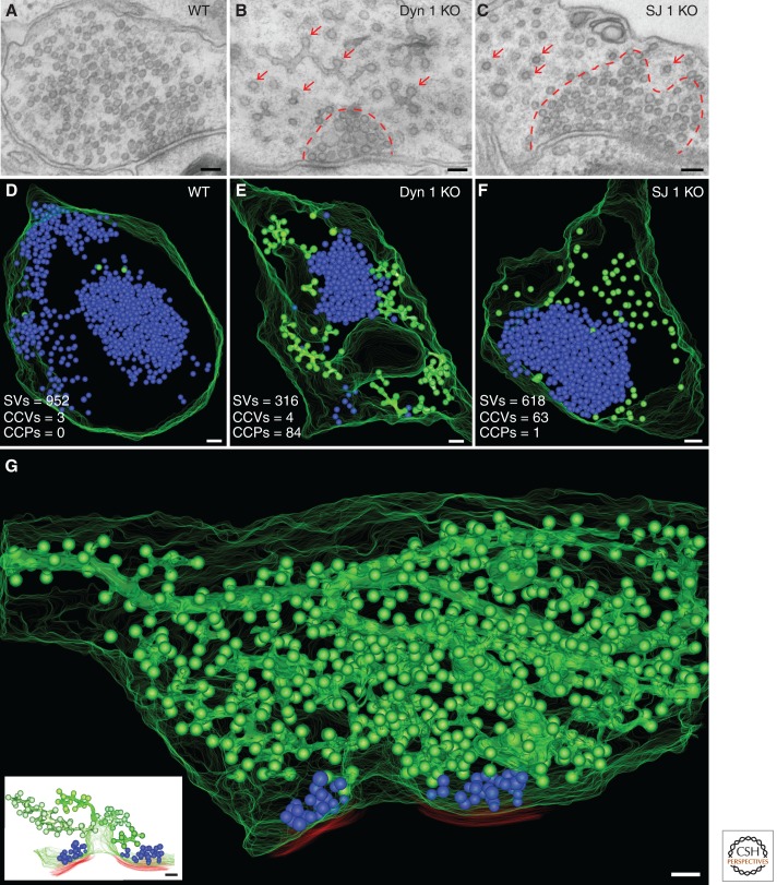
Neurons can sustain high rates of synaptic transmission without exhausting their supply of synaptic vesicles. This property relies on a highly efficient local endocytic recycling of synaptic vesicle membranes, which can be reused for hundreds, possibly thousands, of exo-endocytic cycles. Morphological, physiological, molecular, and genetic studies over the last four decades have provided insight into the membrane traffic reactions that govern this recycling and its regulation. These studies have shown that synaptic vesicle endocytosis capitalizes on fundamental and general endocytic mechanisms but also involves neuron-specific adaptations of such mechanisms. Thus, investigations of these processes have advanced not only the field of synaptic transmission but also, more generally, the field of endocytosis. This article summarizes current information on synaptic vesicle endocytosis with an emphasis on the underlying molecular mechanisms and with a special focus on clathrin-mediated endocytosis, the predominant pathway of synaptic vesicle protein internalization.


Figures

Figure 1.
Figure 1.
Pathways of synaptic vesicle recycling and examples of methods to monitor synaptic vesicle endocytosis at living synapses. (A) Schematic diagram of membrane traffic in axon terminals illustrating endocytosis of synaptic vesicle (SV) membranes via clathrin-coated pits (CCP) from the plasma membrane and its deep infoldings (1 and 1a), “kiss and run” (2), and bulk endocytosis (3) followed by vesicle formation via unknown mechanisms (?) from endocytic intermediates (EI). This recycling traffic is interconnected with housekeeping membrane recycling (4) involving clathrin-mediated endocytosis and canonical early endosomes (EE) as well as with traffic to the cell body (5) via late endosomes (LE) and multivesicular bodies (MVBs). (B) Compensatory endocytosis of synaptic vesicle membranes at the frog neuromuscular junction following stimulation by depolarization in high potassium, visualized by the uptake of the fluorescent styryl dye FM1-43. Partial and full dye loading are shown in the middle and lower panels (Panel B is from Betz and Bewick 1992; reprinted, with permission, from Science © 1992). (C) Time course of exo- and endocytosis in response to a 10-Hz, 30-sec stimulus at synapses of cultured mouse cortical neurons as monitored by the increase and decrease in the fluorescence of synaptopHluorin (spH), a fusion protein of pHluorin to synaptobrevin/VAMP2. Bafilomycin (Baf), a membrane-permeable blocker of the V-type ATPase that is required for vesicle reacidification, was used to trap vesicles at neutral pH after endocytosis, thus allowing spH to remain fluorescent even after endocytosis. Control runs (black) consist of the sum of exo- and endocytosis, and Baf runs (green) monitor all the exocytic events. The difference between the two traces allows estimating total endocytosis occurring during the experiment. (Panel C is from Nicholson-Tomishima and Ryan 2004; reprinted, with permission, from Proc Natl Acad Sci © 2004.) (D) Endocytic recovery of the increase in surface area produced by a secretory burst at a calyx of Held giant nerve terminal as monitored by the measurement of membrane capacitance (Cm, blue trace). Secretion was evoked by a single 20-msec (+10 mV) presynaptic pulse that elicited the Ca2+ current shown in the top trace. Membrane conductance (Gm) and series conductance (Gs) did not change during the recording. (Panel D is from Lou et al. 2008; reprinted, with permission, from the author.)
Figure 2.
Figure 2.
Sequential recruitment of endocytic proteins during clathrin-mediated synaptic vesicle endocytosis. Assembly and maturation of endocytic clathrin-coated pits involves the sequential action of a variety of protein factors. Putative sites of action of various factors are indicated (see text for discussion). PI(4,5)P2 (green), which is generated at the synaptic plasma membrane primarily by PIP kinase type 1γ (PIPK1γ), plays a critical role both in exocytosis and in the recruitment of endocytic factors.
Figure 3.
Figure 3.
Presence of actin at periactive zones of synapses of the giant reticulospinal axon of the lamprey. Fluorescence image of a giant axon microinjected with Alexa Fluor 488-phalloidin. Fluorescent rings represent actin at periactive zones surrounding the vesicle cluster. The top inset is from an axon preincubated with an FM dye (red) to produce an endocytic labeling of synaptic vesicles, then fixed and labeled with phalloidin (green) to reveal actin. Note the actin ring nesting the vesicle cluster. The bottom inset is a schematic diagram of the synapses of the giant axon illustrating the vesicle cluster and the periactive zone (J Morgan, V Pieribone, and P De Camilli, unpubl.).
Figure 4.
Figure 4.
Ultrastructure and 3D models of wild-type (WT), dynamin 1 knockout (KO), and synaptojanin 1 KO synapses demonstrating the accumulation of clathrin-coated intermediates in the KO synapses. (AC) Conventional electron micrographs of synapses from mouse primary cortical cultures of the indicated genotypes. Note the abundance of clathrin-coated profiles (arrows) in dynamin 1 and synaptojanin 1 KO nerve terminals and the less dense packing of these structures relative to the packing of synaptic vesicles (SVs) (enclosed by dashed red lines). (DF) 3D models from the EM tomograms of 250-nm-thick sections of WT and mutant nerve terminals showing that the bulk of coated profiles (green) are buds that are located that at the tip of branched tubules in dynamin 1 KO synapses and free vesicles in synaptojanin 1 KO synapses. Synaptic vesicles are shown in blue. (G) 3D model from the EM tomogram of an 800-nm-thick volume of a dynamin 1 KO nerve terminal. Synaptic vesicles are blue spheres, the plasma membrane is outlined by a series of green curves, and red lines indicate postsynaptic membranes. The overwhelming majority of coated structures (green) are buds originating from tubular plasma membrane invaginations (four such invaginations are shown separately in the inset). (Scale bars, 100 nm.) (Panel G from Hayashi et al. 2008; reprinted, with permission, from the author. Bottom inset from Ferguson et al. 2007; reprinted, with permission, from Science © 2007.)

Similar articles

Cited by

References

    1. Albillos A, Dernick G, Horstmann H, Almers W, Alvarez de Toledo G, Lindau M 1997. The exocytotic event in chromaffin cells revealed by patch amperometry. Nature 389: 509–512 - PubMed
    1. Andersson F, Jakobsson J, Low P, Shupliakov O, Brodin L 2008. Perturbation of syndapin/PACSIN impairs synaptic vesicle recycling evoked by intense stimulation. J Neurosci 28: 3925–3933 - PMC - PubMed
    1. Anggono V, Smillie KJ, Graham ME, Valova VA, Cousin MA, Robinson PJ 2006. Syndapin I is the phosphorylation-regulated dynamin I partner in synaptic vesicle endocytosis. Nat Neurosci 9: 752–760 - PMC - PubMed
    1. Antonin W, Riedel D, von Mollard GF 2000. The SNARE Vti1a-β is localized to small synaptic vesicles and participates in a novel SNARE complex. J Neurosci 20: 5724–5732 - PMC - PubMed
    1. Antonny B 2006. Membrane deformation by protein coats. Curr Opin Cell Biol 18: 386–394 - PubMed

Publication types

MeSH terms

LinkOut - more resources