. 2012 Aug;139(16):2926-34.
doi: 10.1242/dev.082396. Epub 2012 Jul 4.

Identification of a novel developmental mechanism in the generation of mesothelia

Affiliations

Identification of a novel developmental mechanism in the generation of mesothelia

Nichelle I Winters et al. Development. 2012 Aug.

Abstract

Mesothelium is the surface layer of all coelomic organs and is crucial for the generation of their vasculature. Still, our understanding of the genesis of this essential cell type is restricted to the heart where a localized exogenous population of cells, the proepicardium, migrates to and envelops the myocardium supplying mesothelial, vascular and stromal cell lineages. Currently it is not known whether this pattern of development is specific to the heart or applies broadly to other coelomic organs. Using two independent long-term lineage-tracing studies, we demonstrate that mesothelial progenitors of the intestine are intrinsic to the gut tube anlage. Furthermore, a novel chick-quail chimera model of gut morphogenesis reveals these mesothelial progenitors are broadly distributed throughout the gut primordium and are not derived from a localized and exogenous proepicardium-like source of cells. These data demonstrate an intrinsic origin of mesothelial cells to a coelomic organ and provide a novel mechanism for the generation of mesothelial cells.


Figures

Fig. 1.
Fig. 1.
A trilaminar gut tube was generated by HH15. (A) HH13 splanchnopleure is composed of two layers. (B,C) Boxed regions shown in A. The splanchnic mesoderm appears stratified and is underlain by a basement membrane (yellow arrow). The endoderm has its own basement membrane (white arrow). Arrowheads in C indicate a single mesenchymal cell. (C’) The endoderm, but not the splanchnic mesoderm, is cytokeratin positive at HH13. (D-F) At HH15, a mesenchymal layer resides between the aforementioned basement membranes (arrows). (F’) The outer epithelium is not cytokeratin positive at HH15. (G-I) At HH19, the mesenchymal layer has expanded (space between two arrows) and the basement membrane of the outer epithelium has fragmented (yellow arrow). (I’) The endoderm, but not the outer epithelium, is cytokeratin positive. E, endoderm; Me, mesenchyme; NT, neural tube; OE, outer epithelium; S, somite; So, somatic mesoderm; Sp, splanchnic mesoderm.
Fig. 2.
Fig. 2.
Definitive intestinal mesothelium is present at HH29 (Day 6). (A) At day 6, a simple squamous, cytokeratin-positive (green) mesothelium is present surrounding the intestine. (B) A basement membrane underlies the mesothelium (red, yellow arrow). White arrow indicates endodermal basement membrance. (C) Merge of A and B. (D) Higher magnification of boxed region shown in C. E, endoderm; Me, mesenchyme; OE, outer epithelium.
Fig. 3.
Fig. 3.
Electroporation of the splanchnic mesoderm at HH14 demonstrates labeling of the outer epithelium and mesenchyme. (A) Schematic demonstrating injection of the GFP reporter plasmid into the right lateral cavity of an embryo in ovo. (B) Whole-mount image of the ventral surface of an embryo electroporated at HH14 and then incubated for 6 hours. Electrodes were placed near the vitelline artery. GFP was observed in the region near the vitelline artery and was restricted to the lateral plates (arrows). (C) GFP-positive cells localized to the splanchnic mesoderm. (D) Boxed area shown in C. GFP-positive cells were found primarily within the outer epithelium (arrows) with a few cells within the mesenchymal layer (arrowheads). No GFP-positive cells were identified in the endoderm. (E) Merge of D with TOPRO-3. BV, blood vessel; GN, glass needle; H, heart; LC, lateral cavity; LP, lateral plate; Me, mesenchymal layer; N, notochord; NT, neural tube; OE, outer epithelium; S, somite, VA, vitelline artery.
Fig. 4.
Fig. 4.
DNA electroporation demonstrates that splanchnic mesoderm harbors mesothelial progenitors. Sections through gut tubes of embryos electroporated at HH15-HH17 and incubated for 8 days. (A-D) GFP-positive cells (arrows) were identified within the squamous mesothelial layer of the intestine associated closely with the basement membrane (laminin, red). (E) GFP-positive cells were also identified within the forming α-smooth muscle actin (SMA)-positive muscularis externa (boxed region) and into the submucosa (arrowhead). (F-H) Higher magnification of the boxed region. GFP-positive cells within the muscularis externus were not SMA positive (arrows). ME, muscularis externa; Mu, mucosa; SM, submucosa.
Fig. 5.
Fig. 5.
Long-term retroviral lineage tracing of splanchnic mesoderm. Whole-mount images of intestine from embryos infected with virus between HH14 and HH17 and analyzed 14 days later. (A) High magnification of intestinal surface demonstrated cells resembling mesothelium with prominent nuclei and broad cell processes (arrows). (B) Bright-field image of gut tube demonstrating the vasculature (arrows). (B′) GFP fluorescence of gut tube pictured in B. GFP-positive cells surrounded the vasculature within the mesentery and intestine (arrows). (C) GFP-positive cells were also found distributed deeply in the intestine (arrows). GT, gut tube; VA, vitelline artery
Fig. 6.
Fig. 6.
Lineage tracing of splanchnic mesoderm reveals mesothelial, perivascular and mesenchymal derivatives. (A-H) Sections of intestine from embryos infected between HH13-14 and isolated 14 days later. (A) Squamous GFP-positive cells frequently populated the mesothelium (arrowheads) closely associating with the basement membrane (red, laminin). (B) High magnification of boxed area in A. (C) GFP-positive cells associated with large mesenteric blood vessels. (D) High magnification of boxed area in C demonstrates GFP-positive vascular smooth muscle cells (arrow) and perivascular cells (arrowhead). (E) GFP-positive cells were identified within the muscularis externa. (F-H) High magnification of boxed area shown in E. A rare population of GFP-positive cells found within the muscularis externus were spindle shaped and SMA positive (arrowheads). (I-L) Sections of intestine from embryos infected between HH15 and HH17, and isolated 14 days later. (I) Squamous GFP-positive cells populated the mesothelium (arrowheads), closely associating with the basement membrane (red, laminin). (J) SMA-negative mesenchymal cells within the muscularis externa layer (arrowheads). (K) GFP-positive vascular smooth muscle cells (arrowheads). (L) Submucosal GFP-positive, SMA-negative cells. M, mesothelium; ME, muscularis externus; Mes, mesentery; Mu, mucosa, SM, submucosa.
Fig. 7.
Fig. 7.
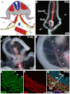
Transplanted splanchnopleure forms a highly structured gut tube. (A) Transplants were generated by cutting along the dorsal aspect of the splanchnopleure (1) and the ventral edges near the vitelline veins (2). The splanchnopleure was then cut along the AP axis (3) to generate six or seven pieces for transplantation. (B) A representative graft-derived gut tube 8 days after transplantation. The graft had generated a tube and attached to the mesentery of the host gut tube. (C) A representative graft-derived gut tube 14 days after transplantation (G, bracketed). The graft-derived gut tube was attached to the host (H) via a mesentery. (D) The mesentery of the graft-derived gut tube contained a regular arrangement of blood vessels (arrowheads). (E-G) Sections through the graft-derived gut tube demonstrated normal morphogenesis with villi (arrowheads), submucosa (SM) and a SMA-positive muscularis externus layer. All layers were derived from quail cells (QCPN positive, green). E, endoderm; Ec, ectoderm; G, graft-derived gut tube; H, host gut tube; LC, lateral cavity; M, mesothelium; ME, muscularis externa; Mu, mucosa; NT, neural tube; S, somite; So, somatic mesoderm; Sp, splanchnic mesoderm; SM, submucosa; VA, vitelline artery; VV, vitelline vein.
Fig. 8.
Fig. 8.
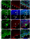
Graft mesothelium is quail derived. (A-C) Section of graft-derived gut tube generated from tissue isolated from the anterior splanchnopleure of a HH16 quail donor. Co-staining for QCPN and cytokeratin demonstrated that the mesothelial cells lining the graft were quail derived (arrowheads). (D-F) Section of a graft-derived gut tube generated from the posterior splanchnopleure of a HH14 quail donor. QCPN staining demonstrates the mesenteric mesothelium is quail derived (arrowheads). (G-I) Host-derived cells (8F3-positive) were also identified within the graft (arrows). However, 8F3-positive chick cells were only rarely (6%) identified within the mesothelial layer (arrowheads) of the graft-derived gut tube. (J-L) Staining of a chick (host) gut tube reveals mesothelial cells (arrowheads) robustly label with the chick marker 8F3. GT, gut tube; Mes, mesentery.

References

    1. Acencio M. M., Vargas F. S., Marchi E., Carnevale G. G., Teixeira L. R., Antonangelo L., Broaddus V. C. (2007). Pleural mesothelial cells mediate inflammatory and profibrotic responses in talc-induced pleurodesis. Lung 185, 343–348 - PubMed
    1. Aroeira L. S., Aguilera A., Selgas R., Ramírez-Huesca M., Pérez-Lozano M. L., Cirugeda A., Bajo M. A., del Peso G., Sánchez-Tomero J. A., Jiménez-Heffernan J. A., et al. (2005). Mesenchymal conversion of mesothelial cells as a mechanism responsible for high solute transport rate in peritoneal dialysis: role of vascular endothelial growth factor. Am. J. Kidney Dis. 46, 938–948 - PubMed
    1. Asahina K., Zhou B., Pu W. T., Tsukamoto H. (2011). Septum transversum-derived mesothelium gives rise to hepatic stellate cells and perivascular mesenchymal cells in developing mouse liver. Hepatology 53, 983–995 - PMC - PubMed
    1. Braun N., Alscher D. M., Fritz P., Edenhofer I., Kimmel M., Gaspert A., Reimold F., Bode-Lesniewska B., Ziegler U., Biegger D., et al. (2011). Podoplanin-positive cells are a hallmark of encapsulating peritoneal sclerosis. Nephrol. Dial. Transplant. 26, 1033–1041 - PubMed
    1. Chegini N. (2008). TGF-beta system: the principal profibrotic mediator of peritoneal adhesion formation. Semin. Reprod. Med. 26, 298–312 - PubMed

Publication types

MeSH terms

Substances