. 2012 Sep;53(9):1932-43.
doi: 10.1194/jlr.M028563. Epub 2012 Jul 4.

Molecular characterization of proprotein convertase subtilisin/kexin type 9-mediated degradation of the LDLR

Affiliations

Molecular characterization of proprotein convertase subtilisin/kexin type 9-mediated degradation of the LDLR

Yan Wang et al. J Lipid Res. 2012 Sep.

Abstract

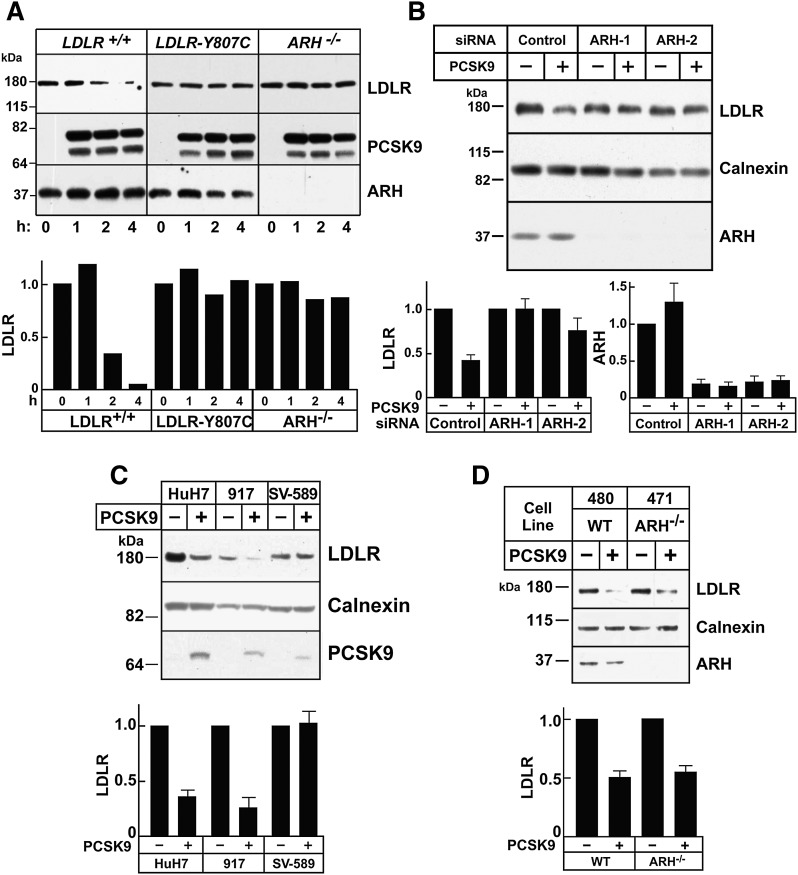
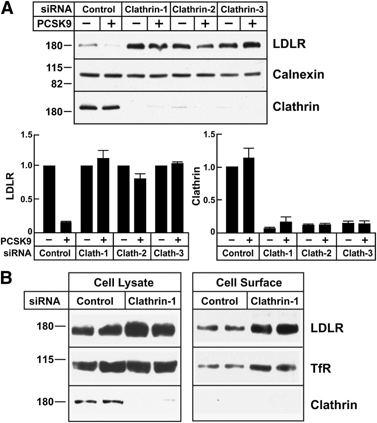
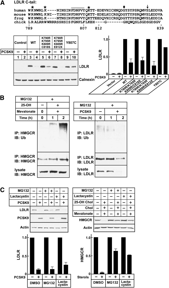
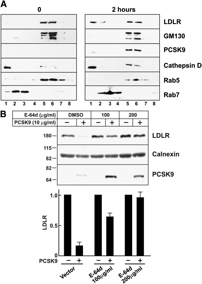
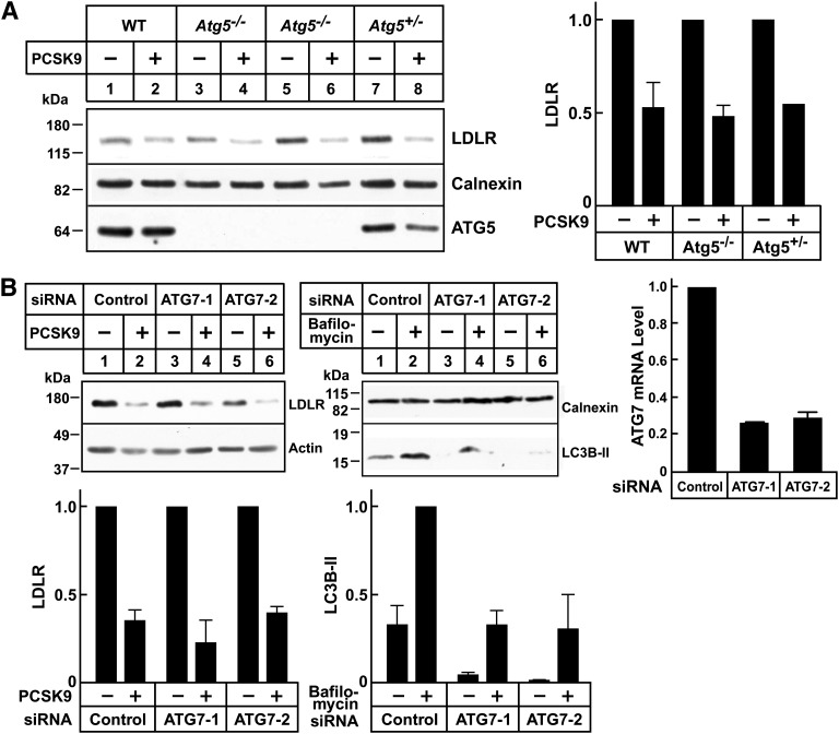
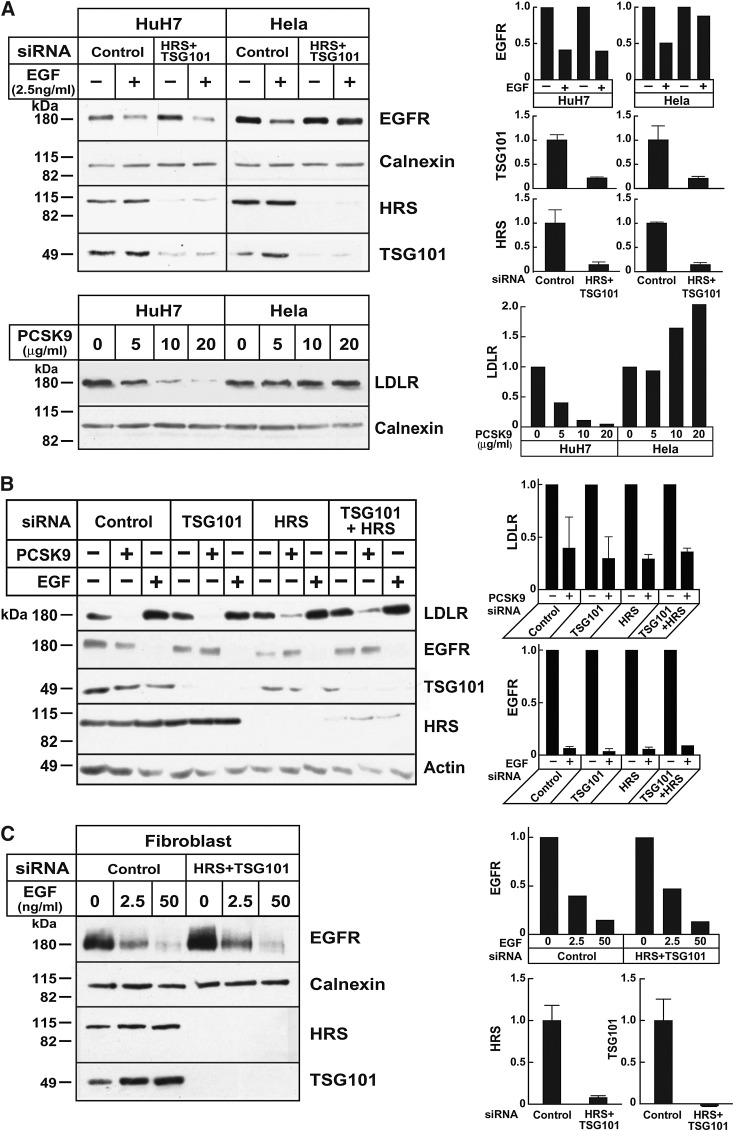
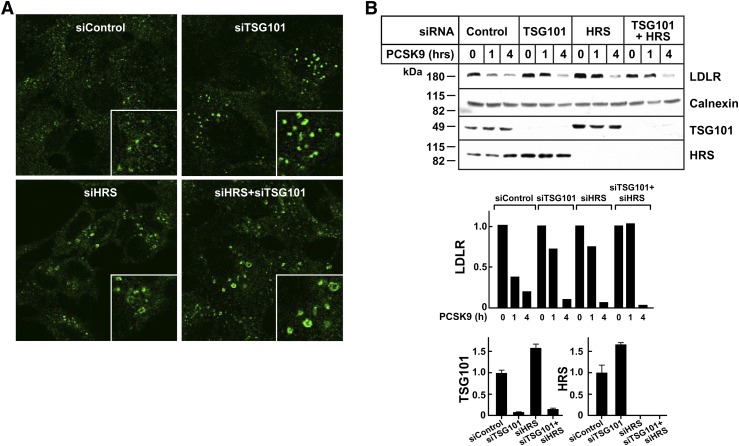
Proprotein convertase subtilisin/kexin type 9 (PCSK9) is a secreted protein that promotes degradation of cell surface LDL receptors (LDLRs) in selected cell types. Here we used genetic and pharmacological inhibitors to define the pathways involved in PCSK9-mediated LDLR degradation. Inactivating mutations in autosomal recessive hypercholesterolemia (ARH), an endocytic adaptor, blocked PCSK9-mediated LDLR degradation in lymphocytes but not in fibroblasts. Thus, ARH is not specifically required for PCSK9-mediated LDLR degradation. Knockdown of clathrin heavy chain with siRNAs prevented LDLR degradation. In contrast, prevention of ubiquitination of the LDLR cytoplasmic tail, inhibition of proteasomal activity, or disruption of proteins required for lysosomal targeting via macroautophagy (autophagy related 5 and 7) or the endosomal sorting complex required for trafficking (ESCRT) pathway (hepatocyte growth factor-regulated Tyr-kinase substrate and tumor suppressor gene 101) failed to block PCSK9-mediated LDLR degradation. These findings are consistent with a model in which the LDLR-PCSK9 complex is internalized via clathrin-mediated endocytosis and then routed to lysosomes via a mechanism that does not require ubiquitination and is distinct from the autophagy and proteosomal degradation pathways. Finally, the PCSK9-LDLR complex appears not to be transported by the canonical ESCRT pathway.


Figures

Fig. 1.
Fig. 1.
PCSK9-mediated LDLR degradation requires ARH in lymphocytes and hepatocytes but not in fibroblasts. A: Immortalized lymphocytes (5 × 10⁵ cells/ml) from a normal subject, a patient homozygous for a mutation in the LDLR that blocks LDL internalization (LDLR-Y807C) (61), and a patient with ARH deficiency were cultured at 37°C. PCSK9 (5 μg/ml) was added to the medium for the indicated times. After cells were washed in ice-cold PBS, lysates were prepared and subjected to immunoblotting. The graph represents quantification of the data shown. B: HuH7 cells (1 × 10⁵ cells/60 mm dish) were grown for 24 h and transfected with two separate anti-ARH siRNAs (ARH-1 and ARH-2) or a control siRNA (Control). After 3 days, PCSK9 (10 μg/ml) was added to the medium. After 4 h at 37°C, cells were processed as described in A. C: Primary human fibroblasts (917), immortalized fibroblasts (SV-589), and hepatocytes (HuH7) were treated with PCSK9 (10 μg/ml) for 4 h, and cell lysates were processed as described in A. D: Primary human fibroblasts from normal (480) or ARH-deficient (471) individuals were treated with PCSK9 and whole cell lysate were analyzed as described in A. All experiments were performed at least twice with similar results. Proteins were quantified as described in Materials and Methods. Each graph represents the mean ± SEM for three independent experiments, except for A, which represents the data shown.
Fig. 2.
Fig. 2.
PCSK9-mediated LDLR degradation in clathrin-depleted HuH7 cells. A: HuH7 cells were transfected with three different anti-clathrin heavy chain (Clathrin-1, Clathrin-2, and Clathrin-3) or control siRNAs as indicated in Fig. 1. After 3 days, PCSK9 (10 μg/ml) was added to the medium for 4 h. B: HuH7 cells were transfected with control siRNA or siRNA targeting clathrin heavy chain for 72 h as indicated in Fig. 1. Cell surface proteins were isolated by biotinylation as described in Materials and Methods. Whole cell lysates and streptavidin-precipitated proteins were analyzed by immunoblotting. The immunoreactive proteins were quantified as described in Materials and Methods. Graphs represent the means ± SEM from three independent experiments. TfR, transferrin receptor.
Fig. 3.
Fig. 3.
PCSK9-mediated LDLR degradation and the ubiquitin-proteosome pathway. A: Alignment of the amino acid sequence of the LDLR cytoplasmic tail from four species is shown. Mouse Hepalclc7 cells (plated at 4 × 10⁵ cells/well in 6-well plates) were grown in 2 ml MEMα containing 10% FCS (Day 0). On Day 1, cells were transfected with empty vector or with plasmids expressing wild-type (WT) or mutant LDLR using Lipofectamine 2000. On Day 2, the medium was switched to MEMα plus 10% NCLPPS, cholesterol (10 μg/ml), and 25-hydroxycholesterol (1 μg/ml). On Day 3, PCSK9-D374Y (2.0 μg/ml) was added. After 4 h at 37°C, lysates were prepared and immunoblotted with HL1, a monoclonal antibody against human LDLR that does not recognize mouse LDLR. B: HuH7 cells were cultured as described and treated as indicated in B. HMGCR and LDLR were immunoprecipitated from cell lysates using antibodies 804c and 3143, respectively. Immunoprecipitates were size-fractionated on SDS-PAGE gels and probed with anti-ubiquitin (P4D1), anti-HMGCR (IgG-A9), and anti-LDLR (HL1) antibodies. C: HuH7 cells (plated at 3 × 10⁵/well in 6-well plates) were grown in medium C (Day 0). On Day 1, medium was switched to hDMEM containing 10% NCLPPS plus compactin (10 µM), mevalonate (50 µM) for HMGCR degradation assays, and hDMEM containing 10% NCLPPS for LDLR degradation. After 16 h, medium containing MG132 (10 μM), lactacystin(10 μM), or DMSO alone was added. After 1 h, cells were treated with 25-hydroxycholesterol (1 μg/ml), cholesterol (10 μg/ml), and mevalonate (10 mM) or with PCSK9 (5 μg/ml) for 4 h. Cell lysates were subjected to immunoblot analysis. All experiments were performed twice with similar results. Proteins were quantified as described in Materials and Methods. Graphs represent the means ± SEM for two independent experiments.
Fig. 4.
Fig. 4.
PCSK9-mediated LDLR degradation and lysosome function. A: Confluent HuH7 cells were treated with PCSK9 (10 μg/ml) for 0 or 2 h. Cell lysates were fractionated on Percoll gradients as described in Materials and Methods. Proteins were precipitated from the gradient fractions, size-fractionated on 4–12% gradient SDS-polyacrylamide gels, and visualized by immunoblot analysis. B: Confluent HuH7 cells were treated with the lysosome inhibitor E-64d at the doses indicated or with DMSO alone for 30 min at 37°C in medium D. PCSK9 (10 μg/ml) was added to the medium, and cells were allowed to grow for another 4 h. Cells were harvested, and cell lysates were subjected to immunoblot analysis. Proteins were quantified as described in Materials and Methods. Graphs represent the means ± SEM for three independent experiments.
Fig. 5.
Fig. 5.
PCSK9-mediated LDLR degradation in autophagy-deficient hepatocytes. A: Primary hepatocytes were isolated from WT Atg5−/− or Atg5+/− mouse embryos. After 48 h, cells were treated with PCSK9 (10 μg/ml) for 4 h at 37°C. Cell lysates were subjected to immunoblot analysis. B: HuH7 cells were transfected with control or anti-ATG7 siRNA oligos as described in Fig. 1. After 72 h, cells were treated with PCSK9 (10 μg/ml) for 4 h or with bafilomycin A1 (100 nM) for 2 h at 37°C. To visualize LC3B protein, cell lysates were fractionated on 15% SDS-PAGE gels and transferred to polyvinylidene difluoride membranes for immunoblot analysis. The efficiency of ATG7 knockdown was evaluated by qRT-PCR as described in Materials and Methods. Data were normalized to control dishes, and GAPDH was used as an internal standard. Similar results were obtained in at least two independent experiments. Proteins were quantified as described in Materials and Methods. Graphs represent the means ± SEM from two independent experiments.
Fig. 6.
Fig. 6.
The ESCRT pathway and PCSK9-mediated LDLR degradation. A, upper panel: HuH7 and Hela cells were cultured as indicated in Materials and Methods, and siRNA transfection was performed as described in Fig. 1. The medium was changed to serum-free medium 72 h after transfection, and after 2 h, EGF was added to the medium. Cells were collected after another 4 h. A, lower panel: Confluent HuH7 and Hela cells were treated with the indicated amounts of PCSK9 at 37°C for 4 h. Cell lysates were subjected to immunoblot analysis. Graphs represent the mean ± SEM for two independent experiments. B: HuH7 cells were transfected with control siRNA or with siRNAs against TSG101, HRS, or both. After 72 h, cells were switched to hDMEM without serum. Cells were incubated for 24 h before the addition of PCSK9 (10 μg/ml) or EGF (50 ng/ml) for 4 h at 37°C. Cells lysates were subjected to immunoblot analysis. Data represent the mean ± SEM for three independent experiments. C: Human skin fibroblasts (917) were plated at a density of 2 × 10⁴ cells per 60 mm dish in DMEM with 10% FCS and 100 units/ml penicillin G/streptomycin. After 24 h (day 1), cells were transfected with siRNA targeting HRS and TSG101 or with a control siRNA. On day 3, cells were split (1:3) and plated in new 60 mm dishes. On day 4, cells were transfected with the same siRNAs. On day 7, cells were incubated in serum-free medium for 2 h and treated with EGF. After treatment, cell lysates were subjected to immunoblot analysis as indicated. All experiments were performed at least twice with similar results. Proteins were quantified as described in Materials and Methods. Graphs represent the data from the experiment shown.
Fig. 7.
Fig. 7.
The ESCRT pathway and PCSK9-mediated LDLR degradation in HEK293 cells. A: HEK293 cells were cultured on cover slides as indicated in Materials and Methods, and siRNA transfection was performed as described in Fig. 1. After 72 h, cells were fixed and stained with anti-EEA1 antibody as indicated in Materials and Methods. B: HEK293 cells were cultured, and siRNA transfection was performed as described in Fig. 1. After 72 h, cells were treated with PCSK9 (10 μg/ml) for the indicated times. Cell lysates were subjected to immunoblot analysis. Graphs represent the quantitative results from this experiment. The experiments were performed twice with similar results. Graphs represent the data shown in the experiment.

References

    1. Brown M. S., Goldstein J. L. 1986. A receptor-mediated pathway for cholesterol homeostasis. Science. 232: 34–47. - PubMed
    1. Goldstein J. L., Brown M. S. 2009. The LDL receptor. Arterioscler. Thromb. Vasc. Biol. 29: 431–438. - PMC - PubMed
    1. Park S. W., Moon Y. A., Horton J. D. 2004. Post-transcriptional regulation of low density lipoprotein receptor protein by proprotein convertase subtilisin/kexin type 9a in mouse liver. J. Biol. Chem. 279: 50630–50638. - PubMed
    1. Benjannet S., Rhainds D., Essalmani R., Mayne J., Wickham L., Jin W., Asselin M. C., Hamelin J., Varret M., Allard D., et al. 2004. NARC-1/PCSK9 and its natural mutants: zymogen cleavage and effects on the LDLR and LDL-cholesterol. J. Biol. Chem. 279: 48865–48875. - PubMed
    1. Maxwell K. N., Breslow J. L. 2004. Adenoviral-mediated expression of Pcsk9 in mice results in a low-density lipoprotein receptor knockout phenotype. Proc. Natl. Acad. Sci. USA. 101: 7100–7105. - PMC - PubMed

Publication types