. 2012 Jul 4;4(141):141ra90.
doi: 10.1126/scitranslmed.3003985.

Pharmacological rescue of mitochondrial deficits in iPSC-derived neural cells from patients with familial Parkinson's disease

Affiliations

Pharmacological rescue of mitochondrial deficits in iPSC-derived neural cells from patients with familial Parkinson's disease

Oliver Cooper et al. Sci Transl Med. .

Abstract

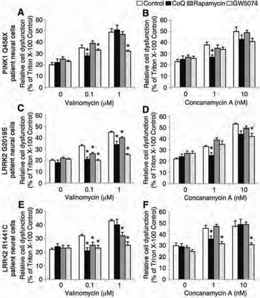
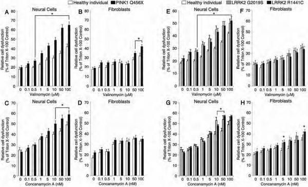
Parkinson's disease (PD) is a common neurodegenerative disorder caused by genetic and environmental factors that results in degeneration of the nigrostriatal dopaminergic pathway in the brain. We analyzed neural cells generated from induced pluripotent stem cells (iPSCs) derived from PD patients and presymptomatic individuals carrying mutations in the PINK1 (PTEN-induced putative kinase 1) and LRRK2 (leucine-rich repeat kinase 2) genes, and compared them to those of healthy control subjects. We measured several aspects of mitochondrial responses in the iPSC-derived neural cells including production of reactive oxygen species, mitochondrial respiration, proton leakage, and intraneuronal movement of mitochondria. Cellular vulnerability associated with mitochondrial dysfunction in iPSC-derived neural cells from familial PD patients and at-risk individuals could be rescued with coenzyme Q(10), rapamycin, or the LRRK2 kinase inhibitor GW5074. Analysis of mitochondrial responses in iPSC-derived neural cells from PD patients carrying different mutations provides insight into convergence of cellular disease mechanisms between different familial forms of PD and highlights the importance of oxidative stress and mitochondrial dysfunction in this neurodegenerative disease.


Figures

Figure 1
Figure 1. Cellular stressors used to treat PD patient iPSC-derived neural cells
To analyze cellular vulnerability and dysfunctional oxidative phosphorylation relevant to PD, ten different cellular stressors were administered to cells derived from PD patients and asymptomatic individuals carrying PINK1 or LRRK2 genetic mutations, and healthy subjects who were not carrying these PD-associated mutations. The cellular stressors were known to affect pathways implicated in PD pathogenesis, such as oxidative phosphorylation, the autophagy-lysosomal pathway or the ubiquitin-proteasome system (UPS).
Figure 2
Figure 2. Effects of cellular stressors on iPSC-derived neural cells with a PINK1 mutation
PINK1 Q456X homozygote PD patient neural cells were treated with low concentrations of valinomycin and mitochondrial reactive oxygen species (mROS) and GSH concentrations were measured. (A–C) After 24 hours of incubation in vehicle, 0.1 μM or 1 μM valinomycin, patient specific neural cells were labeled with a fluorescent indicator of mitochondrial reactive oxygen species and analyzed by fluorescence-activated cell sorting (FACS). (A) Few fluorescent cellular events were recorded from healthy subject neural cells exposed to 0.1 μM valinomycin relative to unstained cultures. In contrast, many fluorescent cellular events representing increased mROS production were recorded from PINK1 patient neural cells exposed to 0.1 μM valinomycin. (B) Quantification of fluorescent events revealed that 0.1 μM valinomycin increased the percentage of fluorescent PINK1 patient specific neural cells. (C) Parallel incubations of healthy subject and patient-specific neural cells with 1 nM or 10 nM hydrogen peroxide (H2O2) showed dose-dependent increases in the percentage of fluorescent cellular events independent of genotype, confirming the specificity of the assay for ROS levels. (D–I) After exposure to valinomycin (0.1 or 1 μM, D), concanamycin A (10 nM, E), MPP+ (5 μM, F), or H2O2 (10 μM, G), PINK1 patient neural cells (black bars) showed reduced GSH levels relative to healthy subject neural cells (white bars). In contrast, low concentrations of 6OHDA (1 or 10 μM, H) or MG132 (1 or 10 μM, I) did not change GSH levels in PINK1 patient or healthy subject neural cells. Data are represented as Mean ± SEM, N=3, * p<0.05 ANOVA PINK1 versus Healthy subject, δ p<0.05 ANOVA chemical stressor versus vehicle.
Figure 3
Figure 3. Respiration in PD patient iPSC-derived neural cells
(A) iPSC-derived neural cells from healthy subjects (black) or PD patients carrying the PINK1 Q456X mutation (red) were administered oligomycin (which inhibits ATP synthesis), FCCP (which induces maximum respiratory capacity) and rotenone (which inhibits total mitochondrial respiration) sequentially and the oxygen consumption rate (OCR), indicative of oxidative phosphorylation, was measured in real time. (B) Quantification of the OCR for each cellular stressor demonstrated an increased basal respiration rate and reduced sensitivity to oligomycin of PINK1 Q456X patient neural cells. (C) Subtraction of the rotenone-induced OCR from the oligomycin OCR demonstrated an increase in proton leakage from PINK1 Q456X PD patient neural cells. (D) iPSC-derived neural cells from healthy subjects (black) and PD patient carrying the LRRK2 G2019S mutation (blue) were treated with oligomycin, FCCP and rotenone sequentially and the OCR was measured in real time. (E) iPSC-derived neural cells from a PD patient carrying the LRRK2 G2019S mutation exhibited a reduced basal respiration rate and increased sensitivity to FCCP. (F) Subtraction of the rotenone-induced OCR from the oligomycin OCR demonstrated that the levels of proton leakage from LRRK2 G2019S patient neural cells were similar to those for healthy subject neural cells. (G) iPSC-derived neural cells from healthy subjects (black) or individuals carrying the LRRK2 R1441C mutation (green) were administered oligomycin, FCCP and rotenone sequentially and the OCR was measured in real time. (H) LRRK2 R1441C patient neural cells exhibited a reduced basal respiration rate and increased sensitivity to both oligomycin and rotenone. (I) Subtraction of the rotenone-induced OCR from the oligomycin OCR demonstrated that the levels of proton leakage from LRRK2 R1441C patient neural cells were similar to those for healthy subject neural cells. Data are represented as Mean ± SEM, N=3, * p<0.05 ANOVA.
Figure 4
Figure 4. Mobility of mitochondria in the proximal axon of PD patient iPSC-derived neurons
(A) Analysis of live cell imaging showed that the mitochondria in the proximal axons of neurons from individuals carrying LRRK2 G2019S and R1441C mutations moved more randomly than similarly located mitochondria in healthy control neurons. Further analysis showed that mitochondrial movements in the proximal axons of LRRK2 G2019S patient neurons were more bidirectional (both anterograde and retrograde) than mitochondrial movements in healthy control neurons. *p<0.05 ANOVA. (B) The axons of neurons from healthy subjects and individuals carrying PINK1 Q456X, LRRK2 G2019S and LRRK2 R1441C mutations the same length (Kolmogorov–Smirnov test). (C) However, the mitochondria in neurons from individuals carrying the LRRK2 R1441C mutation were shorter than mitochondria from healthy control neurons. *p<0.05, Kolmogoro–Smirnov test. (D) Representative kymographs of mitochondria labeled with mitoDendra fluorescent protein in proximal axons from neurons of healthy subjects and from individuals carrying PINK1 Q456X, LRRK2 G2019S and LRRK2 R1441C mutations taken every 5 seconds for 5 minutes. (E) Representative images of mitochondria labeled with mitoDendra fluorescent protein in the proximal axons of neurons from healthy subjects and from individuals carrying PINK1 Q456X, LRRK2 G2019S and LRRK2 R1441C mutations. Data are represented as Mean ± SD, N=3.
Figure 5
Figure 5. Pharmacological treatment of iPSC-derived neural cells from PD patients
iPSC-derived neural cells from individuals with PINK1 and LRRK2 mutations were treated with Coenzyme Q10, rapamycin or GW5074 after exposure to low doses of valinomycin or concanamycin A and LDH release was measured. (A–F) The administration of either valinomycin (0.1 μM and 1 μM) or concanamycin A (1 nM and 10 nM) increased LDH release from PINK1 Q456X, LRRK2 G2019S and LRRK2 R1441C PD patient neural cells (white bars). (A) Treatment with 1 μM Coenzyme Q10 (black bars) or 1 μM GW5074 (light grey bars) but not 1 μM rapamycin (dark grey bars) reduced LDH release by PINK1 PD patient neural cells induced by valinomycin (control, white bars). (B) Treatment with Coenzyme Q10 but not rapamycin or GW5074 reduced LDH release from PINK1 Q456X PD patient neural cells induced by concanamycin A (control, white bars). (C) Treatment with Coenzyme Q10, rapamycin or GW5074 reduced LDH release by LRRK2 G2019S PD patient neural cells induced by valinomycin (control, white bars). (D) Treatment with Coenzyme Q10 or GW5074 but not rapamycin reduced LDH release by LRRK2 G2019S PD patient neural cells induced by concanamycin A (control, white bars). (E) Treatment with Coenzyme Q10, rapamycin or GW5074 reduced LDH release by neural cells from individuals carrying the LRRK2 R1441C mutation induced by valinomycin (control, white bars). (F) Treatment with Coenzyme Q10 or GW5074 but not rapamycin reduced LDH release by neural cells from individuals carrying the LRRK2 R1441C mutation induced by concanamycin A (control, white bars). The relative cell dysfunction was calculated from LDH release values as a percentage of untreated cells completely lysed by incubation with Triton X-100. Data are represented as Mean ± SEM, N=3, * p<0.05 ANOVA.
Figure 6
Figure 6. Cell type specific vulnerability to cellular stressors and oxidative stress
iPSC-derived neural cells from individuals carrying PINK1 and LRRK2 mutations are more vulnerable to both valinomycin and concanamycin A than primary fibroblasts taken from PD patients. (A) In response to a range of valinomycin concentrations, PINK1 patient neural cells (white bars) released more LDH than healthy control neural cells (black bars). (B) Similarly, PINK1 PD patient fibroblasts (white bars) released more LDH than fibroblasts from healthy subjects (black bars) in response to valinomycin, albeit at greater concentrations of valinomycin than PINK1 PD patient neural cells. (C) In response to a range of concentrations of concanamycin A, PINK1 PD patient neural cells (white bars) released more LDH than healthy subject neural cells (black bars). (D) In contrast, PINK1 PD patient (white bars) and healthy subject (black bars) fibroblasts released similar amounts of LDH in response to concanamycin A. (E) In response to a range of concentrations of valinomycin, neural cells from individuals carrying LRRK2 G2019S (grey bars) and LRRK2 R1441C (black bars) mutations released more LDH than healthy subject neural cells (white bars). (F) In contrast, LRRK2 G2019S PD patient (grey bars), LRRK2 R1441C PD patient (black bars) and healthy subject (white bars) fibroblasts released similar levels of LDH in response to valinomycin. (G) In response to a range of concentrations of concanamycin A, neural cells from individuals carrying LRRK2 G2019S mutation (grey bars) and LRRK2 R1441C mutation (black bars) released more LDH than healthy subject neural cells (white bars). (H) Similarly, LRRK2 G2019S PD patient fibroblasts (grey bars) and LRRK2 R1441C patient fibroblasts (black bars) released more LDH than healthy subject fibroblasts (white bars) in response to concanamycin A, but the LDH levels were lower than for neural cells from individuals with LRRK2 G2019S and R1441C mutations. The relative cell dysfunction was calculated from LDH release values as a percentage of untreated cells completely lysed by incubation with Triton X-100. Data are represented as Mean ± SEM, N=3, * p<0.05 ANOVA.

Similar articles

Cited by

References

    1. Chung CY, Seo H, Sonntag KC, Brooks A, Lin L, Isacson O. Cell type-specific gene expression of midbrain dopaminergic neurons reveals molecules involved in their vulnerability and protection. Hum Mol Genet. 2005 Jul 1;14:1709. - PMC - PubMed
    1. Guzman JN, Sanchez-Padilla J, Wokosin D, Kondapalli J, Ilijic E, Schumacker PT, Surmeier DJ. Oxidant stress evoked by pacemaking in dopaminergic neurons is attenuated by DJ-1. Nature. 2010 Dec 2;468:696. - PMC - PubMed
    1. Lesnick TG, Papapetropoulos S, Mash DC, Ffrench-Mullen J, Shehadeh L, de Andrade M, Henley JR, Rocca WA, Ahlskog JE, Maraganore DM. A genomic pathway approach to a complex disease: axon guidance and Parkinson disease. PLoS Genet. 2007 Jun;3:e98. - PMC - PubMed
    1. Langston JW, Ballard P, Tetrud JW, Irwin I. Chronic parkinsonism in humans due to a product of meperidine analog synthesis. Science. 1983;219:979. - PubMed
    1. Gorell JM, Johnson CC, Rybicki BA, Peterson EL, Richardson RJ. The risk of Parkinson's disease with exposure to pesticides, farming, well water, and rural living. Neurology. 1998;50(5):1346–50. - PubMed

Publication types

MeSH terms