. 2012 Jul 4;32(27):9182-93.
doi: 10.1523/JNEUROSCI.1461-12.2012.

Modulation of circuit feedback specifies motor circuit output

Affiliations

Modulation of circuit feedback specifies motor circuit output

Dawn M Blitz et al. J Neurosci. .

Abstract

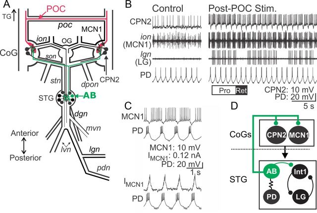
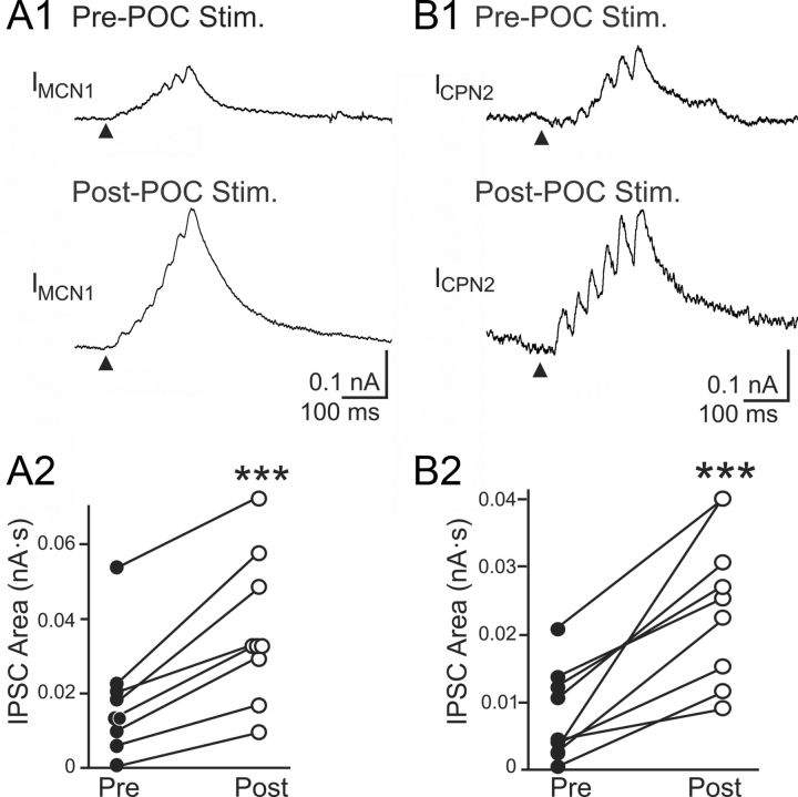
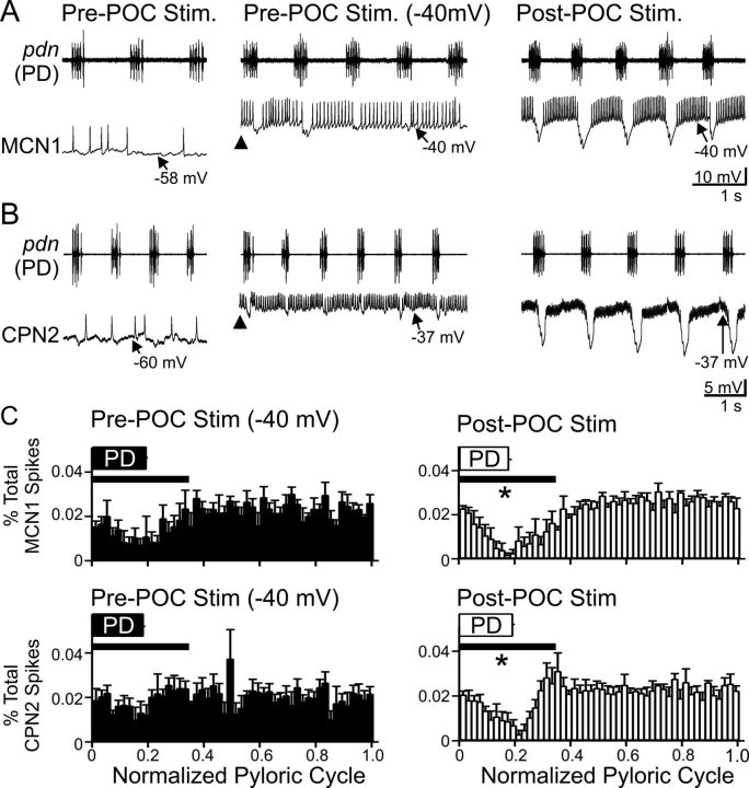
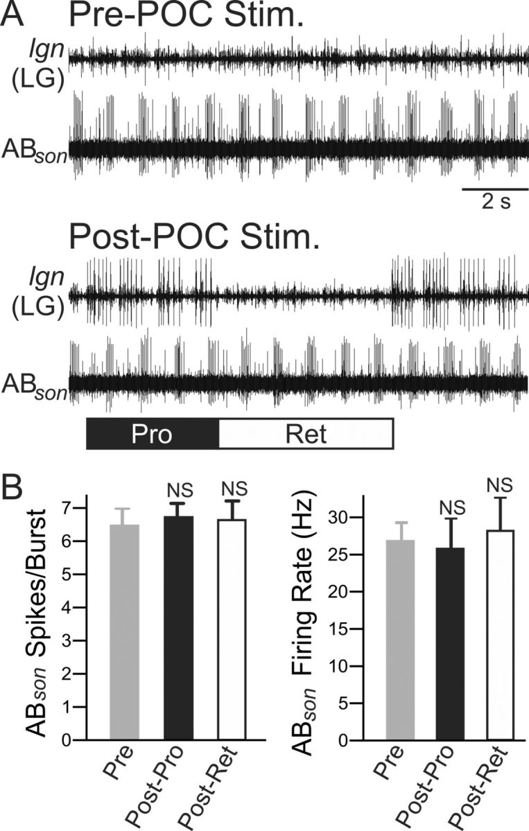
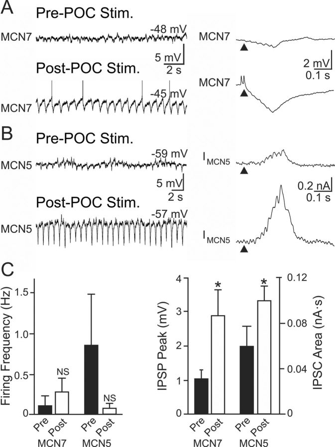
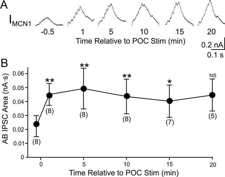
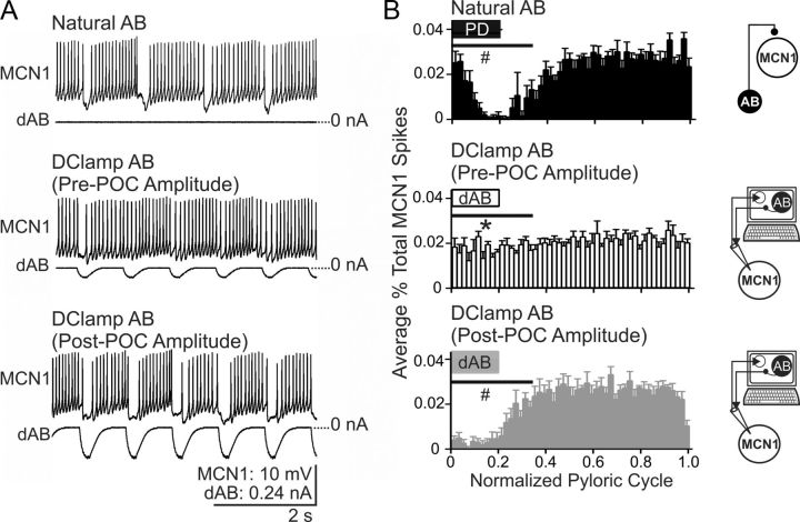
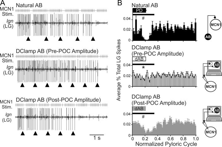
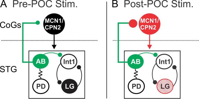
Bidirectional communication (i.e., feedforward and feedback pathways) between functional levels is common in neural systems, but in most systems little is known regarding the function and modifiability of the feedback pathway. We are exploring this issue in the crab (Cancer borealis) stomatogastric nervous system by examining bidirectional communication between projection neurons and their target central pattern generator (CPG) circuit neurons. Specifically, we addressed the question of whether the peptidergic post-oesophageal commissure (POC) neurons trigger a specific gastric mill (chewing) motor pattern in the stomatogastric ganglion solely by activating projection neurons, or by additionally altering the strength of CPG feedback to these projection neurons. The POC-triggered gastric mill rhythm is shaped by feedback inhibition onto projection neurons from a CPG neuron. Here, we establish that POC stimulation triggers a long-lasting enhancement of feedback-mediated IPSC/Ps in the projection neurons, which persists for the same duration as POC-gastric mill rhythms. This strengthened CPG feedback appears to result from presynaptic modulation, because it also occurs in other projection neurons whose activity does not change after POC stimulation. To determine the function of this strengthened feedback synapse, we compared the influence of dynamic-clamp-injected feedback IPSPs of pre- and post-POC amplitude into a pivotal projection neuron after POC stimulation. Only the post-POC amplitude IPSPs elicited the POC-triggered activity pattern in this projection neuron and enabled full expression of the POC-gastric mill rhythm. Thus, the strength of CPG feedback to projection neurons is modifiable and can be instrumental to motor pattern selection.


Figures

Figure 1.
Figure 1.
POC neurons activate MCN1 and CPN2 to trigger a particular version of the gastric mill rhythm. A, Schematic of the STNS illustrating the axon pathways of projection (MCN1, CPN2), CPG feedback (AB), and extrinsic input (POC) neurons. There is a single MCN1 and CPN2 in each CoG, ∼100 POC axons entering each CoG, and a single AB neuron in the STG. For clarity, the complete axonal pathways of MCN1 and CPN2 are only illustrated unilaterally. Abbreviations: Ganglia: CoG, Commissural ganglion; OG, oesophageal ganglion; STG, stomatogastric ganglion. Nerves: dgn, dorsal gastric nerve; dpon, dorsal posterior oesophageal nerve; ion, inferior oesophageal nerve; lgn, lateral gastric nerve; lvn, lateral ventricular nerve; mvn, medial ventricular nerve; pdn, pyloric dilator nerve; poc, post-oesophageal commissure; son, superior oesophageal nerve; stn, stomatogastric nerve. Neurons: AB, anterior burster; CPN2, commissural projection neuron 2; MCN1, modulatory commissural neuron 1; POC, post-oesophageal commissure. B, Left, During control conditions, MCN1 and CPN2 are weakly active, there is an ongoing pyloric rhythm (PD neuron) and there is no gastric mill rhythm (LG neuron silent in the lgn recording). Right, POC stimulation triggers a gastric mill rhythm that is characterized by rhythmic interruptions in the activity of MCN1, CPN2, and the gastric mill protractor motor neuron LG that are time-locked to the ongoing pyloric rhythm (e.g., PD neuron) (Blitz et al., 2008). The white (“PRO”) and black (“RET”) bars indicate protraction and retraction phases of one cycle of the gastric mill rhythm. C, Pyloric (PD neuron)-timed inhibition is evident in a MCN1 recording as barrages of IPSPs that transiently eliminate MCN1 activity (top) and IPSCs in voltage-clamp recording mode (bottom). MCN1 recordings in both panels are from the same preparation. D, Schematic highlighting the feedback inhibitory synapse from AB onto MCN1 and CPN2 in the CoGs (Blitz and Nusbaum, 2008). The AB soma is in the STG, from which it projects an axon to innervate each CoG and cause glutamatergic inhibition of projection neurons, including MCN1 and CPN2 (Blitz and Nusbaum, 1999). The dotted line indicates spatial separation of the CoGs from the STG. AB is electrically coupled to both PD neurons, causing them to fire during the same phase of the pyloric rhythm (Marder and Eisen, 1984a). Thus, activity of the more readily accessible PD neuron is often used as a marker of AB activity.
Figure 2.
Figure 2.
POC stimulation triggers an increased AB IPSC area in MCN1 and CPN2. Average IPSC barrage in MCN1 (A1) and CPN2 (B1) is shown before and after POC stimulation. Fifty AB bursts were averaged for MCN1, and 10–13 AB bursts were averaged for CPN2. The arrowheads indicate onset of AB/PD activity. Recordings in A and B are from separate preparations. A2, B2, The average area of the AB-mediated IPSC barrage per preparation in MCN1 (n = 9) (A2) and CPN2 (n = 9) (B2) is plotted before and after POC stimulation. Each line connects the corresponding data points for a single preparation. ***p < 0.001.
Figure 3.
Figure 3.
CPG feedback to projection neurons is strengthened by POC stimulation. A, B, Left, At their resting potentials before POC stimulation, AB inhibition is weak in MCN1 and CPN2. Middle, Before POC stimulation, MCN1 and CPN2 were depolarized via intracellular current injection to approximately −40 mV (arrowheads). Under this condition, AB inhibition elicits small-amplitude hyperpolarizations in MCN1 and CPN2 that modestly regulate their firing rate. Right, POC stimulation depolarizes MCN1 and CPN2 to approximately −40 mV, during which time AB inhibition elicits larger amplitude hyperpolarizations and longer duration pauses in their firing, despite their firing rate being increased (MCN1) or comparable (CPN2) relative to their activity during the current injections before POC stimulation (see Results). MCN1 and CPN2 recordings are from different preparations. C, The percentage of MCN1 (top) and CPN2 (bottom) action potentials per bin (2% of pyloric cycle per bin) and the phase of AB/PD activity (PD box) are plotted against the normalized pyloric cycle (see Materials and Methods) before (left) and after (right) POC stimulation. The pre-POC stimulation data were obtained while MCN1/CPN2 was depolarized by continual current injection to approximately −40 mV. After POC stimulation, there is a larger decrease in the percentage of MCN1/CPN2 spikes during the initial 35% of the normalized pyloric cycle (bar), relative to pre-POC stimulation. Means ± SEM per bin are plotted. MCN1: n = 4; CPN2: n = 4. *p < 0.05.
Figure 4.
Figure 4.
POC stimulation does not trigger a change in AB activity. A, Suction electrode recordings from the AB axon in the son illustrate that the activity of AB appears similar pre- and post-POC stimulation. Before POC stimulation, there was no gastric mill rhythm (no LG activity in lgn), but there was a pyloric rhythm (rhythmic AB bursting). Note that POC stimulation triggered a gastric mill rhythm, represented by the LG neuron bursting (lgn), but did not evidently change AB firing rate or burst duration. B, Quantification of the average number of action potentials per pyloric-timed AB burst (left) and AB intraburst firing frequency (right) indicate no difference between pre- and post-POC stimulation. After POC stimulation, AB activity was quantified separately during the protraction (Post-Pro) and retraction (Post-Ret) phases of the gastric mill rhythm. Error bars indicate SEM. NS, p > 0.05.
Figure 5.
Figure 5.
POC stimulation triggers increased AB IPSP/C amplitude in nonactivated CoG projection neurons. A, B, Left, The activity of the CoG projection neurons MCN7 (A) and MCN5 (B) is not altered after POC stimulation. Right, However, the average AB-mediated IPSP amplitude in MCN7 (10 cycles) and IPSC amplitude in MCN5 (20 cycles) increases after POC stimulation. C, There was no change in MCN7 or MCN5 firing frequency after POC stimulation. However, the AB-mediated IPSP amplitude measured in MCN7 and IPSC area measured in MCN5 both increased after POC stimulation. Error bars indicate SEM. *p < 0.05; NS, p > 0.05.
Figure 6.
Figure 6.
The POC modulation of AB feedback to CoG projection neurons is long-lasting. A, Average traces (10 cycles/trace) of AB IPSCs in MCN1 at several time points relative to POC stimulation. B, The AB IPSC area is plotted against time relative to POC stimulation. Numbers in brackets indicate the number of preparations contributing to each data point. Error bars indicate SEM. **p < 0.01; *p < 0.05; NS, p > 0.05.
Figure 7.
Figure 7.
The pre-POC stimulation amplitude of AB inhibition is not sufficient to elicit pyloric-timed activity in MCN1 after POC stimulation. A, After POC stimulation, natural AB inhibition suppresses MCN1 activity, resulting in a pyloric-timed MCN1 firing pattern. Replacing the natural AB inhibition of MCN1 with dynamic-clamp AB (dAB) inhibition injected at the pre-POC amplitude is not sufficient to reliably suppress MCN1 firing, although it does modulate the MCN1 firing frequency. Using the post-POC amplitude, dAB inhibition injected into MCN1 does successfully suppress MCN1 firing, causing pyloric-timed MCN1 activity similar to that occurring in response to natural AB inhibition. B, The percentage of MCN1 action potentials per bin and the phase of AB/PD activity (box) are plotted against the normalized pyloric cycle for three conditions: natural AB inhibition of MCN1, plus pre- and post-POC amplitude dAB inhibition of MCN1. In response to natural AB inhibition and post-POC amplitude dAB inhibition, there is a decrease in the percentage of MCN1 spikes occurring during the initial 35% of the normalized pyloric cycle relative to pre-POC dAB inhibition. The effect of dAB inhibition begins earlier in the normalized pyloric cycle than the natural AB inhibition because there is only a brief delay (∼5 ms) from the onset of the dAB burst in the dynamic-clamp software to the injection of dAB inhibition into MCN1. In contrast, there is a conduction delay of ∼25 ms for the natural AB spikes to travel from the STG to each CoG (Blitz and Nusbaum, 2008). The horizontal bars indicate the region of statistical analysis. Schematics to the right of each panel indicate whether natural AB inhibition was present or dAB inhibition was injected into MCN1. The arrows indicate that MCN1 voltage is read into, and dAB current is sent out from the dynamic-clamp software. Error bars indicate SEM. *p < 0.05 compared with natural AB; #p < 0.05 compared with pre-POC dAB.
Figure 8.
Figure 8.
POC-modulation of the AB feedback synapse to MCN1 is necessary for the POC-triggered gastric mill motor pattern. A, MCN1 was stimulated (MCN1 Stim.) using the activity patterns recorded in response to natural AB feedback (top), pre-POC amplitude dAB (middle), and post-POC amplitude dAB (bottom) (see Materials and Methods). The pre-POC amplitude dynamic-clamp stimulation pattern did not elicit pyloric-timed interruptions in the LG bursts, whereas the other two conditions did elicit pyloric-timed pauses in LG firing such as are characteristic of the POC-gastric mill rhythm. The arrowheads indicate onset of natural or dynamic-clamp AB activity. B, The average percentage of LG action potentials per bin and the phase of AB/PD activity are plotted against the normalized pyloric cycle for three conditions: top, natural AB; middle, pre-POC amplitude dAB; bottom, post-POC amplitude dAB. MCN1 stimulation patterns based on the MCN1 activity pattern during natural AB inhibition and post-POC amplitude dAB inhibition resulted in a smaller percentage of LG spikes during the initial 35% of the normalized pyloric cycle, relative to the MCN1 pattern occurring during the pre-POC amplitude dAB inhibition. The horizontal bars indicate the region of statistical analysis. Schematics to the right of each panel indicate whether natural AB inhibition was present or dAB inhibition was injected into MCN1. The arrows are as in Figure 7. Error bars indicate SEM. *p < 0.05 compared with natural AB; #p < 0.05 compared with pre-POC dAB.
Figure 9.
Figure 9.
POC stimulation triggers a specific version of the gastric mill rhythm via postsynaptic activation of MCN1 and CPN2 and presynaptic modulation of the AB feedback synapse to these projection neurons. A, Before POC stimulation, AB inhibition of MCN1 and CPN2 is weak, plus the projection neurons and LG are weakly active or silent and there is no gastric mill rhythm (black circles). B, POC stimulation triggers a long-lasting activation of MCN1 and CPN2 (red) that includes a pyloric rhythm-timed activity pattern due to a long-lasting strengthening of the AB inhibition of these projection neurons (red) without a change in AB activity (green). Note that there is not a concomitant strengthening of AB inhibition within the STG (green). The presynaptic and postsynaptic POC actions on MCN1 and CPN2 are both necessary for the POC version of the gastric mill rhythm, which is characterized by pyloric-timed activity patterns in the projection neurons and the protraction neuron LG (red stripes). The dotted line indicates spatial separation of the CoGs from the STG.

Similar articles

Cited by

References

    1. Antri M, Auclair F, Albrecht J, Djeudjang N, Dubuc R. Serotoninergic modulation of sensory transmission to brainstem reticulospinal cells. Eur J Neurosci. 2008;28:655–667. - PubMed
    1. Antri M, Fénelon K, Dubuc R. The contribution of synaptic inputs to sustained depolarizations in reticulospinal neurons. J Neurosci. 2009;29:1140–1151. - PMC - PubMed
    1. Arshavsky YI, Orlovsky GN, Perret C. Activity of rubrospinal neurons during locomotion and scratching in the cat. Behav Brain Res. 1988;28:193–199. - PubMed
    1. Barrière G, Simmers J, Combes D. Multiple mechanisms for integrating proprioceptive inputs that converge on the same motor pattern-generating network. J Neurosci. 2008;28:8810–8820. - PMC - PubMed
    1. Bartos M, Nusbaum MP. Intercircuit control of motor pattern modulation by presynaptic inhibition. J Neurosci. 1997;17:2247–2256. - PMC - PubMed

Publication types