. 2012 Jul 4;32(27):9429-37.
doi: 10.1523/JNEUROSCI.0394-12.2012.

Otx2 binding to perineuronal nets persistently regulates plasticity in the mature visual cortex

Affiliations

Otx2 binding to perineuronal nets persistently regulates plasticity in the mature visual cortex

Marine Beurdeley et al. J Neurosci. .

Abstract

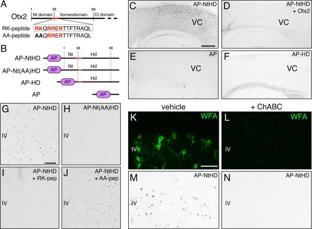
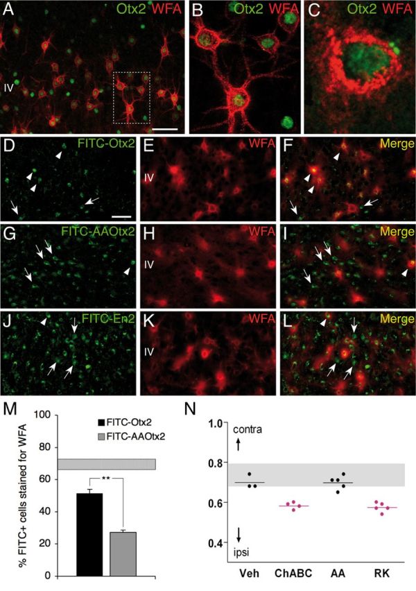
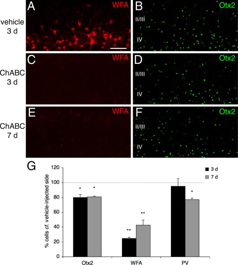
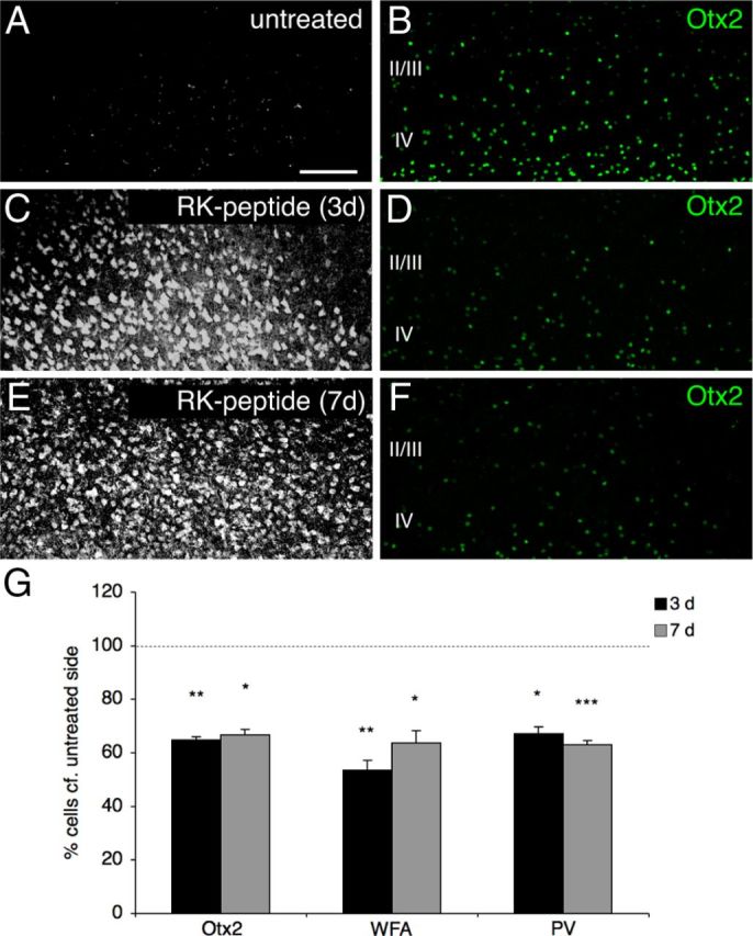
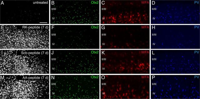
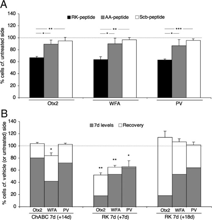
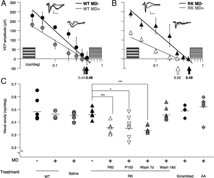
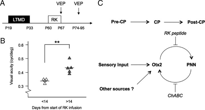
Specific transfer of (orthodenticle homeobox 2) Otx2 homeoprotein into GABAergic interneurons expressing parvalbumin (PV) is necessary and sufficient to open, then close, a critical period (CP) of plasticity in the developing mouse visual cortex. The accumulation of endogenous Otx2 in PV cells suggests the presence of specific Otx2 binding sites. Here, we find that perineuronal nets (PNNs) on the surfaces of PV cells permit the specific, constitutive capture of Otx2. We identify a 15 aa domain containing an arginine-lysine doublet (RK peptide) within Otx2, bearing prototypic traits of a glycosaminoglycan (GAG) binding sequence that mediates Otx2 binding to PNNs, and specifically to chondroitin sulfate D and E, with high affinity. Accordingly, PNN hydrolysis by chondroitinase ABC reduces the amount of endogenous Otx2 in PV cells. Direct infusion of RK peptide similarly disrupts endogenous Otx2 localization to PV cells, reduces PV and PNN expression, and reopens plasticity in adult mice. The closure of one eye during this transient window reduces cortical acuity and is specific to the RK motif, as an Alanine-Alanine variant or a scrambled peptide fails to reactivate plasticity. Conversely, this transient reopening of plasticity in the adult restores binocular vision in amblyopic mice. Thus, one function of PNNs is to facilitate the persistent internalization of Otx2 by PV cells to maintain CP closure. The pharmacological use of the Otx2 GAG binding domain offers a novel, potent therapeutic tool with which to restore cortical plasticity in the mature brain.


Figures

Figure 1.
Figure 1.
An Otx2 motif necessary for binding to cortical sections. A, Putative GAG-binding motif (in red) in the Otx2 N-terminal region bears an RK doublet just before the homeodomain. RK and AA peptide sequences are indicated. B, Schematic representation of AP fusion proteins used in binding experiments: AP-NtHD contains amino-acids 1–98 including the RK doublet and homeodomain; AP-Nt(AA)HD, in which the RK doublet is mutated to an alanine doublet; and AP-HD composed of AP fused directly to the homeodomain (aa 38–98) without the RK doublet. Nonfused AP was used as a negative control. C, E, F, AP staining on fresh-frozen sections reveals AP-NtHD (C) but not AP (E) or AP-HD (F) binding to cells in the posterior cortex (V1). D, Full-length Otx2 disrupts AP-NtHD cortical binding (D). G, H, Higher magnification of V1 sections shows AP-NtHD binding (G) but not AP-Nt(AA)HD binding (H). I, J, RK (I), but not AA (J), peptide antagonizes AP-NtHD binding. K–N, ChABC treatment of frozen sections abolishes WFA staining (K, L) and prevents AP-NtHD binding (M, N). Scale bars: (in C) CF, 350 μm; (in G) GJ, 100 μm; (in K) K, L, 100 μm.
Figure 2.
Figure 2.
Preferential Otx2 capture by PV cells requires the RK doublet. A–C, Codetection of WFA and Otx2 in layer IV of P60 mouse V1b. Inset in A is magnified in B. C, Staining performed under nonpermeabilizing conditions reveals extracellular/membranous Otx2 staining. D, G, J, Distribution of FITC-labeled Otx2 (D), AA-Otx2 (G), and En2-FITC (J) recombinant proteins after injection into P60 mice. E, F, H, I, K, L, WFA detection (E, H, K) and merged FITC-Otx2 images reveal preferential Otx2 capture by WFA-labeled cells (F) compared with AA-Otx2 (I) and En2 (L). Arrowheads, Double-stained cells; arrows, FITC-positive cells not stained with WFA. Scale bars: A, 50 μm; (in D) DL, 50 μm. M, Percentage of FITC-positive cells costained for WFA after FITC-Otx2 or AA-Otx2 injection. Striped bar, endogenous percentage of Otx2-positive cells costained for WFA (mean ± SEM). N, CBI of single-unit recordings from mice treated with ChABC, RK, or AA peptide after a period of brief MD (4 d) in adulthood. Both ChABC and RK peptide infusion similarly reactivate visual cortical plasticity (decrease of CBI compared with non-MD), which is not typically seen in saline- or AA peptide-infused adult mice.
Figure 3.
Figure 3.
PNNs facilitate the preferential capture of endogenous Otx2 by PV cells. A, C, E, ChABC injection into adult V1 disrupts PNNs, as revealed by the near absence of WFA staining at 3 or 7 d postinjection (C, E) compared with the contralateral, vehicle-injected side (A). B, D, F, PNN hydrolysis decreases the number of Otx2-positive cells, respectively, at 3 d (D) and 7 d (F) postinjection compared with the contralateral, vehicle-injected side (B). G, Percentages of Otx2-, WFA-, and PV-positive cells in supragranular layers of V1b (ChABC-treated compared with the contralateral vehicle side) 3 and 7 d postinjection (*p < 0.05, **p < 0.01, paired t test; four mice per group; 700 × 200 μm area). Error bars indicate SEM. Scale bar: (in A) AF, 100 μm .
Figure 4.
Figure 4.
RK peptide reduces endogenous Otx2 localization in V1. B, D, F, Otx2 immunostaining in layers II-IV of V1b from an untreated P60 mouse (B) or a P60 mouse infused for 3 d (D) or 7 d (F) with biotinylated RK peptide. A, C, E, Peptides revealed by Alexa Fluor 633-conjugated streptavidin. G, Percentages of Otx2-, WFA-, and PV-positive cells (infused compared with contralateral, untreated side) in supragranular layers of V1b (*p < 0.05, **p < 0.01, ***p < 0.001, paired t test; four mice per group; 700 × 450 μm area). Error bars indicated SEM. Scale bar: (in A) AF, 100 μm.
Figure 5.
Figure 5.
Specific, reversible control of PV cell Otx2 content and maturational state by RK peptide. A–P, Immunostaining for Otx2, WFA, and PV in layers II-IV of V1b from P60 mice untreated (A–D) or infused for 7 d, respectively, with RK (E–H), Scb (I–L), or AA peptide (M–P). A, E, I, M, Peptides revealed by Alexa Fluor 633-conjugated streptavidin. Scale bar: (in A) AP, 100 μm.
Figure 6.
Figure 6.
Quantification of RK peptide specificity and recovery. A, Percentages of Otx2-, WFA-, and PV-positive cells (infused compared with contralateral, untreated side) in supragranular layers of V1b (*p < 0.05, ***p < 0.001, paired t test; four mice per group; 700 × 450 μm area). Error bars, SEM. B, Percentages of Otx2-, WFA-, and PV-positive cells (infused compared with contralateral, untreated side) in supragranular layers of V1b. Gray bars, 7 d after ChABC injection or RK peptide infusion. Open bars, extended recovery period after ChABC treatment (ChABC 7d + 14d) or RK peptide infusion (RK 7d + 7d and RK 7d + 18d), respectively.
Figure 7.
Figure 7.
Reactivation of plasticity by RK peptide in adulthood: loss of cortical acuity by MD. A, No acuity loss after brief MD (4 d, gray line) compared with nondeprived wild-type (WT) animals (black line) by averaged VEP amplitudes (mean ± SEM) of first negative peak (inset traces; scale: 20 mV, 0.1 s). B, Significant acuity reduction upon brief MD (white symbols) paired with RK peptide infusion (black symbols) into adult mouse V1. C, Cortical acuity in adult WT mice is unaltered by brief MD (4 d) paired with saline, Scb, or AA peptide. A brief window of plasticity persists <18 d after RK peptide treatment ends. The mean value for each group is represented by a horizontal bar.
Figure 8.
Figure 8.
Functional recovery from amblyopia by RK peptide. A, Amblyopia rescue paradigm: long-term MD (LTMD) spanning the CP in mice (P19–P33) is followed by eye reopening and subsequent RK infusion (7 d) at P60. VEP recordings are performed at different stages after RK infusion to assess cortical acuity. B, Amblyopic mice are not rescued by 7 d RK infusion alone (VEP at P67) but are significantly corrected after >14 d of concurrent binocular vision (VEP at P74–P95; **p < 0.01, Student's t test; five mice per group). C, Otx2 accumulation in PV cells driven by sensory experience (Sugiyama et al., 2008) and potentially other sources initially triggers a CP for plasticity, which eventually closes as PNNs condense around them. This, in turn, maintains a stable post-CP state by persistently attracting Otx2 throughout life in a positive feedback loop. Blocking Otx2 transfer (RK peptide) or PNN removal (ChABC) resets PV cells to reactivate CP plasticity.

Similar articles

Cited by

References

    1. Balmer TS, Carels VM, Frisch JL, Nick TA. Modulation of perineuronal nets and parvalbumin with developmental song learning. J Neurosci. 2009;29:12878–12885. - PMC - PubMed
    1. Bavelier D, Levi DM, Li RW, Dan Y, Hensch TK. Removing brakes on adult brain plasticity: from molecular to behavioral interventions. J Neurosci. 2010;30:14964–14971. - PMC - PubMed
    1. Berardi N, Pizzorusso T, Maffei L. Extracellular matrix and visual cortical plasticity: freeing the synapse. Neuron. 2004;44:905–908. - PubMed
    1. Brunet I, Weinl C, Piper M, Trembleau A, Volovitch M, Harris W, Prochiantz A, Holt C. The transcription factor Engrailed-2 guides retinal axons. Nature. 2005;438:94–98. - PMC - PubMed
    1. Brunet I, Di Nardo AA, Sonnier L, Beurdeley M, Prochiantz A. The topological role of homeoproteins in the developing central nervous system. Trends Neurosci. 2007;30:260–267. - PubMed

Publication types

Substances