. 2012 Oct;53(10):1756-64.
doi: 10.1111/j.1528-1167.2012.03570.x. Epub 2012 Jul 5.

Memory in frontal lobe epilepsy: an fMRI study

Affiliations

Memory in frontal lobe epilepsy: an fMRI study

Maria Centeno et al. Epilepsia. 2012 Oct.

Abstract

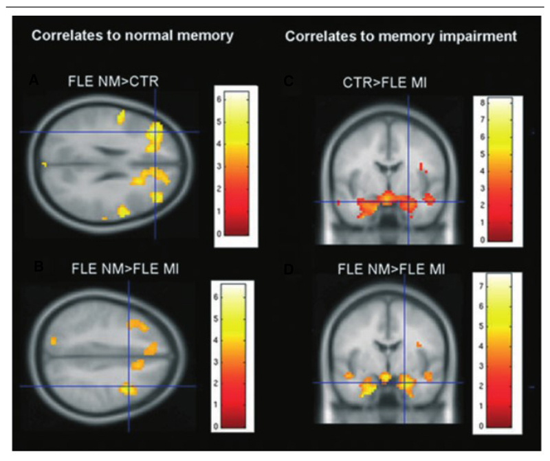
Purpose: Focal epilepsies are often associated with structural and functional changes that may extend beyond the area of seizure onset. In this study we investigated the functional anatomy of memory in patients with frontal lobe epilepsy (FLE), focusing on the local and remote effects of FLE on the networks supporting memory encoding.

Methods: We studied 32 patients with drug-resistant FLE and 18 controls using a functional magnetic resonance imaging (fMRI) memory encoding paradigm.

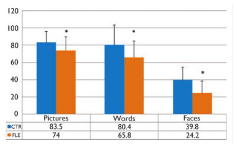
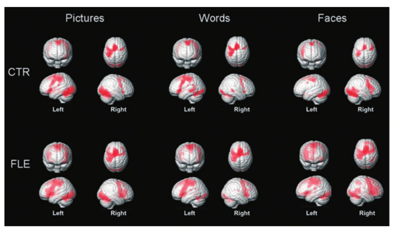
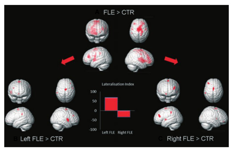
Key findings: During encoding of stimuli, patients with FLE recruited more widely distributed areas than healthy controls, in particular within the frontal lobe contralateral to the seizure onset. Normal memory performance was associated with increased recruitment of frontal areas, and conversely a poor performance was associated with an absence of this increased recruitment and decreased activation in mesial temporal lobe areas.

Significance: In patients with FLE, recruitment of wider areas, particularly in the contralateral frontal lobe, appears to be an effective compensatory mechanism to maintain memory function. Impaired hippocampal activation is relatively rare and, in turn, associated with poor recognition memory.


Figures

Figure 1
Figure 1
Recognition accuracy. Recognition accuracy measures the proportion of correctly remembered items minus proportion of falsely recognized items. Patients with FLE have a significantly decreased recognition accuracy compared to controls for all categories. Mean RA = average of recognition accuracy for the three type of stimuli. Error bars represent I standard deviation. CTR = healthy controls. FLE = patients with frontal lobe epilepsy. *Significant difference of means at a p-value < 0.05.
Figure 2
Figure 2
Verbal/nonverbal performance and laterality of seizure focus. Side of epileptic focus does not have an effect on the recognition accuracy of verbal and nonverbal material. Patients with right FLE show a tendency to perform poorer on the recognition and learning tasks for verbal and nonverbal items. Error bars represent I standard deviation.
Figure 3
Figure 3
Activation maps for the effect of encoding the different stimuli in controls and patients with FLE. Activations are located in dorsolateral and ventrolateral prefrontal cortex, visual areas, and mesial temporal lobe areas (not shown). Lateralization of activations in frontal lobes is dependent on the stimuli type. Activations are right lateralized for faces and left lateralized for words and pictures in both patients and controls. Left central and bilateral medial supplementary motor area activation is induced by joystick response and similar for all stimuli. CTR = healthy controls. FLE = patients with frontal lobe epilepsy.
Figure 4
Figure 4
Effect of frontal lobe epilepsy and focus lateralization. (A) Areas of increased activation in patients with FLE relative to controls (FLE > CTR) collapsed for all item types are located within the frontal lobe areas involved in the task. Increased activation was lateralized differently for patients with left FLE and patients with right FLE (patients with left FLE showed increased activations in the right hemisphere (B) and patients with right in the left hemisphere (C). Bar chart shows the values of lateralization index (LI) of the maps (B, C). LI values range from 1 to –1; positive values indicate a right hemispheric lateralization, whereas negative values indicate a left lateralization. In left FLE, increased activations are lateralized to the right and the inverse for right FLE.
Figure 5
Figure 5
Functional correlates of different performances. (A,B) Patients with FLE with normal memory (FLE NM) showed increased frontal activation when compared to controls (CTR) (FLE NM > CTR) and to patients with memory impairment (FLE Ml) (FLE NM > FLE Ml). (C,D) Patients with FLE with impaired memory showed decreased amygdala and hippocampal activation when compared to CTR (CTR > FLE Ml) and to patients with normal memory (FLE NM > FLE Ml). Scaling bars show T scores for the activations.

Similar articles

Cited by

References

    1. Bastin C, Van der Linden M, Lekeu F, Andres P, Salmon E. Variability in the impairment of recognition memory in patients with frontal lobe lesions. Cortex. 2006;42:983–994. - PubMed
    1. Bernhardt BC, Worsley KI, Besson P, Concha L, Lerch JP, Evans AC, Bernasconi N. Mapping limbic network organization in temporal lobe epilepsy using morphometric correlafions: insights on the relation between mesiotemporal connectivity and cortical atrophy. Neuroimage. 2008;42:515–524. - PubMed
    1. Blumenfeld RS, Ranganath C. Prefrontal cortex and long-term memory encoding: an integrative review of findings from neuropsychology and neuroimaging. Neuroscientist. 2007;13:280–291. - PubMed
    1. Blumenfeld RS, Parks CM, Yonelinas AP, Ranganath C. Putting the pieces together: the role of dorsolateral prefrontal cortex in relational memory encoding. J Cogn Neurosci. 2010;23:257–265. - PMC - PubMed
    1. Bonelli SB, Powell RH, Yogarajah M, Samson RS, Symms MR, Thompson PJ, Koepp MJ, Duncan JS. Imaging memory in temporal lobe epilepsy: predicting the effects of temporal lobe resection. Brain. 2010;133:1186–1199. - PMC - PubMed

Publication types