. 2012 Sep;124(3):411-24.
doi: 10.1007/s00401-012-1009-1. Epub 2012 Jul 6.

Reduced astrocytic NF-κB activation by laquinimod protects from cuprizone-induced demyelination

Affiliations

Reduced astrocytic NF-κB activation by laquinimod protects from cuprizone-induced demyelination

Wolfgang Brück et al. Acta Neuropathol. 2012 Sep.

Abstract

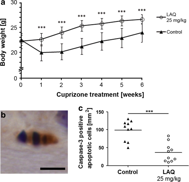
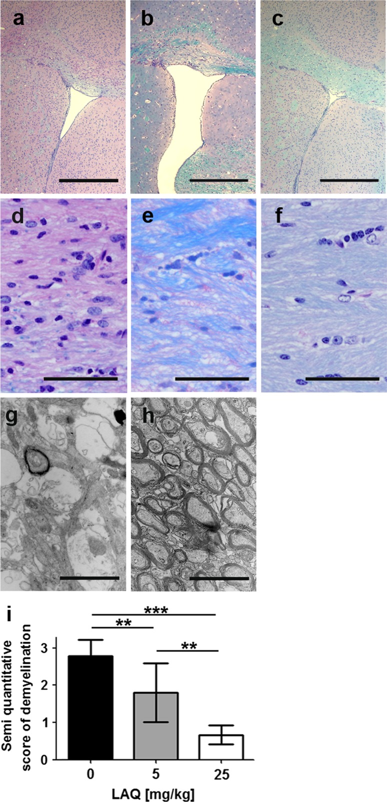
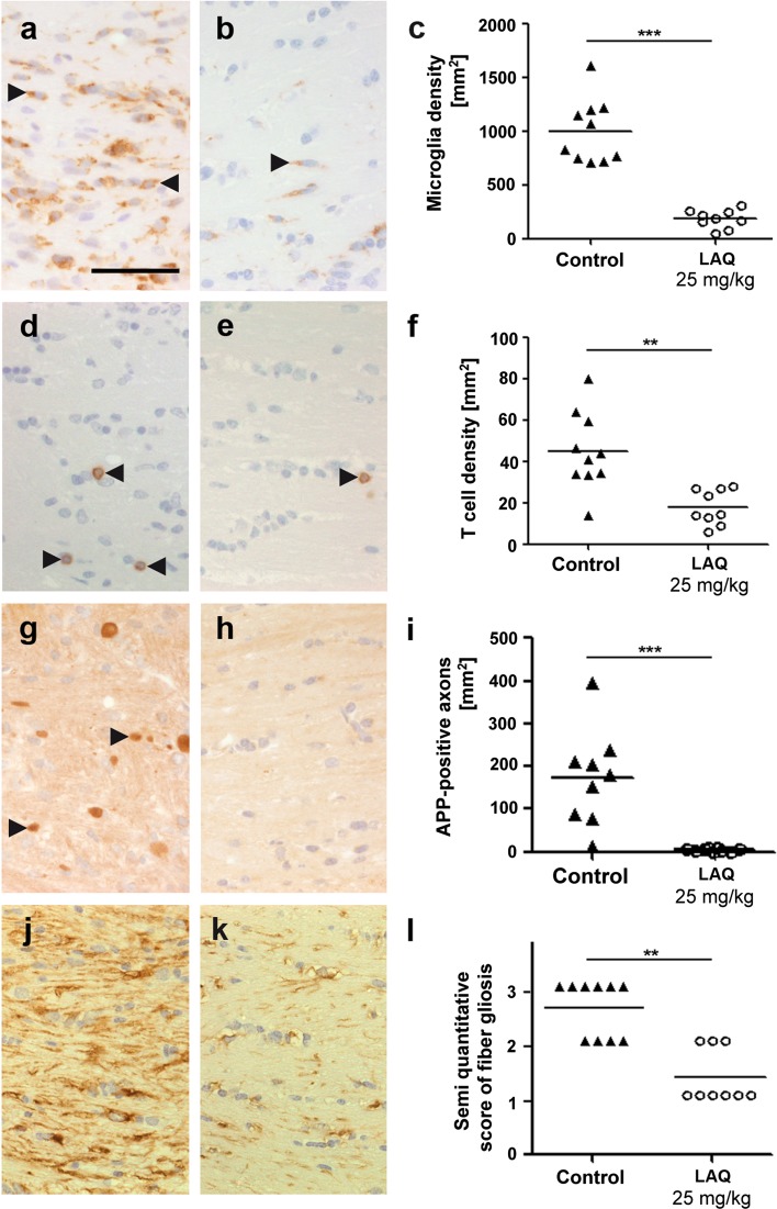
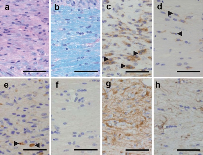
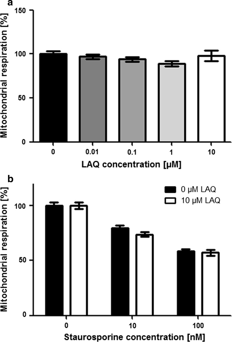
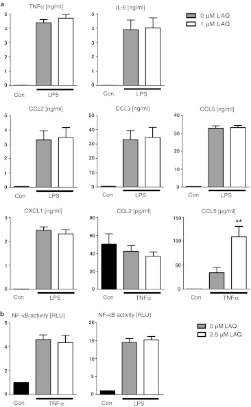
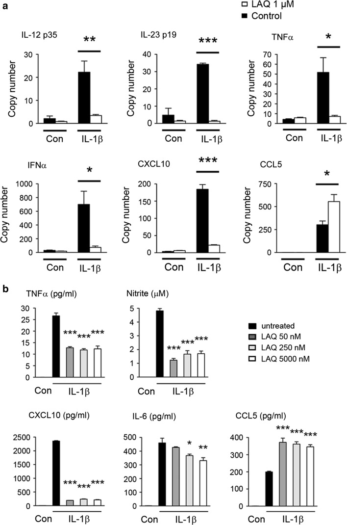
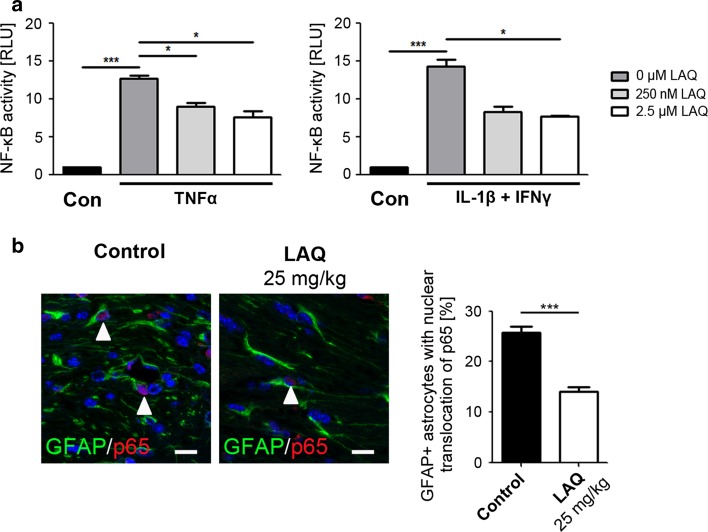
Laquinimod (LAQ) is a new oral immunomodulatory compound that reduces relapse rate, brain atrophy and disability progression in multiple sclerosis (MS). LAQ has well-documented effects on inflammation in the periphery, but little is known about its direct activity within the central nervous system (CNS). To elucidate the impact of LAQ on CNS-intrinsic inflammation, we investigated the effects of LAQ on cuprizone-induced demyelination in mice in vivo and on primary CNS cells in vitro. Demyelination, inflammation, axonal damage and glial pathology were evaluated in LAQ-treated wild type and Rag-1-deficient mice after cuprizone challenge. Using primary cells we tested for effects of LAQ on oligodendroglial survival as well as on cytokine secretion and NF-κB activation in astrocytes and microglia. LAQ prevented cuprizone-induced demyelination, microglial activation, axonal transections, reactive gliosis and oligodendroglial apoptoses in wild type and Rag-1-deficient mice. LAQ significantly decreased pro-inflammatory factors in stimulated astrocytes, but not in microglia. Oligodendroglial survival was not affected by LAQ in vitro. Astrocytic, but not microglial, NF-κB activation was markedly reduced by LAQ as evidenced by NF-κB reporter assay. LAQ also significantly decreased astrocytic NF-κB activation in cuprizone-treated mice. Our data indicate that LAQ prevents cuprizone-induced demyelination by attenuating astrocytic NF-κB activation. These effects are CNS-intrinsic and not mediated by peripheral immune cells. Therefore, LAQ downregulation of the astrocytic pro-inflammatory response may be an important mechanism underlying its protective effects on myelin, oligodendrocytes and axons. Modulation of astrocyte activation may be an attractive therapeutic target to prevent tissue damage in MS.


Figures

Fig. 1
Fig. 1
No weight loss and fewer oligodendroglial apoptoses in LAQ-treated mice. a At all time points, mice treated with 25 mg/kg LAQ (n = 9) display higher body weights than controls (n = 10) during 0.25 % cuprizone challenge (p < 0.001). Data are expressed as mean values with standard error of the mean (SEM). b Apoptotic cells staining positively for active caspase-3 and displaying apoptotic bodies are detected in both groups (scale bar 5 μm, shown for an untreated control), but c shows that LAQ-treated mice (n = 10) display significantly fewer caspase-3-positive apoptoses in the corpus callosum than controls (n = 10) after 1 week of 0.25 % cuprizone (*** p < 0.001)
Fig. 2
Fig. 2
Dose-dependent reduction of callosal demyelination under LAQ after 6 weeks of cuprizone. Demyelination is extensive in controls (n = 9) (a, d, g) compared to moderate and minimal demyelination in mice treated with 5 mg/kg (n = 10) (b, e) and 25 mg/kg (n = 9) (c, f, h) LAQ, respectively, on LFB-PAS-stained sections (af) and electron microscopic images (g, h). The semi-quantitative scores for demyelination (i) are significantly higher in controls than in both treatment groups. Mice treated with 25 mg/kg LAQ display significantly lower scores than animals treated with 5 mg/kg LAQ (*p < 0.01, ***p < 0.001). Scale bars ac 500 μm, d–f 50 μm, g, h 2 μm
Fig. 3
Fig. 3
Reduced inflammation, axonal damage and gliosis after LAQ treatment. Compared to untreated controls (n = 10) (left column), LAQ-treated animals (n = 9) (middle column) display significantly reduced Mac3-positive microglia (ac) and CD3-positive T cell (df) infiltration, APP-positive axonal spheroids (gi) and GFAP-positive fibrillary gliosis (jl) in the corpus callosum (scale bars 50 μm). Black arrowheads mark exemplary Mac3-positive microglia (a, b), CD3-positive T cells (d, e) and APP-positive axonal spheroids (g) (**p < 0.01, ***p < 0.001)
Fig. 4
Fig. 4
Reduced demyelination, microglia infiltration, acute axonal damage and gliosis in LAQ-treated Rag1−/− mice (n = 9) compared to untreated controls (n = 10) after 6 weeks of cuprizone. Compared to untreated Rag1−/− mice (a, c, e, g) LAQ-treated Rag1−/− mice (b, d, f, h) show significantly less demyelination (a, b), fewer Mac3-positive microglia (c, d), fewer APP-positive axonal spheroids (e, f) and less extensive fiber gliosis (g, h) in the corpus callosum (scale bars 50 μm). Black arrowheads mark exemplary Mac3-positive microglia (c, d) and APP-positive axonal spheroids (e)
Fig. 5
Fig. 5
No effect of LAQ on oligodendroglial viability and cell death. a OPCs treated with 0, 0.01, 0.1, 1 and 10 μM LAQ for 48 h display similar mitochondrial respiration determined by MTT assays. b Treatment with staurosporine for 12 h results in a dose-dependent decrease in mitochondrial respiration indicating cell death. The decrease in mitochondrial respiration was similar in treated (10 μM LAQ) and untreated cells. Graphs show mean values with SEM from three independent experiments
Fig. 6
Fig. 6
No marked effect of LAQ on microglial cytokine secretion (a) and NF-κB activation (b). a Microglial cultures were exposed to LPS (10 ng/ml) or TNFα (10 ng/ml) for 18 h in the absence or presence of 1 μM LAQ. LAQ-treated cells also received a pre-incubation with LAQ for 2 h. Cyto- and chemokines were determined in the supernatant. LPS and TNFα stimulation significantly increased the cytokine levels compared to unstimulated controls (p < 0.05 for TNFα-induced CCL5, p < 0.001 for remaining cytokines shown). Cells with and without LAQ (pre-) treatment showed similar levels of LPS-induced TNFα, IL-6, CXCL1, CCL2, CCL3 and CCL5. After TNFα stimulation, LAQ-treated cells showed higher CCL5 levels (*p < 0.01), but similar CCL2 release. Graphs show mean values with SEM from two independent experiments. b Treatment with 2.5 μM LAQ has no effect on NF-κB activity in stimulated primary mouse microglia assessed by a NF-κB reporter assay. Cells stimulated with LPS or TNFα were pre-treated with and without LAQ. NF-κB activity was measured luminometrically (RLU relative light units). Cells exposed to these cytokines displayed a significant NF-κB activation that was not reduced by the (pre-) treatment with LAQ. Graphs show mean values with SEM from three experiments
Fig. 7
Fig. 7
Down-modulation of inflammatory factors by LAQ treatment in vitro. LAQ reduces astrocytic inflammatory factors assessed on mRNA level by qPCR (a) and on protein level by ELISA and nitrite assay in supernatants (b). Cultures were pre-treated with LAQ and then exposed to cytokines for 24 h. For factors including TNFα, IFNα, IL-23 p19, IL-12 p35 and CXCL10, LAQ strongly reduced induction by IL-1β/IFNγ (a, b). Nitrite levels were significantly decreased by LAQ treatment (*p < 0.05, **p < 0.01, ***p < 0.001). Graphs show mean values with SEM from three independent experiments
Fig. 8
Fig. 8
Reduction of astrocytic NF-κB activation by LAQ in vitro and in vivo. a Pre-treatment with LAQ reduces NF-κB activity in stimulated primary mouse astrocytes assessed by NF-κB reporter assay. Transfected cells were stimulated with IL-1β and IFNγ or TNFα alone and pre-treated with 0, 250 nM and 2.5 μM LAQ. Unstimulated cells served as controls. NF-κB activity was measured luminometrically (RLU relative light units). Both cytokine treatments without LAQ markedly increase NF-κB activation compared to unstimulated controls. Pre-treatment with LAQ significantly reduces NF-κB activation after cytokine stimulation. Graphs show mean values with SEM from three independent experiments. b Double immunofluorescence with antibodies to p65 (red) and GFAP (green) shows that the proportion of callosal astrocytes with nuclear p65 immunoreactivity (marked by white arrowheads) is significantly reduced in LAQ-treated mice (n = 8) (14.0 ± 0.9 %) in comparison to controls (n = 9) (25.8 ± 1.1 %) after 6 weeks of cuprizone (scale bars 10 μm) (*p < 0.05, ***p < 0.001)

Comment in

References

    1. Acarin L, Gonzalez B, Castellano B. Triflusal posttreatment inhibits glial nuclear factor-kappaB, downregulates the glial response, and is neuroprotective in an excitotoxic injury model in postnatal brain. Stroke. 2001;32:2394–2402. doi: 10.1161/hs1001.097243. - DOI - PubMed
    1. Bjartmar C, Trapp BD. Axonal and neuronal degeneration in multiple sclerosis: mechanisms and functional consequences. Curr Opin Neurol. 2001;14:271–278. doi: 10.1097/00019052-200106000-00003. - DOI - PubMed
    1. Blakemore WF. Observations on oligodendrocyte degeneration, the resolution of status spongiosus and remyelination in cuprizone intoxication in mice. J Neurocytol. 1972;1:413–426. doi: 10.1007/BF01102943. - DOI - PubMed
    1. Brambilla R, Bracchi-Ricard V, Hu WH, Frydel B, Bramwell A, Karmally S, Green EJ, Bethea JR. Inhibition of astroglial nuclear factor kappaB reduces inflammation and improves functional recovery after spinal cord injury. J Exp Med. 2005;202:145–156. doi: 10.1084/jem.20041918. - DOI - PMC - PubMed
    1. Brambilla R, Persaud T, Hu X, Karmally S, Shestopalov VI, Dvoriantchikova G, Ivanov D, Nathanson L, Barnum SR, Bethea JR. Transgenic inhibition of astroglial NF-kappa B improves functional outcome in experimental autoimmune encephalomyelitis by suppressing chronic central nervous system inflammation. J Immunol. 2009;182:2628–2640. doi: 10.4049/jimmunol.0802954. - DOI - PMC - PubMed

Publication types

MeSH terms

LinkOut - more resources