

BPIFB1 (LPLUNC1) is upregulated in cystic fibrosis lung disease
- PMID: 22767025
- PMCID: PMC3470695
- DOI: 10.1007/s00418-012-0990-8
BPIFB1 (LPLUNC1) is upregulated in cystic fibrosis lung disease
Abstract
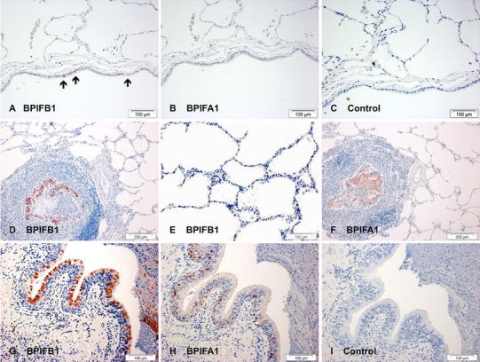
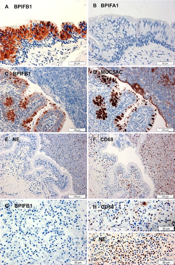
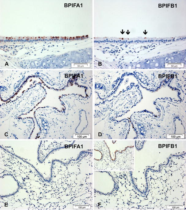
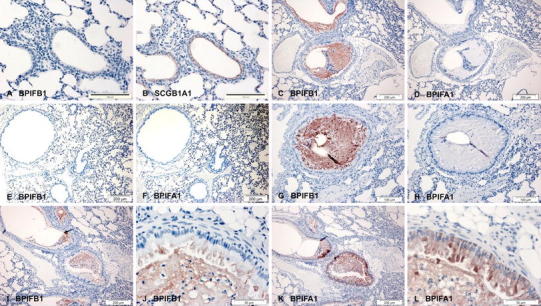
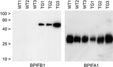
Although the biology the PLUNC (recently renamed BPI fold, BPIF) family of secreted proteins is poorly understood, multiple array based studies have suggested that some are differentially expressed in lung diseases. We have examined the expression of BPIFB1 (LPLUNC1), the prototypic two-domain containing family member, in lungs from CF patients and in mouse models of CF lung disease. BPIFB1 was localized in CF lung samples along with BPIFA1, MUC5AC, CD68 and NE and directly compared to histologically normal lung tissues and that of bacterial pneumonia. We generated novel antibodies to mouse BPIF proteins to conduct similar studies on ENaC transgenic (ENaC-Tg) mice, a model for CF-like lung disease. Small airways in CF demonstrated marked epithelial staining of BPIFB1 in goblet cells but staining was absent from alveolar regions. BPIFA1 and BPIFB1 were not co-localised in the diseased lungs. In ENaC-Tg mice there was strong staining of both proteins in the airways and luminal contents. This was most marked for BPIFB1 and was noted within 2 weeks of birth. The two proteins were present in distinct cells within epithelium. BPIFB1 was readily detected in BAL from ENaC-Tg mice but was absent from wild-type mice. Alterations in the expression of BPIF proteins is associated with CF lung disease in humans and mice. It is unclear if this elevation of protein production, which results from phenotypic alteration of the cells within the diseased epithelium, plays a role in the pathogenesis of the disease.
Figures
Similar articles
-
Association of innate defense proteins BPIFA1 and BPIFB1 with disease severity in COPD.Int J Chron Obstruct Pulmon Dis. 2017 Dec 19;13:11-27. doi: 10.2147/COPD.S144136. eCollection 2018. Int J Chron Obstruct Pulmon Dis. 2017. PMID: 29296079 Free PMC article.
-
Polymorphisms associated with expression of BPIFA1/BPIFB1 and lung disease severity in cystic fibrosis.Am J Respir Cell Mol Biol. 2015 Nov;53(5):607-14. doi: 10.1165/rcmb.2014-0182OC. Am J Respir Cell Mol Biol. 2015. PMID: 25574903
-
Differential epithelial expression of the putative innate immune molecule SPLUNC1 in cystic fibrosis.Respir Res. 2007 Nov 7;8(1):79. doi: 10.1186/1465-9921-8-79. Respir Res. 2007. PMID: 17988392 Free PMC article.
-
Molecular biology of BPIFB1 and its advances in disease.Ann Transl Med. 2020 May;8(10):651. doi: 10.21037/atm-20-3462. Ann Transl Med. 2020. PMID: 32566588 Free PMC article. Review.
-
Does epithelial sodium channel hyperactivity contribute to cystic fibrosis lung disease?J Physiol. 2013 Sep 15;591(18):4377-87. doi: 10.1113/jphysiol.2012.240861. Epub 2013 Jul 22. J Physiol. 2013. PMID: 23878362 Free PMC article. Review.
Cited by
-
Attached stratified mucus separates bacteria from the epithelial cells in COPD lungs.JCI Insight. 2018 Sep 6;3(17):e120994. doi: 10.1172/jci.insight.120994. eCollection 2018 Sep 6. JCI Insight. 2018. PMID: 30185674 Free PMC article.
-
Evaluation of polyhexamethylene guanidine-induced lung injuries by chest CT, pathologic examination, and RNA sequencing in a rat model.Sci Rep. 2021 Mar 18;11(1):6318. doi: 10.1038/s41598-021-85662-z. Sci Rep. 2021. PMID: 33737587 Free PMC article.
-
Short palate, lung, and nasal epithelial clone-1 is a tightly regulated airway sensor in innate and adaptive immunity.Am J Respir Cell Mol Biol. 2013 Jun;48(6):717-24. doi: 10.1165/rcmb.2012-0072OC. Am J Respir Cell Mol Biol. 2013. PMID: 23470624 Free PMC article.
-
Differential localisation of BPIFA1 (SPLUNC1) and BPIFB1 (LPLUNC1) in the nasal and oral cavities of mice.Cell Tissue Res. 2012 Dec;350(3):455-64. doi: 10.1007/s00441-012-1490-9. Epub 2012 Sep 18. Cell Tissue Res. 2012. PMID: 22986921 Free PMC article.
-
Proteomic networks and related genetic variants associated with smoking and chronic obstructive pulmonary disease.BMC Genomics. 2024 Sep 2;25(1):825. doi: 10.1186/s12864-024-10619-1. BMC Genomics. 2024. PMID: 39223457 Free PMC article.
References
-
- Bewley MA, Marriott HM, Tulone C, Francis SE, Mitchell TJ, Read RC, Chain B, Kroemer G, Whyte MK, Dockrell DH. A cardinal role for cathepsin d in co-ordinating the host-mediated apoptosis of macrophages and killing of pneumococci. PLoS Pathog. 2011;7(1):e1001262. doi: 10.1371/journal.ppat.1001262. - DOI - PMC - PubMed
Publication types
MeSH terms
Substances
Grants and funding
LinkOut - more resources
Full Text Sources
Medical
Miscellaneous
