. 2012 Dec;8(4):763-77.
doi: 10.1007/s11302-012-9323-6. Epub 2012 Jul 6.

P2Y receptor subtypes evoke different Ca2+ signals in cultured aortic smooth muscle cells

Affiliations

P2Y receptor subtypes evoke different Ca2+ signals in cultured aortic smooth muscle cells

Sriram Govindan et al. Purinergic Signal. 2012 Dec.

Abstract

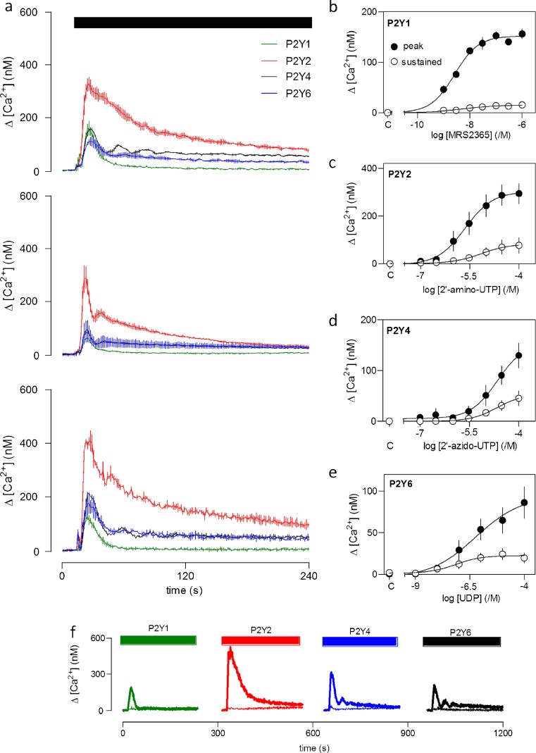
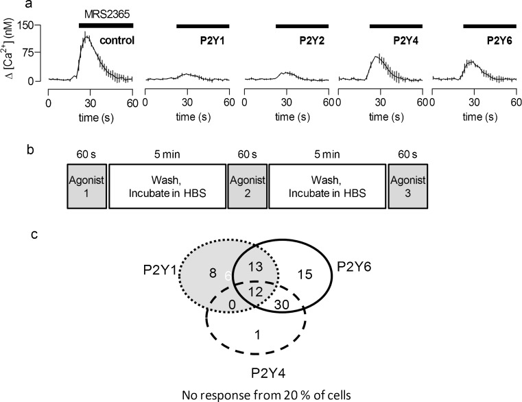
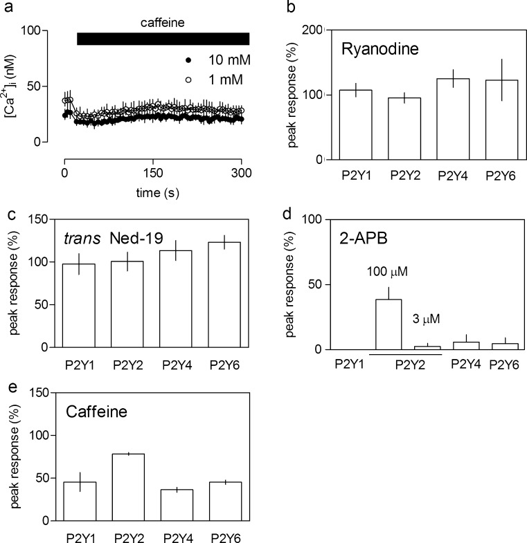
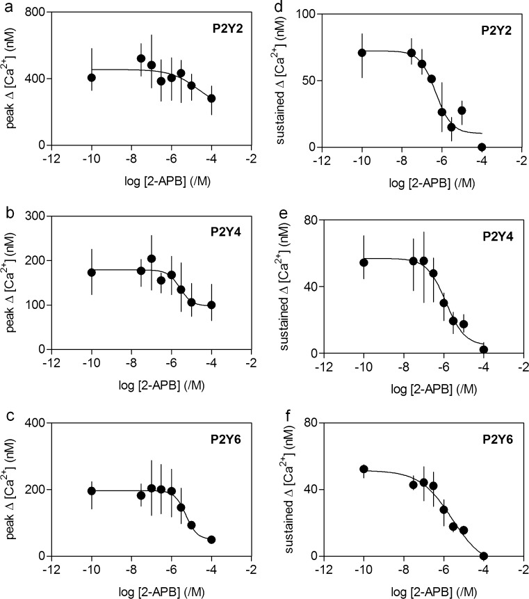
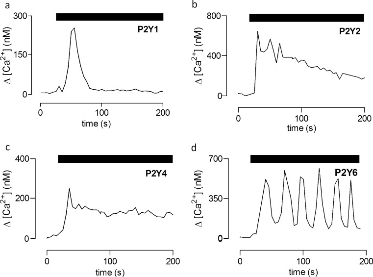
Adenine and uridine nucleotides evoke Ca(2+) signals via four subtypes of P2Y receptor in cultured aortic smooth muscle cells, but the mechanisms underlying the different patterns of these Ca(2+) signals are unresolved. Cytosolic Ca(2+) signals were recorded from single cells and populations of cultured rat aortic smooth muscle cells, loaded with a fluorescent Ca(2+) indicator and stimulated with agonists that allow subtype-selective activation of P2Y1, P2Y2, P2Y4, or P2Y6 receptors. Activation of P2Y1, P2Y2, and P2Y6 receptors caused homologous desensitisation, while activation of P2Y2 receptors also caused heterologous desensitisation of the other subtypes. The Ca(2+) signals evoked by each P2Y receptor subtype required activation of phospholipase C and release of Ca(2+) from intracellular stores via inositol 1,4,5-trisphosphate (IP(3)) receptors, but they were unaffected by inhibition of ryanodine or nicotinic acid adenine dinucleotide phosphate (NAADP) receptors. Sustained Ca(2+) signals were independent of the Na(+)/Ca(2+) exchanger and were probably mediated by store-operated Ca(2+) entry. Analyses of single cells established that most cells express P2Y2 receptors and at least two other P2Y receptor subtypes. We conclude that four P2Y receptor subtypes evoke Ca(2+) signals in cultured aortic smooth muscle cells using the same intracellular (IP(3) receptors) and Ca(2+) entry pathways (store-operated Ca(2+) entry). Different rates of homologous desensitisation and different levels of receptor expression account for the different patterns of Ca(2+) signal evoked by each P2Y receptor subtype.


Figures

Fig. 1
Fig. 1
Ca2+ signals evoked by different P2Y receptors in populations of cultured rat ASMC. a Typical results (each showing mean ± range from two wells) show the Ca2+ signals evoked by activation of each of the four P2Y receptors subtypes. Three examples are shown from different experiments using cells from different passages and aorta preparations. The stimuli used to achieve maximal activation of each P2Y receptor subtype here and in all subsequent figures were: P2Y1, MRS2365 (1 μM); P2Y2, 2'-amino-UTP (100 μM); P2Y4, 2'-azido-UTP (100 μM), and P2Y6, UDP (100 μM). b–e Concentration-dependent effects of the subtype-selective agonists on peak Ca2+ signals, which result largely from release of Ca2+ from intracellular stores [10], and the sustained signal measured 260–300 s after agonist addition. Results are means ± SEM from three independent experiments. f Ca2+ signals evoked by maximal activation of the four P2Y receptors subtypes in the presence of the inhibitor of PLC, U73122 (20 μM, thin traces), or its inactive analogue, U73343 (20 μM, thick traces), each added 5 min before and then during the stimulation. Results are typical of those from three independent plates
Fig. 2
Fig. 2
Homologous desensitisation of the Ca2+ signals evoked by P2Y receptors. a The protocol is designed to avoid errors arising from the small loss of cells during washing of plates between stimuli (see “Materials and methods” section). Cell populations were first stimulated for 60 s with either a maximally effective concentration of the subtype-selective agonist (as described in the legend to Fig. 1a) or HBS alone. The wells were then washed and incubated in HBS for a further 5–60 min before addition of the same agonist and assessment of the Ca2+ responses. The relative amplitudes of the Ca2+ signals evoked during the second addition (shaded area) provide the measure of desensitisation. b Typical traces (means ± SEM of three replicates from a single experiment, each repeated at least three times) show the Ca2+ signals evoked by each P2Y receptor subtype in control cells (pre-treated with HBS, thick traces) and in cells pre-treated with the homologous agonist (thin traces). The interval between the pre-treatments and measurement of the Ca2+ signals was 5 min. c Summary data show the peak Ca2+ signals for cells pre-treated with the homologous agonist, expressed as percentages of the response from control cells (pre-treated with HBS). The interval between the end of the pre-treatment and measurement of the Ca2+ signals is indicated. Results are means ± SEM from three independent experiments. ND not determined
Fig. 3
Fig. 3
Heterologous desensitisation of responses to P2Y receptor subtypes. a The protocol is similar to that described in Fig. 2a but with the second stimulus different from that used for the first stimulation. The interval between stimuli (each applied for 60 s) was 5 min. b Typical results (means ± range of duplicates from a single experiment) are shown for cell populations in which the second stimulus activated P2Y1 receptors and the first stimulus activated the receptors shown. Summary data are presented in Table. 1
Fig. 4
Fig. 4
Distribution of functional P2Y receptor subtypes between individual ASMC. a Populations of cells were stimulated in Ca2+-free HBS using a protocol similar to that shown in Fig. 2a but with different subtype-selective agonists used for the first and second stimulation. Typical results (means ± range of duplicates from a single experiment) are shown for cells in which the second stimulus activated P2Y1 receptors and the first stimulus activated the receptors shown. The interval between stimuli (each applied for 60 s) was 5 min. The data are summarised in Table 2. b In a second series of experiments, Ca2+ signals were recorded from fields of single cells in normal HBS according to the stimulus regime shown. c The diagram shows the distribution of cells (percent, derived from analysis of 158 cells from 7 fields) that responded to activation of the indicated P2Y receptor subtypes. P2Y2 receptors are omitted from the diagram because most cells (95 ± 2 %) responded to activation of P2Y2 receptors
Fig. 5
Fig. 5
IP3 receptors are alone responsible for the Ca2+ release evoked by P2Y receptors. a Populations of cells were stimulated with caffeine in HBS (1 or 10 mM) for the period shown. Results are means ± SEM for triplicate determinations from a single experiment, which was repeated three times. be Peak increases in [Ca2+]i evoked by maximal activation of the indicated P2Y receptor subtypes (details in legend to Fig. 1a) are shown as percentages of control responses for cell populations treated with ryanodine (b 100 μM, pretreatment for 30 min), trans Ned-19 (c 100 nM, pretreatment for 3 min), 2-APB (d 300 μM, pretreatment for 3 min) and caffeine (e 10 mM, pretreatment for 3 min). Results (be) are means ± SEM from three independent experiments. In d, P2Y2 receptors were stimulated either sub-maximally or maximally with 3 μM or 100 μM 2'-amino-UTP, respectively
Fig. 6
Fig. 6
Ca2+ entry evoked by P2Y2, P2Y4 and P2Y6 receptors. ac Effects of nimodipine (100 nM, applied 40 s before the stimulus, a, KB-R7943 (10 μM, applied) 15 min before the stimulus), b or replacement of extracellular Na+ with N-methyl-d-glucamine (c) on the sustained Ca2+ signals (measured 260-300 s after agonist addition) evoked by maximal activation of each of the P2Y receptor subtypes. Results show means ± SEM from three independent experiments. The agonists used and their concentrations are defined in the legend to Fig. 1a. d Restoration of extracellular Ca2+ (1.5 mM) to cells incubated in Ca2+-free HBS alone (thin trace) or after pre-incubation with thapsigargin (1 μM, 30 min in Ca2+-free HBS) to empty intracellular stores (thick trace). Results are from a single experiment, which was repeated three times. ef Concentration-dependent effects of Gd3+ (e applied 40 s before) and 2-APB (f applied 3 min before) on Ca2+ signals evoked by restoration of extracellular Ca2+ to cells incubated with thapsigargin (1 μM, 30 min) in Ca2+-free HBS. Results (e and f) from populations of ASMC are means ± SEM from three independent experiments
Fig. 7
Fig. 7
Effects of 2-APB on the Ca2+ signals evoked by P2Y receptors. af Concentration-dependent effects of 2-APB on peak (ac) and sustained (df) Ca2+ signals evoked by maximally effective concentrations of subtype-selective agonists of P2Y receptors (details in Fig. 1). Sustained responses were measured 250–400 s after addition of agonists. 2-APB was present for 3 min before and then during the stimulation. Results from cell populations are means ± SEM from three independent experiments
Fig. 8
Fig. 8
Ca2+ signals evoked by activation of different P2Y receptor subtypes in single cells. ad Traces show typical Ca2+ signals recorded from single cells after maximal activation of each of the four P2Y receptor subtypes. Details of the agonists used and their concentrations are given in the legend to Fig. 1. Results are typical of responses measured from 7 (a, c and d) or 4 (b) different coverslips. Only P2Y6 receptors (72 ± 3 % of cells from 7 coverslips) reproducibly evoked Ca2+ oscillations

Similar articles

Cited by

References

    1. House SJ, Potier M, Bisaillon J, Singer HA, Trebak M. The non-excitable smooth muscle: calcium signaling and phenotypic switching during vascular disease. Pflugers Arch. 2008;456:769–785. doi: 10.1007/s00424-008-0491-8. - DOI - PMC - PubMed
    1. Owens GK, Kumar MS, Wamhoff BR. Molecular regulation of vascular smooth muscle cell differentiation in development and disease. Physiol Rev. 2004;84:767–801. doi: 10.1152/physrev.00041.2003. - DOI - PubMed
    1. Vallot O, Combettes L, Jourdon P, Inamo J, Marty I, Claret M, Lompre AM. Intracellular Ca2+ handling in vascular smooth muscle cells is affected by proliferation. Pflugers Arch. 2000;20:1225–1235. - PubMed
    1. Berra-Romani R, Mazzocco-Spezzia A, Pulina MV, Golovina VA. Ca2+ handling is altered when arterial myocytes progress from a contractile to a proliferative phenotype in culture. Am J Physiol. 2008;295:C779–C790. doi: 10.1152/ajpcell.00173.2008. - DOI - PMC - PubMed
    1. Thyberg J. Differentiated properties and proliferation of arterial smooth muscle cells in culture. Int Rev Cytol. 1996;169:183–265. doi: 10.1016/S0074-7696(08)61987-7. - DOI - PubMed

Publication types

LinkOut - more resources