. 2012 Oct;26(10):4230-42.
doi: 10.1096/fj.12-207969. Epub 2012 Jul 5.

α-Actinin2 is required for the lateral alignment of Z discs and ventricular chamber enlargement during zebrafish cardiogenesis

Affiliations

α-Actinin2 is required for the lateral alignment of Z discs and ventricular chamber enlargement during zebrafish cardiogenesis

Jingchun Yang et al. FASEB J. 2012 Oct.

Abstract

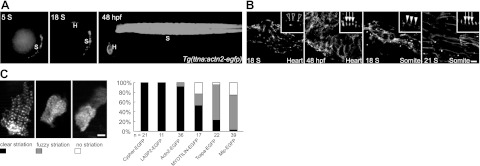
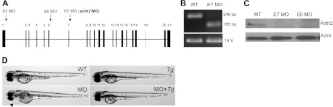
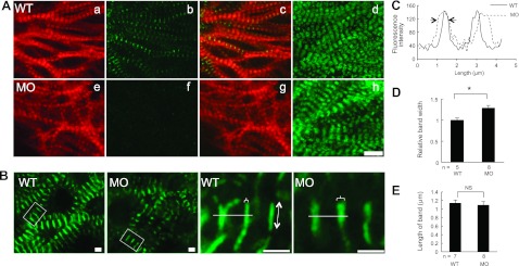
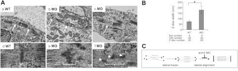
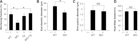
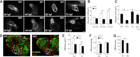
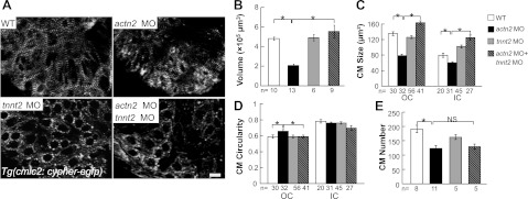
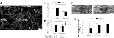
α-Actinin2 (Actn2) is a predominant protein in the sarcomere Z disc whose mutation can lead to cardiomyopathy. However, the function of Actn2 in Z-disc assembly and cardiomyopathy in vertebrates remains elusive. We leveraged genetic tools in zebrafish embryos to elucidate the function of Actn2. We identified a single Actn2 homologue expressed in the zebrafish heart and conducted loss-of-function studies by antisense morpholino technology. Although zebrafish Actn2 assembles early into the Z disc, depletion of actn2 did not affect the early steps of sarcomere assembly. Instead, Actn2 is required for Z bodies to register laterally, forming well-aligned Z discs. Presumably as a consequence to this structural defect in the sarcomere, the depletion of Actn2 resulted in reduced cardiac function, primarily characterized as a reduced end-diastolic diameter. The depletion of actn2 also significantly reduced the ventricle chamber size, due to both reduced cardiomyocyte (CM) size and CM number. Interestingly, reduced CM size can be rescued by the cessation of heart contractions. The genetic studies of zebrafish uncovered a function for actn2 in lateral registration of Z body. In actn2 morphant fish, the Z-disc defect sequentially affects cardiac function, which leads to morphological changes in the ventricle through a mechanical force-dependent mechanism.


Figures

Figure 1.
Figure 1.
Actn2-GFP recapitulates the localization of endogenous Actn2 and assembles early into the Z disc. A) In Tg(ttna:actn2-egfp) fish, a ttna enhancer drives the expression of EGFP-tagged Actn2 in the somites starting at 5 S and in the heart starting at 18 S. EGFP signal can be detected in both the heart and somites at 48 hpf. H, heart; B) Images of dissected hearts or somites from Tg(ttna:actn2-egfp) fish. Actn2-egfp is localized to Z bodies as irregular dots (empty arrowhead) at 18 S and periodic Z discs (arrow) at 48 hpf in the heart. In the somites, Actn2-egfp forms Z bodies as regular dots (solid arrowhead) at 18 S and striated Z discs (arrow) at 21 S. C) Left panel: examples of 3 types of CMs exhibiting different striation that may coexist in embryos injected with constructs encoding EGFP-tagged Z-disc proteins. Right panel: quantification of the percentage of CMs that exhibit different degrees of striation at 48 hpf. Compared to MYOTILIN-EGFP, Tcapa-EGFP, and Mlp-EGFP, injection of Actn2-EGFP resulted in a higher percentage of CMs exhibiting clear striation. n, CM number. Scale bars = 10 μm (B); 5 μm (C).
Figure 2.
Figure 2.
actn2 MO effectively knocks down the actn2 gene and induces a heart-specific phenotype. A) Schematic of the gene structure of actn2 and the exons that are targeted by the three splice donor MOs E1 MO, E6 MO, and E7 MO. actn2 MO (bold) refers to E7 MO. B) Injection of actn2 MO induces an exon-skipping event, as revealed by DNA gel electrophoresis after RT-PCR using a primer pair spanning exons 6 and 8. The 246-bp band detected in the WT fish is shifted to 165 bp in the actn2 MO fish, indicating the deletion of exon 7. C) Western blot analysis indicates a strong reduction in Actn2 protein in hearts dissected from embryos that were injected with either E7 MO or E6 MO. D) Lateral views of zebrafish embryos show that embryos injected with actn2 MO exhibit a cardiac-specific phenotype. Pericardial edema (arrowhead) can be rescued by Tg(ttna:actn2-egfp).
Figure 3.
Figure 3.
Depletion of Actn2 leads to broader Z band without affecting other myofibril substructures, including I bands and A bands. A) Images of dissected ventricles from WT and acnt2 MO fish at 48 hpf with phalloidin staining (a, e), immunostaining using an anti-Actn2 antibody (b, f), merged images of phalloidin (red) and Actn2 antibody staining (green) (c, g), and immunostaining using an anti-myosin antibody (d, h). In actn2 morphant fish, Actn2 immunoreactivity is essentially undetectable (b, f), but the striated pattern of thin and thick filaments remain normal. B) Subcellular localization of Cypher in Tg(cmlc2:cypher-egfp) fish is shown in low and high magnification (boxed area in low magnification). Cypher-EGFP exhibits as striated bands in both WT and actn2 MO fish, but the bands are wider in actn2 MO fish heart than in WT. Brackets indicate bandwidth; line with double-ended arrow indicates band length. C) Pseudo-line scan analysis of the fluorescence intensity change along the white line in B. Arrows indicate that actn2 MO fish has wider cypher-GFP band than WT fish. D, E) Measurement of the width (D) and length (E) of Cypher-EGFP bands shows that the width is significantly increased in actn2 MO fish heart, while the length of the bands is not affected. MO, fish injected with actn2 MO; NS, not significant. Scale bars = 10 μm (A); 2 μm (B). *P < 0.05.
Figure 4.
Figure 4.
Depletion of Actn2 leads to lateral alignment defects in the Z discs. A) a–c) Electron micrographs of CMs of 52-hpf embryos at low magnification. Compared to WT fish, actn2 morphants exhibit organized myofibrils but disrupted Z-disc structures (arrows). d–f) High-magnification images of boxed regions panels a–c. In contrast to the laterally aligned Z disc in WT fish (d), the electron-dense Z discs in actn2 morphants are misaligned, exhibiting as either zigzagged lines (e) or dispersed dots (f). Arrowheads indicate dispersed electron-dense dots. Brackets indicate Z-disc width. Scale bars =1 μm (a–c); 200 nm (d–f). B) Measurement of Z-disc width based on electron microscopy data. C) Schematic of the Z-disc defects in actn2 morphants. Injection of actn2 MO blocks the lateral alignment but not the lateral fusion of the Z discs. MO, fish injected with actn2 MO. *P < 0.05.
Figure 5.
Figure 5.
Injection of actn2 MO leads to reduced cardiac function. A) Injection of actn2 MO reduces the shortening fraction, which is rescued by transgenic actn2 in Tg(ttna:actn2-egfp) fish. B) End-diastolic diameter is reduced in actn2 morphants. C) End-systolic diameter is comparable between WT fish and actn2 morphants. D) Heart rate is not affected by injection of actn2 MO. MO, actn2 MO fish; Tg, Tg(ttna:actn2-egfp) fish; MO+Tg, Tg(ttna:actn2-egfp) fish injected with actn2 MO; n, fish number; NS, not significant. *P < 0.05.
Figure 6.
Figure 6.
Injection of actn2 MO leads to reduced size of the ventricular chamber and individual CMs. A) Ventricle enlargement is halted by actn ctn2 MO. Images show individual fish hearts from living Tg(ttna:megfp) fish embryos with or without actn2 MO injection. Dorsal view and ventral view with the head facing up is shown for embryos at 24 and 44 hpf, respectively; embryos at 52 and 72 hpf are shown in lateral views with the head to the left. V, ventricle. B) Quantification of ventricular volume reveals a significantly reduced ventricular chamber size in MO fish at both 52 and 72 hpf. C) Reduced ventricular chamber sizes in MO fish can be rescued by Tg(ttna:actn2-egfp). D) Image of a ventricle after immunostaining with Mef2 (red) and β-catenin (green) to define the nuclei and the outline of the CMs. Typical OC and IC cells are encircled with solid and dashed lines, respectively. E, F) Quantification of CM size and circularity shows that CM size is significantly reduced in both the OC and IC regions, while CM circularity is only increased in the OC region. G) CM number in the ventricle is reduced in actn2 morphant fish. MO, fish injected with actn2 MO; Tg, Tg(ttna:actn2-egfp) fish; MO+Tg, Tg(ttna:actn2-egfp) fish injected with actn2 MO, OC, outer curvature; IC, inner curvature; n, number of fish (B, C, G) or CMs (E, F). Scale bars =20 μm (A); 40 μm (D). *P < 0.05.
Figure 7.
Figure 7.
Cessation of heart contraction rescues the ventricle chamber size defect in actn2 MO fish. A) Sarcomere integrity is disrupted by a tnnt2 MO, as revealed by Tg(cmlc2:cypher-egfp). Bundling of the Z discs is disrupted in embryos injected with either tnnt2 MO or tnnt2, actn2 double MO but is intact in WT and actn2 MO fish embryos. Scale bar = 10 μm. B) Quantification of the ventricular volume in embryos at 72 hpf. Ventricular volume is significantly reduced in actn2 morphant fish, but this defect can be rescued by coinjection of a tnnt2 MO. C) Compared to WT fish, both OC and IC cell size are reduced in actn2 morphant fish, but this defect can be rescued by coinjection of a tnnt2 MO. D) Compared to WT fish, the circularity of OC cells is reduced in actn2 MO fish, but this defect can be rescued by coinjection of a tnnt2 MO. E) Compared to WT fish, CM number is significantly reduced in actn2 morphant fish. However, there is no significant rescue of CM number by coinjection of a tnnt2 MO. n, number of fish (B, E) or CMs (C, D); NS, not significant. *P < 0.05.
Figure 8.
Figure 8.
Ventricle chamber size reduction in actn2 morphants is rescued by cessation of contraction via blebbistatin or nifedipine treatment. A) Sarcomere integrity is disrupted in embryos treated with blebbistatin, as revealed by Tg(cmlc2:cypher-egfp). Compared to the long Z discs in DMSO-treated WT or actn2 MO fish, the Z discs in blebbistatin-treated WT or actn2 MO fish is significantly shorter, indicating disrupted Z-disc bundling. Double-ended arrow indicates the Z-disc length. B) Quantification of the Z-disc length. n, number of fish. C) Electron micrographs show that cessation of heart contraction via blebbistatin treatment cannot rescue the disrupted Z disc in actn2 MO fish heart. Bracket indicates Z-disc width. D) Quantification of the Z-disc width, which cannot be rescued by cessation of heart contraction. E) Measurement of the ventricular volume in DMSO and blebbistatin treated fish at 52 hpf. WT and actn2 morphant embryos were bathed in egg water containing 8 μM blebbistatin or 60 μM nifedipine from 37 to 52 hpf. Ventricular volume is significantly reduced in MO fish, but this defect can be rescued by either blebbistatin or nifedipine treatment. MO, actn2 MO-injected fish; Ble, blebbistatin-treated WT fish; MO Ble, blebbistatin-treated actn2 MO fish; NS, not significant. Scale bars = 10 μm (A); 200 nm (C). *P < 0.05.

References

    1. Clark K. A., McElhinny A. S., Beckerle M. C., Gregorio C. C. (2002) Striated muscle cytoarchitecture: an intricate web of form and function. Annu. Rev. Cell Dev. Biol. 18, 637–706 - PubMed
    1. Frank D., Kuhn C., Katus H. A., Frey N. (2006) The sarcomeric Z-disc: a nodal point in signalling and disease. J. Mol. Med. 84, 446–468 - PubMed
    1. Pyle W. G., Solaro R. J. (2004) At the crossroads of myocardial signaling: the role of Z-discs in intracellular signaling and cardiac function. Circ. Res. 94, 296–305 - PubMed
    1. Hoshijima M. (2006) Mechanical stress-strain sensors embedded in cardiac cytoskeleton: Z disk, titin, and associated structures. Am. J. Physiol. Heart Circ. Physiol. 290, H1313–H1325 - PMC - PubMed
    1. Keren A., Syrris P., McKenna W. J. (2008) Hypertrophic cardiomyopathy: the genetic determinants of clinical disease expression. Nat. Clin. Pract. Cardiovasc. Med. 5, 158–168 - PubMed

Publication types

MeSH terms