. 2012 Oct;14(5):829-838.
doi: 10.1007/s10544-012-9662-7.

Three-dimensional scaffolding to investigate neuronal derivatives of human embryonic stem cells

Affiliations

Three-dimensional scaffolding to investigate neuronal derivatives of human embryonic stem cells

Pranav Soman et al. Biomed Microdevices. 2012 Oct.

Abstract

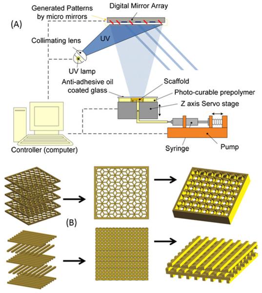
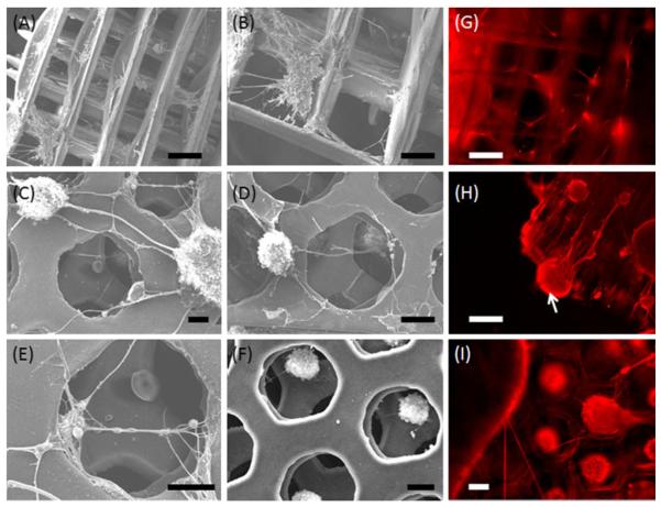
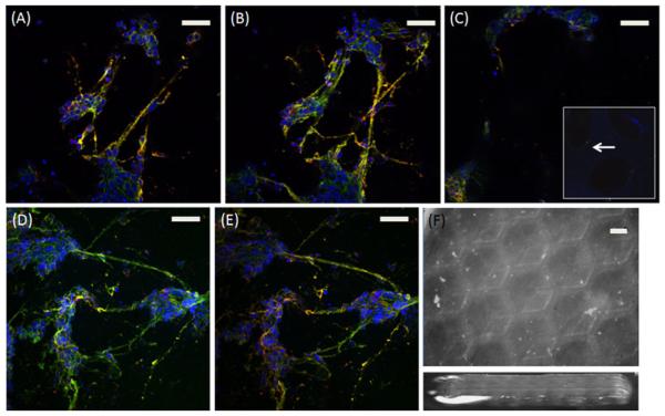
Access to unlimited numbers of live human neurons derived from stem cells offers unique opportunities for in vitro modeling of neural development, disease-related cellular phenotypes, and drug testing and discovery. However, to develop informative cellular in vitro assays, it is important to consider the relevant in vivo environment of neural tissues. Biomimetic 3D scaffolds are tools to culture human neurons under defined mechanical and physico-chemical properties providing an interconnected porous structure that may potentially enable a higher or more complex organization than traditional two-dimensional monolayer conditions. It is known that even minor variations in the internal geometry and mechanical properties of 3D scaffolds can impact cell behavior including survival, growth, and cell fate choice. In this report, we describe the design and engineering of 3D synthetic polyethylene glycol (PEG)-based and biodegradable gelatin-based scaffolds generated by a free form fabrication technique with precise internal geometry and elastic stiffnesses. We show that human neurons, derived from human embryonic stem (hESC) cells, are able to adhere to these scaffolds and form organoid structures that extend in three dimensions as demonstrated by confocal and electron microscopy. Future refinements of scaffold structure, size and surface chemistries may facilitate long term experiments and designing clinically applicable bioassays.


Figures

Fig. 1
Fig. 1
(a) Schematic of the digital micromirror device based projection printing system (DMD-PP) used for fabricating multi-layer biomaterial scaffolds having log-pile and hexagonal internal architectures. The DMD-PP method produced precise features using a system of dynamic masks generated by a digital light controlling chip and photocurable biomaterials. (b) Schematic of the assembled structures; the layers of cross-linked photopolymer can be stacked to form scaffolds having log-pile and hexagonal micro-architecture
Fig. 2
Fig. 2
Mechanical properties of DMD-PP fabricated scaffolds having 3D log-pile and hexagonal structures using soft (19 %) PEGDA, stiff (95 %) PEGDA and gelatin methyacrylate (GelMA). (a) A characteristic stress–strain curve displaying low strain and high strain compressive moduli was obtained for every sample tested (b) Comparison of compressive modulus of various scaffolds at low and high strains. (c) Magnified angled SEM demonstrates the 3D log-pile scaffold. Inset depicts the top-view of the scaffold with precise pore geometries and sharp features. (d, e) Optical images of log-pile and hexagonal scaffolds (f) Side-view of 3D scaffolds showing a 5-layer structure. Black star denotes that the stiffness values are adapted from our earlier work (Gauvin et al. 2012). Scale: C=100 μm; D–F=500 μm
Fig. 3
Fig. 3
SEM images of PEG scaffolds with (a, b) log-pile and (c–f) hexagonal internal architecture support survival and transition of neurospheres to neuronal morphology, spreading from adherent cell clusters, and forming 2D and 3D intercellular projections. Neurospheres evenly penetrate and adhere to hexagonal PEGDA scaffolds. Larger neurospheres are largely excluded from the interior of the scaffold. (g–i) Neurons form 3D lattices on log-pile and hexagonal PEGDA scaffolds labeled for Tuj1. White arrow in (h) denotes a cell cluster adhered to the side of the log-pile scaffold with neural connections along the struts. Scale: 100 μm
Fig. 4
Fig. 4
Confocal images of the stiff PEGDA scaffold with hexagonal (a–c) and log-pile (d–f) architecture. Images selected from selected z-planes demonstrate the formation of neural connections in 3D labeled with Tuj1, a marker of early neurons. White arrows in (f) denotes the log-pile structure. Scale: 100 μm
Fig. 5
Fig. 5
Confocal images of soft PEGDA scaffold with hexagonal (a–c) geometry from selected z-sections labeled with Tuj1(green), MAP2 (red) and nucleus (DAPI-blue). (d) 3D reconstruction of the hexagonal scaffold. (e–g) High resolution image of the soft PEGDA scaffolds show clusters of neural cells formatting 3D neural connections between two hexagonal geometries. White arrows in (e) denote the hexagonal structure. (h–k) SEM images of the soft PEGDA scaffold. Scale: A–I=100 μm; J,K=10 μm
Fig. 6
Fig. 6
Confocal z-stack images of gelatin methacrylate (GelMA) scaffolds with hexagonal (a–c) geometry labeled with Tuj1(green), MAP2 (red) and nucleus (blue). White arrows in inset (c) denote the degraded hexagonal structure after three-week of cell culture. (d–e) The hexagonal structure is not observed after four-week of culture. (f) Optical images of the GelMA scaffold with hexagonal structures before cell culture. Scale: 100 μm

References

    1. Bini TB, Gao S, Wang S, Ramakrishna S. Poly(l-lactide-co-glycolide) biodegradable microfibers and electrospun nanofibers for nerve tissue engineering: an in vitro study. J. Mater. Sci. 2006;41(19):6453–6459.
    1. Björn Carlberg MZA, Ulf Nannmark, Johan Liu, Georg Kuhn H. Electrospun polyurethane scaffolds for proliferation and neuronal differentiation of human embryonic stem cells. Biomed. Mater. 2009;4(4):045004. - PubMed
    1. Chambers SM, Fasano CA, Papapetrou EP, Tomishima M, Sadelain M, Studer L. Highly efficient neural conversion of human ES and iPS cells by dual inhibition of SMAD signaling. Nat. Biotechnol. 2009;27(3):275–280. - PMC - PubMed
    1. Chandran PL, Barocas V. Affine versus non-affine fibril kinematics in collagen networks: theoretical studies of network behavior. J. Biomech. Eng. 2006;128(2):259–270. - PubMed
    1. Deleyrolle LP, Reynolds BA. Isolation, expansion, and differentiation of adult Mammalian neural stem and progenitor cells using the neurosphere assay. Methods Mol. Biol. 2009;549:91–101. - PubMed

Publication types

LinkOut - more resources