. 2012 Oct 1;125(Pt 19):4609-19.
doi: 10.1242/jcs.107995. Epub 2012 Jul 5.

Schwann cell myelination requires integration of laminin activities

Affiliations

Schwann cell myelination requires integration of laminin activities

Karen K McKee et al. J Cell Sci. .

Abstract

Laminins promote early stages of peripheral nerve myelination by assembling basement membranes (BMs) on Schwann cell surfaces, leading to activation of β1 integrins and other receptors. The BM composition, structural bonds and ligands needed to mediate this process, however, are not well understood. Mice hypomorphic for laminin γ1-subunit expression that assembled endoneurial BMs with reduced component density exhibited an axonal sorting defect with amyelination but normal Schwann cell proliferation, the latter unlike the null. To identify the basis for this, and to dissect participating laminin interactions, LAMC1 gene-inactivated dorsal root ganglia were treated with recombinant laminin-211 and -111 lacking different architecture-forming and receptor-binding activities, to induce myelination. Myelin-wrapping of axons by Schwann cells was found to require higher laminin concentrations than either proliferation or axonal ensheathment. Laminins that were unable to polymerize through deletions that removed critical N-terminal (LN) domains, or that lacked cell-adhesive globular (LG) domains, caused reduced BMs and almost no myelination. Laminins engineered to bind weakly to α6β1 and/or α7β1 integrins through their LG domains, even though they could effectively assemble BMs, decreased myelination. Proliferation depended upon both integrin binding to LG domains and polymerization. Collectively these findings reveal that laminins integrate scaffold-forming and cell-adhesion activities to assemble an endoneurial BM, with myelination and proliferation requiring additional α6β1/α7β1-laminin LG domain interactions, and that a high BM ligand/structural density is needed for efficient myelination.


Figures
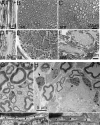
Fig. 1.
Fig. 1.
Sciatic nerve. (A-F) Methylene-blue-stained semi-thin longitudinal (A,D) and cross (B,C,E,F) sections of sciatic nerve (sn, A,B,D,E) and sciatic nerve root (snr, C,F) of Lmγ1 flneo/+ (A-C) and Lmγ1 flneo/− (D-F) adult (12-week-old) mice. In the flneo/− nerve and root the distribution of unmyelinated axons is patchy, but is most pronounced in the root. In addition, the myelin had many infoldings (inset, arrows). (G-J) Electron micrograph of Lmγ1 flneo/− (H) nerve reveals bundles of naked axons and nearby and adjacent non-myelinating Schwann cells (arrows) not seen in flneo/+ littermate nerve (G). Endoneurial BMs are continuous and of similar thickness in flneo/+ (I) and Lmγ1 flneo/− (J) Schwann cells.
Fig. 2.
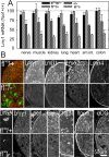
Fig. 2.
Laminin expression. (A) Comparison of laminin mRNA by quantitative real-time RT-PCR revealed Lmγ1 flneo/− mRNA was decreased about threefold compared to wild-type controls in nerve, muscle and other tissues. Lesser reductions were detected in LAMC1+/− tissues (plots are average and standard deviation of experiments performed in triplicate using RNA from five mice of each genotype). (B) Immunofluorescence images of flneo/− adult sciatic nerve reveal decreases in laminin subunits, perlecan (perl), and collagen-IV (ColIV), but not integrin subunits (Itgβ1, Itgα6) or α-dystroglycan (αDG) compared with flneo/+ littermates.
Fig. 3.
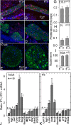
Fig. 3.
Schwann cell proliferation and myelination factors. (A-D) Hindlimb nerve from flneo/+ and flneo/− embryos (E18.5; A,B) and post-natal day 1 littermates (C,D) were labeled with EdU and stained with DAPI to compare DNA synthesis (shown with NFL counter-stain, green). (E,F) Sciatic nerve from 10-week-old flneo/+ (E) and flneo/− (F) littermates were stained with DAPI and NFL antibody. Groups of NFL-stained axons characteristic of the mutant state are seen in F. (G-I) The fraction of EdU-positive (red) DAPI-stained (blue) nuclei in nerves was determined (average and standard deviation of three pairs) at E18.5 (G) and P1 (H). No significant difference was detected. (I) The number of nuclei per nerve section was similar in flneo/+ and flneo/− adults. (J,K) Steady-state mRNA levels of myelination factors. Proximal sciatic nerve segments were isolated from adult (J) and P5 (K) mice followed by RNA extraction and quantitative real-time RT-PCR for mRNA (flneo/− to flneo/+ ratio) of the indicated proteins (number of samples indicated with small digits in graphs). Reductions in mutant nerves were identified for laminin-γ1 (LmC1), Krox-20, MPZ, p75 and neuregulin-1 (Nrg1). Elevations were identified for c-Jun, Oct-6 and Sox2. The patterns were similar at both stages.
Fig. 4.
Fig. 4.
Laminin domains and activities. (A) Map of cell-surface glycolipid attachment sites, integrin-binding, α-dystroglycan (αDG) binding, polymerization (polym.), nidogen (Nd) binding and heparan sulfate (HS) binding activities of Lm111. (B) Schematic diagram of Lmc1 C-terminal sequence (violet) with a critical glutamic acid residue (red) thought to interact with LG domains, followed by the FLAG-tag sequence (yellow) that interferes with integrin binding. (C) Recombinant laminins bearing modifications that reduce and ablate nidogen binding, laminin polymerization, integrin binding through LG domains, and cell-surface adhesion are shown. αLNNd (F1) is a linker protein that binds to Lms at the nidogen-binding locus, adding the α1LN polymerization domain.
Fig. 5.
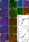
Fig. 5.
Laminin-dependent Schwann cell proliferation and myelination in DRG cultures. LAMC1-flneo/flneo DRGs, isolated from E13.5 pregnant females and rendered laminin-deficient with an adeno-cre recombinase virus, were treated with the different concentrations of rLm111 (0, 3.5, 7, 14 and 28 nM; A-J) followed by evaluation of proliferation (ratio of EdU/DAPI; A-E) at day 0 and myelination (ratio of MBP/NFL+gfp; F-J) at day 6. The corresponding plots for proliferation (L) and myelination induced (M) are shown (average ± s.d. of 3-5 DRGs). (K) Higher magnification views of the DRGs labeled and stained for proliferation (upper panels) and myelination (lower panels) treated with 0 and 14 nM rLm111. Although myelination and proliferation both depended on laminin concentration, proliferation increased at low levels and reached a plateau by 28 nM, whereas myelination continued to increase over the full concentration range.
Fig. 6.
Fig. 6.
Schwann cell adhesion to basement membrane components and integrin binding. (A,B) Schwann cells isolated from DRGs were expanded in culture and evaluated for adhesion to plastic wells coated with laminins and other BM substrates. (A) Adhesion to rLm111 (closed circles), collagen-IV (Col-IV, open inverted triangles), rNd1 (open diamonds), rLmΔα1LG1-3 (open hexagons), and miniagrin (mA, open squares). Data are average ± s.d., n = 4. (B) Adhesion of cells to rLm111 (closed circles) and rLm211 (closed triangles) and corresponding FLAG-tagged rLm111F (open circles) and rLm211F (open triangles). Higher coat concentrations were required to achieve similar adhesion for FLAG-tagged laminins. (C-F) Binding of soluble integrin-ectodomain-dimers to wild-type and FLAG-tagged rLm211 (C), and to correspondingly modified rLm111 (D,E). Five- to tenfold reductions of binding affinity were detected for FLAG-tagged laminins for integrins α7β1 and α6β1. (F) Miniagrin (mA), containing LG cell-adhesion domains, bound to integrin α3β1 (closed circles), α6β1 (open diamonds), αvβ1 (open circles), α7β1X2 (open inverted triangles), α7β1X1 (open squares), α5β1 (open triangles).
Fig. 7.
Fig. 7.
DRG myelination in response to functional modifications of laminins. (A) Composite images of Lm-deficient DRGs cultured with 28 nM recombinant laminins and nidogen, immunostained for MBP (red) and NFL (green). (B) Plot of myelination (ratio of MBP/NFL+gfp; average ± s.d. for the indicated number of DRGs) showing reductions in DRGs treated with C-terminal FLAG-tagged laminins, and ablation of polymerization through deletion of LN-containing segments of either the α1 or β1 subunit eliminated myelination. Myelination was substantially increased by treating rLmΔαLN-L4b with αLNNd. A partial (but not statistically significant, P = 0.1) loss of myelination was seen with laminin unable to bind nidogen. Deletion of either the proximal or distal LG domains ablated myelination. (C) Laminin-deficient DRGs treated with 28 nM rLm111 were sectioned and immunostained for laminin subunits, perlecan and collagen-IV. (D) Comparison of Schwann cell proliferation (average ± s.d., 5 DRGs/condition) in response to rLm211F (closed squares, upper plot) compared with wild-type rLm211 (closed circles), and comparison of cell proliferation in response to rLmΔαLN-L4b (closed squares, lower plot) compared with wild-type rLm111 (closed circles). Presence of the FLAG tag reduced proliferation whereas loss of polymerization completely prevented proliferation above baseline (no added laminin) levels for all concentrations.
Fig. 8.
Fig. 8.
DRG ultrastructure. (A-E) Laminin-deficient DRGs were treated with the indicated laminins. DRG maintained with no added laminin (A), 3.5 nM rLm111 (B), 7 nM rLm111 (C), 28 nM rLm111 (D) and 28 nM rLm211 (E). Nearly all axons were naked (nAx), often in small bundles, in the absence of added laminin. Axons were enveloped (eAx) or myelinated (mAx) following treatment with recombinant laminins, and myelin wrapping was more prominent at higher concentrations. Insets are magnified images to show BM (arrows), gaps (asterisks) and plasma membrane (white arrowheads). (F) Plot of BM coverage on cell surfaces (BM/plasma membrane ratio; average ± s.d. for 8-12 images) after treatment of DRGs with rLm111 reveals increasing BM as laminin concentration increased. (G) Plot of BM/PM ratio for DRGs treated with the laminins bearing domain modifications.

Similar articles

Cited by

References

    1. Ard M. D., Bunge R. P., Bunge M. B. (1987). Comparison of the Schwann cell surface and Schwann cell extracellular matrix as promoters of neurite growth. J. Neurocytol. 16, 539–555 10.1007/BF01668507 - DOI - PubMed
    1. Benninger Y., Thurnherr T., Pereira J. A., Krause S., Wu X., Chrostek–Grashoff A., Herzog D., Nave K. A., Franklin R. J., Meijer D.et al. (2007). Essential and distinct roles for cdc42 and rac1 in the regulation of Schwann cell biology during peripheral nervous system development. J. Cell Biol. 177, 1051–1061 10.1083/jcb.200610108 - DOI - PMC - PubMed
    1. Berti C., Bartesaghi L., Ghidinelli M., Zambroni D., Figlia G., Chen Z. L., Quattrini A., Wrabetz L., Feltri M. L. (2011). Non-redundant function of dystroglycan and β1 integrins in radial sorting of axons. Development 138, 4025–4037 10.1242/dev.065490 - DOI - PMC - PubMed
    1. Bunge R. P., Bunge M. B., Eldridge C. F. (1986). Linkage between axonal ensheathment and basal lamina production by Schwann cells. Annu. Rev. Neurosci. 9, 305–328 10.1146/annurev.ne.09.030186.001513 - DOI - PubMed
    1. Chen Z. L., Strickland S. (2003). Laminin gamma1 is critical for Schwann cell differentiation, axon myelination, and regeneration in the peripheral nerve. J. Cell Biol. 163, 889–899 10.1083/jcb.200307068 - DOI - PMC - PubMed

Publication types

MeSH terms