. 2012 Aug 24;287(35):29713-21.
doi: 10.1074/jbc.M112.393074. Epub 2012 Jul 5.

Phosphatidylinositol 3-kinase (PI3K) activity bound to insulin-like growth factor-I (IGF-I) receptor, which is continuously sustained by IGF-I stimulation, is required for IGF-I-induced cell proliferation

Affiliations

Phosphatidylinositol 3-kinase (PI3K) activity bound to insulin-like growth factor-I (IGF-I) receptor, which is continuously sustained by IGF-I stimulation, is required for IGF-I-induced cell proliferation

Toshiaki Fukushima et al. J Biol Chem. .

Abstract

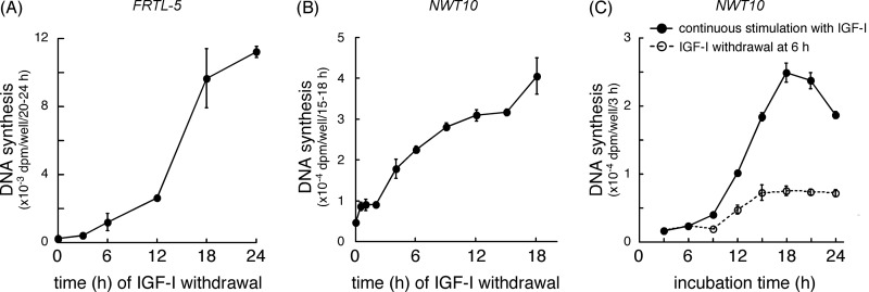
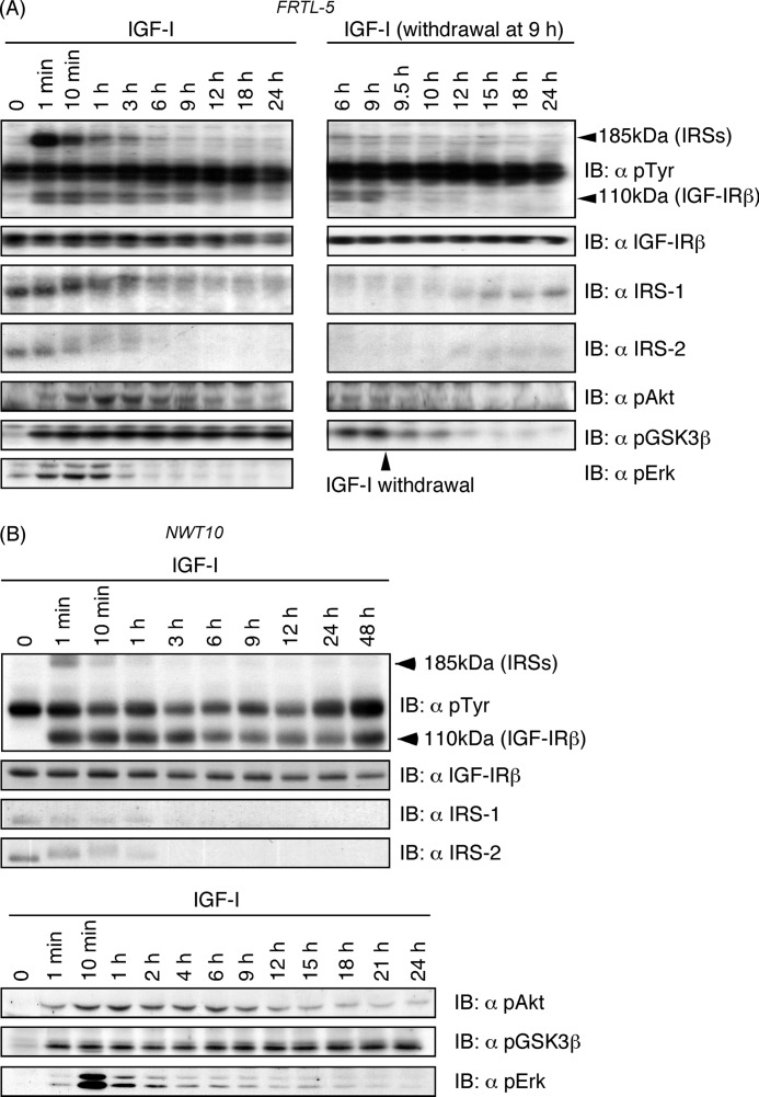
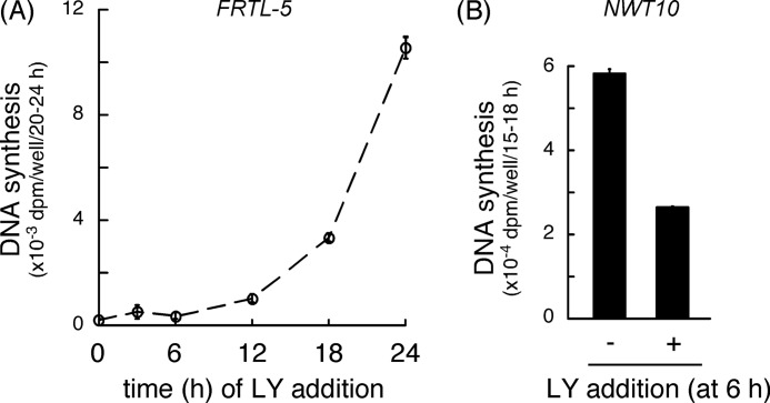
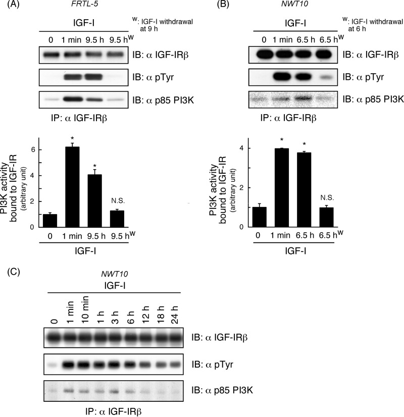
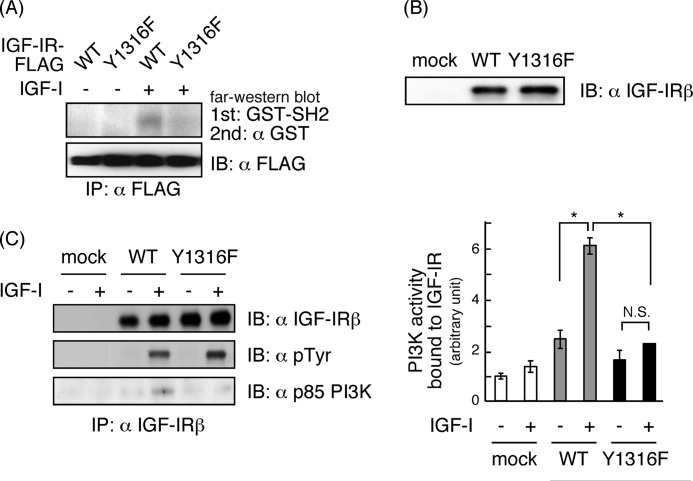
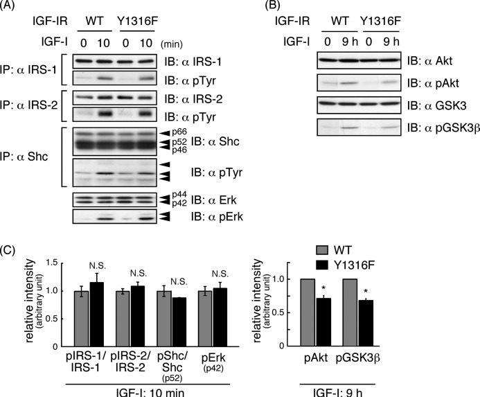
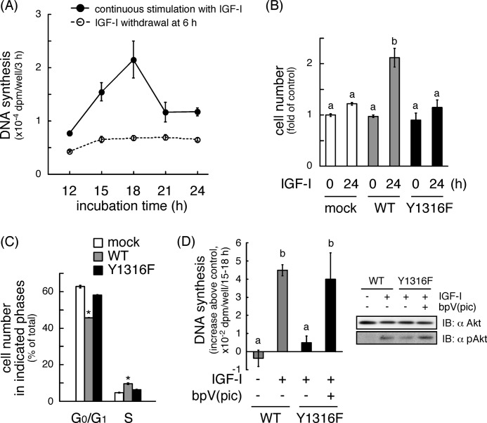
Continuous stimulation of cells with insulin-like growth factors (IGFs) in G(1) phase is a well established requirement for IGF-induced cell proliferation; however, the molecular components of this prolonged signaling pathway that is essential for cell cycle progression from G(1) to S phase are unclear. IGF-I activates IGF-I receptor (IGF-IR) tyrosine kinase, followed by phosphorylation of substrates such as insulin receptor substrates (IRS) leading to binding of signaling molecules containing SH2 domains, including phosphatidylinositol 3-kinase (PI3K) to IRS and activation of the downstream signaling pathways. In this study, we found prolonged (>9 h) association of PI3K with IGF-IR induced by IGF-I stimulation. PI3K activity was present in this complex in thyrocytes and fibroblasts, although tyrosine phosphorylation of IRS was not yet evident after 9 h of IGF-I stimulation. IGF-I withdrawal in mid-G(1) phase impaired the association of PI3K with IGF-IR and suppressed DNA synthesis the same as when PI3K inhibitor was added. Furthermore, we demonstrated that Tyr(1316)-X-X-Met of IGF-IR functioned as a PI3K binding sequence when this tyrosine is phosphorylated. We then analyzed IGF signaling and proliferation of IGF-IR(-/-) fibroblasts expressing exogenous mutant IGF-IR in which Tyr(1316) was substituted with Phe (Y1316F). In these cells, IGF-I stimulation induced tyrosine phosphorylation of IGF-IR and IRS-1/2, but mutated IGF-IR failed to bind PI3K and to induce maximal phosphorylation of GSK3β and cell proliferation in response to IGF-I. Based on these results, we concluded that PI3K activity bound to IGF-IR, which is continuously sustained by IGF-I stimulation, is required for IGF-I-induced cell proliferation.


Figures

FIGURE 1.
FIGURE 1.
Effects of IGF-I withdrawal in G1 phase on DNA synthesis induced by IGF-I. Quiescent FRTL-5 cells were pretreated with 1 mm dibutyryl cAMP for 24 h to prime cells to response to IGF-I, and the cAMP-treated cells were then stimulated with 100 ng/ml IGF-I (A). Quiescent NWT10 cells were stimulated with 100 ng/ml IGF-I (B and C). Culture medium was then changed to that without IGF-I at indicated time points (A and B) or at 6 h (C). Thymidine incorporation into DNA during 20–24 h (A), 15–18 h (B), or the last 3 h of incubation time (C) was measured. Means ± S.E. of triplicate wells are shown.
FIGURE 2.
FIGURE 2.
Effects of IGF-I withdrawal in G1 phase on protein phosphorylation induced by IGF-I. cAMP-treated FRTL-5 cells (A) or quiescent NWT10 cells (B) were stimulated with 100 ng/ml IGF-I. In FRTL-5 cells, culture medium was then changed to that without IGF-I at 9 h (right panel of A). Cells were lysed at indicated time points, and lysates were subjected to immunoblotting (IB). Similar experiments were performed three times, and representative blots are shown.
FIGURE 3.
FIGURE 3.
Effects of addition of PI3K inhibitor in G1 phase on DNA synthesis induced by IGF-I. cAMP-treated FRTL-5 cells (A) or quiescent NWT10 cells (B) were stimulated with 100 ng/ml IGF-I. 50 μm LY294002 (LY) was added into the medium at indicated time points. Thymidine incorporation into DNA during 20–24 (A) or 15–18 h (B) was measured. Means ± S.E. of triplicate wells are shown.
FIGURE 4.
FIGURE 4.
Effects of IGF-I withdrawal in G1 phase on association of PI3K with IGF-IR and PI3K activity bound to IGF-IR. cAMP-treated FRTL-5 cells (A) or quiescent NWT10 cells (B and C) were stimulated with 100 ng/ml IGF-I. In lanes marked with a W, the culture medium was changed to that without IGF-I at 9 (A) or 6 h (B). Cells were lysed at indicated time points, and lysates were subjected to immunoprecipitation (IP) with anti-IGF-IRβ antibody. The immunoprecipitates were subjected to immunoblotting (IB; A–C) and PI3K assay (A and B). Similar experiments were performed three times, and representative blots are shown. In the PI3K assay, means ± S.E. of triplicate assays are shown. *, significantly different from the activity at 0 h; p < 0.05. N.S., not significant.
FIGURE 5.
FIGURE 5.
IGF-I-induced tyrosine phosphorylation of IGF-IR, association of PI3K with IGF-IR, and PI3K activity bound to IGF-IR in R− cells overexpressing IGF-IR or its mutant (Y1316F). R− cells were transfected with plasmids encoding IGF-IR-FLAG (WT) or its mutant (Y1316F) (A), or with plasmids encoding GFP (mock), IGF-IR (WT), or mutant IGF-IR (Y1316F) (B and C). Cells were cultured under serum-free condition to be quiescent, followed by the stimulation with 100 ng/ml IGF-I. A, after 1 min, cells were lysed and subjected to immunoprecipitation (IP) with anti-FLAG antibody-conjugated beads, followed by far Western blot analysis using GST-tagged N-SH2 domain of p85 PI3K as a first probe and anti-GST antibody as a second probe, or by immunoblotting (IB) with anti-FLAG antibody. B, after 18 h, cells were lysed and subjected to immunoblotting. C, after 6.5 h, cells were lysed and subjected to immunoprecipitation with anti-IGF-IRβ antibody, followed by immunoblotting and PI3K assay. Similar experiments were performed three times, and representative blots are shown. In the PI3K assay, means ± S.E. of triplicate assays are shown. *, p < 0.05. N.S., not significant.
FIGURE 6.
FIGURE 6.
IGF-I-induced phosphorylation of IGF-IR receptor substrates, Erk, Akt, and GSK3β in R− cells overexpressing IGF-IR or its mutant (Y1316F). R− cells overexpressing IGF-IR (WT) or mutant IGF-IR (Y1316F) were serum-starved. Cells were stimulated with 100 ng/ml IGF-I for the indicated durations. Cell lysates were subjected to immunoprecipitation (IP) with indicated antibodies. The immunoprecipitates and total cell lysates were subjected to immunoblotting (IB; A and B). Similar experiments were performed three times, and representative blots are shown. Densitometric analyses were performed, and tyrosine phosphorylation of IRS-1, IRS-2, and p52 Shc normalized to their protein levels were calculated. Means ± S.E. of three independent experiments are shown (C). N.S., not significant. *, p < 0.05.
FIGURE 7.
FIGURE 7.
IGF-I-induced increase in cell number and cell cycle progression in R− cells overexpressing IGF-IR or its mutant (Y1316F). A, R− cells overexpressing wild type IGF-IR were serum-starved. Quiescent cells were stimulated with 100 ng/ml IGF-I. Culture medium was then changed to that without IGF-I at 6 h. Thymidine incorporation into DNA during the last 3 h of incubation time was measured. Means ± S.E. of triplicate wells are shown. B, R− cells overexpressing GFP (mock), IGF-IR (WT), or mutant IGF-IR (Y1316F) were serum-starved. Cells were stimulated with 100 ng/ml IGF-I. Cell numbers were counted at 0 and 24 h after IGF-I stimulation. Means ± S.E. of triplicate dishes are shown. There are significant differences between values with different superscript characters (p < 0.05). C, at 18 h after IGF-I stimulation, cells were trypsinized, stained with propidium iodide, and subjected to flow cytometry analysis. Means ± S.E. of ratio of cell numbers in indicated phases from three independent experiments are shown. *, significantly different from others (p < 0.05). D, quiescent cells were stimulated with 100 ng/ml IGF-I in the presence or absence of 200 nm bpV(pic), a phosphatidylinositol-3,4,5-trisphosphate 3-phosphatase inhibitor. Thymidine incorporation into DNA during 15–18 h was measured. Basal thymidine incorporation in mock cells without IGF-I stimulation was subtracted from values of other samples. Means ± S.E. of each well (n = 3–4) are shown. There are significant differences between values with different superscript characters (p < 0.05). To confirm the facilitatory effects of bpV(pic) on the activation of the PI3K pathway, phosphorylation of Akt at 9 h of IGF-I stimulation was measured by immunoblotting (IB; right panel).

Similar articles

Cited by

References

    1. Jones J. I., Clemmons D. R. (1995) Insulin-like growth factors and their binding proteins: biological actions. Endocr. Rev. 16, 3–34 - PubMed
    1. Jones S. M., Kazlauskas A. (2001) Growth-factor-dependent mitogenesis requires two distinct phases of signalling. Nat. Cell Biol. 3, 165–172 - PubMed
    1. Chitnis M. M., Yuen J. S., Protheroe A. S., Pollak M., Macaulay V. M. (2008) The type 1 insulin-like growth factor receptor pathway. Clin. Cancer Res. 14, 6364–6370 - PubMed
    1. Stiles C. D., Capone G. T., Scher C. D., Antoniades H. N., Van Wyk J. J., Pledger W. J. (1979) Dual control of cell growth by somatomedins and platelet-derived growth factor. Proc. Natl. Acad. Sci. U.S.A. 76, 1279–1283 - PMC - PubMed
    1. Leof E. B., Wharton W., van Wyk J. J., Pledger W. J. (1982) Epidermal growth factor (EGF) and somatomedin C regulate G1 progression in competent BALB/c-3T3 cells. Exp. Cell Res. 141, 107–115 - PubMed

Publication types

MeSH terms