. 2012 Aug 24;287(35):29457-67.
doi: 10.1074/jbc.M112.365676. Epub 2012 Jul 5.

Differential function of the two Atg4 homologues in the aggrephagy pathway in Caenorhabditis elegans

Affiliations

Differential function of the two Atg4 homologues in the aggrephagy pathway in Caenorhabditis elegans

Fan Wu et al. J Biol Chem. .

Abstract

The presence of multiple homologues of the same yeast Atg protein endows an additional layer of complexity on the autophagy pathway in higher eukaryotes. The physiological function of the individual genes, however, remains largely unknown. Here we investigated the role of the two Caenorhabditis elegans homologues of the cysteine protease Atg4 in the pathway responsible for degradation of protein aggregates. Loss of atg-4.1 activity causes defective degradation of a variety of protein aggregates, whereas atg-4.2 mutants remove these substrates normally. LGG-1 precursors accumulate in atg-4.1 mutants, but not atg-4.2 mutants. LGG-1 puncta, formation of which depends on lipidation of LGG-1, are present in atg-4.1 and atg-4.2 single mutants, but are completely absent in atg-4.1; atg-4.2 double mutants. In vitro enzymatic analysis revealed that ATG-4.1 processes LGG-1 precursors about 100-fold more efficiently than ATG-4.2. Expression of a mutant form LGG-1, which mimics the processed precursor, rescues the defective autophagic degradation of protein aggregates in atg-4.1 mutants and, to a lesser extent, in atg-4.1; atg-4.2 double mutants. Our study reveals that ATG-4.1 and ATG-4.2 are functionally redundant yet display differential LGG-1 processing and deconjugating activity in the aggrephagy pathway in C. elegans.


Figures
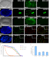
FIGURE 1.
FIGURE 1.
Mutations in atg-4.1 but not atg-4.2 cause defective degradation of autophagy substrates. A and B, SEPA-1::GFP aggregates are undetectable from the comma stage onwards in wild-type embryos. A, Nomarski image of the embryo in B. C, SEPA-1::GFP accumulates into a large number of aggregates in atg-4.1 mutants. D, expression level of SEPA-1::GFP in atg-4.2 mutants is identical to wild-type embryos. E–H, endogenous PGL-3, detected by anti-PGL-3 antibody, is restricted to germ precursor cells in a wild-type embryo (E and F), but ectopically accumulates in somatic cells in atg-4.1 mutant embryos (G). atg-4.2 mutants shows the same distribution of PGL-3 as wild-type embryos (H). E, DAPI image of the embryo shown in F. I–L, SQST-1::GFP is very weakly expressed and diffusely located in the cytoplasm in wild-type embryos (I and J). It accumulates into numerous aggregates in atg-4.1 mutants (K). The expression level of SQST-1::GFP reporter remains unchanged in atg-4.2 mutants (L). I, Nomarski image of the embryo in J. M–P, in atg-4.1(bp501) mutant embryos, SEPA-1 and SQST-1 aggregates, detected by specific antibodies, are largely separable. Q–T, SEPA-1 and SQST-1 aggregates accumulate and are separable in atg-3(bp412) mutant embryos. M and Q, DAPI images of the embryos shown in N–P and R–T, respectively. U, atg-4.1(bp501) mutants show a reduction in the survival of L1 larvae in the absence of food compared with wild type animals (log-rank test, p = 0.000). atg-4.2(tm3948) mutants show no defect in the L1 survival rate. At least 500 wild-type, atg-4.1(bp501) and atg-4.2(tm3948) L1 larvae were scored. V, number of neurons labeled by the touch neuron-specific reporter Pmec-4::gfp. atg-4.1(bp501); mec-4(u231) mutants have more Pmec-4::gfp-labeled neurons (p < 0.05) than mec-4(u231) single mutants. atg-4.2(tm3948); mec-4(u231) shows no significant increase in the neuron number.
FIGURE 2.
FIGURE 2.
Molecular lesions in atg-4.1 mutant alleles. A, phylogenetic tree of Atg4 family proteins. MEGA version 5.05 was used to construct a Neighbor-Joining phylogenetic tree. Bootstrap values are shown in percentages at nodes. The 0.1 scale bar represents 10% change. B, protein sequence of ATG-4.1. Glutamine residues are mutated to stop codons at amino acids 140, 160, and 385 in bp501, bp451 and bp410 mutants, respectively. The tryptophan at amino acid 347 is mutated to a stop codon in bp482. bp504 contains a histidine to proline mutation at amino acid 102. The glycine residue at amino acid 266 and the alanine residue at amino acid 271 are mutated to serine and valine in bp418 and bp321 mutants, respectively. C, schematic structure of atg-4.2. tm3948 deletes 498 nucleotides, introducing an early stop codon and creating a truncated protein with 266 amino acids. Intron-exon boundaries were confirmed by cDNA sequencing. D, crystal structure of the HsAtg4B-LC3 complex. The residues that are mutated in bp504, bp418, and bp321 are labeled. Although HsAtg4B has C74S mutation, Ser-74 is labeled as “C74” in Fig. 2, D–F. E, modeled structure of the HsAtg4B-LGG-1 complex. The LGG-1 tail (green) is recognized by the regulatory loop (blue) and W142 (blue) of HsAtg4B. F, G258S and A263V mutations, found in bp418 and bp321, respectively, disrupt the interaction between Atg4B and LGG-1.
FIGURE 3.
FIGURE 3.
atg-4.1 and atg-4.2 function redundantly in mediating conjugation of LGG-1. A, LGG-1/Atg8 levels in atg-4.1 and atg-4.2 mutants. Western analysis shows accumulation of LGG-1 precursors and also LGG-1-II in atg-4.1 mutants. LGG-1-I is very weakly detected in bp321, bp451, and bp504 mutants and is absent in bp501 mutants. In atg-4.2 mutants, no LGG-1 precursors are detected. LGG-1-II accumulates at a higher level than in wild type animals. B–E, atg-4.1- and atg-4.2-null mutants exhibit the wild-type dynamic immunostaining pattern of LGG-1 during embryogenesis. B, DAPI image of the embryo in C. F, in atg-4.1(bp501); atg-4.2(tm3948) double mutants, SEPA-1 aggregates accumulate at the comma stage. G, no LGG-1 puncta are detected in atg-4.1(bp501); atg-4.2(tm3948) double mutants.
FIGURE 4.
FIGURE 4.
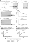
ATG-4.1 and ATG-4. 2 show different processing activity toward LGG-1/Atg8. A, schematic diagram showing cleavage of LGG-1-His6 by ATG-4.1 and ATG-4.2. The cleaved form of LGG-1 lacks the final seven residues as well as the His-tag at its C terminus, resulting in a lower molecular weight than LGG-1-His6 when separated by SDS-PAGE. Bacterially expressed His-tagged LGG-1, ATG-4.1, and ATG-4.2 were purified with Ni-NTA. LGG-1-His6 substrate (0.5 mg/ml) was incubated with vehicle control (first lane), ATG-4.1 (0.0125 mg/ml, second lane), or ATG-4.2 (0.0125 mg/ml and 0.025 mg/ml, third and fourth lanes), in a volume of 20 μl for 1.5 h at 37 °C. The mixture was resolved using SDS-PAGE followed by CBB staining after the reaction was stopped. B, LGG-1(G116tA)-His6 substrate (0.5 mg/ml), in which the conserved glycine is mutated to alanine, was incubated with vehicle control (first lane), ATG-4.1 (0.0125 mg/ml, second lane), or ATG-4.2 (0.025 mg/ml, third lane), under the same reaction conditions as in Fig. 4A. C, ATG-4.1 (0.0125 mg/ml) was incubated with the substrate LGG-1-His6 (0.5 mg/ml) in a volume of 20 μl for the indicated time. The mixture was resolved using SDS-PAGE followed by CBB staining after the reaction was stopped. D, gel was analyzed by Image J and the densities of both substrate and product were measured. About 50% LGG-1-His substrate was cleaved by ATG-4.1 in 45 min. E, ATG-4.2 (0.025 mg/ml) was incubated with the substrate LGG-1-His6 (0.5 mg/ml) in a volume of 20 μl for the indicated time. The mixture was resolved using SDS-PAGE followed by CBB staining. F, gel was analyzed by Image J and the densities of both substrate and product were measured. G–J, decreasing concentrations of the substrate LGG-1-His6 were incubated with fixed amounts of ATG-4.1 (0.0125 mg/ml) (G) and ATG-4.2 (0.025 mg/ml) (H) for 5 min and 300 min, respectively. The mixture was resolved using SDS-PAGE followed by CBB staining. The initial velocity (V, y axis (mm/s), i.e. the change in LGG-1 product concentration/s) was plotted against each substrate concentration (S (mm), x axis) for each reaction. The curves were fitted by the non-linear regression method (GraphPad Prism 5.00). K, kinetic parameters of ATG-4.1 and ATG-4.2 toward LGG-1. The values of Vmax (mol liter−1 s−1) and Km (mol/liter) were derived from curves fitted by the non-linear regression method (GraphPad Prism 5.00). The catalytic constant, kcat (s−1), was calculated by dividing Vmax by the enzyme concentration of each reaction.
FIGURE 5.
FIGURE 5.
The processed form of LGG-1 rescues the defect in atg-4. 1 mutants. A and B, SEPA-1 aggregates, labeled by a gfp reporter, accumulate in 4-fold stage atg-4.1(bp501) mutant embryos. A, Nomarski image of the embryo in B. C, number of SEPA-1::GFP aggregates is dramatically decreased in atg-4.1(bp501) embryos carrying the Plgg-1::LGG-1(G116 end) transgene. D, numerous SEPA-1::GFP aggregates exist in atg-4.1(bp501) embryos carrying the Plgg-1::LGG-1(A116 end) transgene. E and F, SEPA-1::GFP aggregates ectopically accumulate in atg-4.1(bp501); atg-4.2(tm3948) double mutant embryos at the 4-fold stage. E, Nomarski image of the embryo in F. G and H, accumulation of SEPA-1::GFP aggregates is greatly reduced in atg-4.1(bp501); atg-4.2(tm3948); Plgg-1::LGG-1(G116 end) embryos. G, Nomarski image of the embryo in H. I, expression of LGG-1 in wild type, atg-4.1 and atg-4.2 mutants carrying the Plgg-1::LGG-1(G116 end) transgene. LGG-1-II accumulates in wild type and atg-4.2 mutant animals carrying the Plgg-1::LGG-1(G116 end) transgene. In atg-4.1(bp501) mutants carrying this transgene, in addition to LGG-1 precursors and LGG-1-II, the unlipidated form of LGG-1-I also accumulates.
FIGURE 6.
FIGURE 6.
Genetic interactions of atg-4.1 with other autophagy mutants. A–D, in epg-1/Atg13 mutants, LGG-1 forms large puncta in a few cells but is absent in most embryonic cells (B). The morphology and distribution of LGG-1 puncta in atg-4.1; epg-1 double mutants (D) resemble those in epg-1 single mutants. A and C, DAPI images of the embryos shown in B and D, respectively. E–H, SEPA-1 aggregates and LGG-1 puncta form clusters and are largely colocalized in epg-6 mutants. In atg-4.1; epg-6 double mutants, SEPA-1 aggregates are small and are separable from LGG-1 puncta (H), resembling those in atg-4.1 single mutants. E, DAPI image of the embryo in F. Separate images for Fig. 6, F–H are shown in supplemental Fig. S3.

Similar articles

Cited by

References

    1. Xie Z., Klionsky D. J. (2007) Autophagosome formation: core machinery and adaptations. Nat. Cell Biol. 9, 1102–1109 - PubMed
    1. Nakatogawa H., Suzuki K., Kamada Y., Ohsumi Y. (2009) Dynamics and diversity in autophagy mechanisms: lessons from yeast. Nat. Rev. Mol. Cell Biol. 10, 458–467 - PubMed
    1. Ohsumi Y. (2001) Molecular dissection of autophagy: two ubiquitin-like systems. Nat. Rev. Mol. Cell Biol. 2, 211–216 - PubMed
    1. Kirisako T., Ichimura Y., Okada H., Kabeya Y., Mizushima N., Yoshimori T., Ohsumi M., Takao T., Noda T., Ohsumi Y. (2000) The reversible modification regulates the membrane-binding state of Apg8/Aut7 essential for autophagy and the cytoplasm to vacuole targeting pathway. J. Cell Biol. 151, 263–276 - PMC - PubMed
    1. Yoshimori T., Noda T. (2008) Toward unraveling membrane biogenesis in mammalian autophagy. Curr. Opin. Cell Biol. 20, 401–407 - PubMed

Publication types

MeSH terms

Substances

LinkOut - more resources