. 2012 Aug 24;287(35):29887-98.
doi: 10.1074/jbc.M112.371682. Epub 2012 Jul 5.

Aurora B is regulated by the mitogen-activated protein kinase/extracellular signal-regulated kinase (MAPK/ERK) signaling pathway and is a valuable potential target in melanoma cells

Affiliations

Aurora B is regulated by the mitogen-activated protein kinase/extracellular signal-regulated kinase (MAPK/ERK) signaling pathway and is a valuable potential target in melanoma cells

Caroline Bonet et al. J Biol Chem. .

Abstract

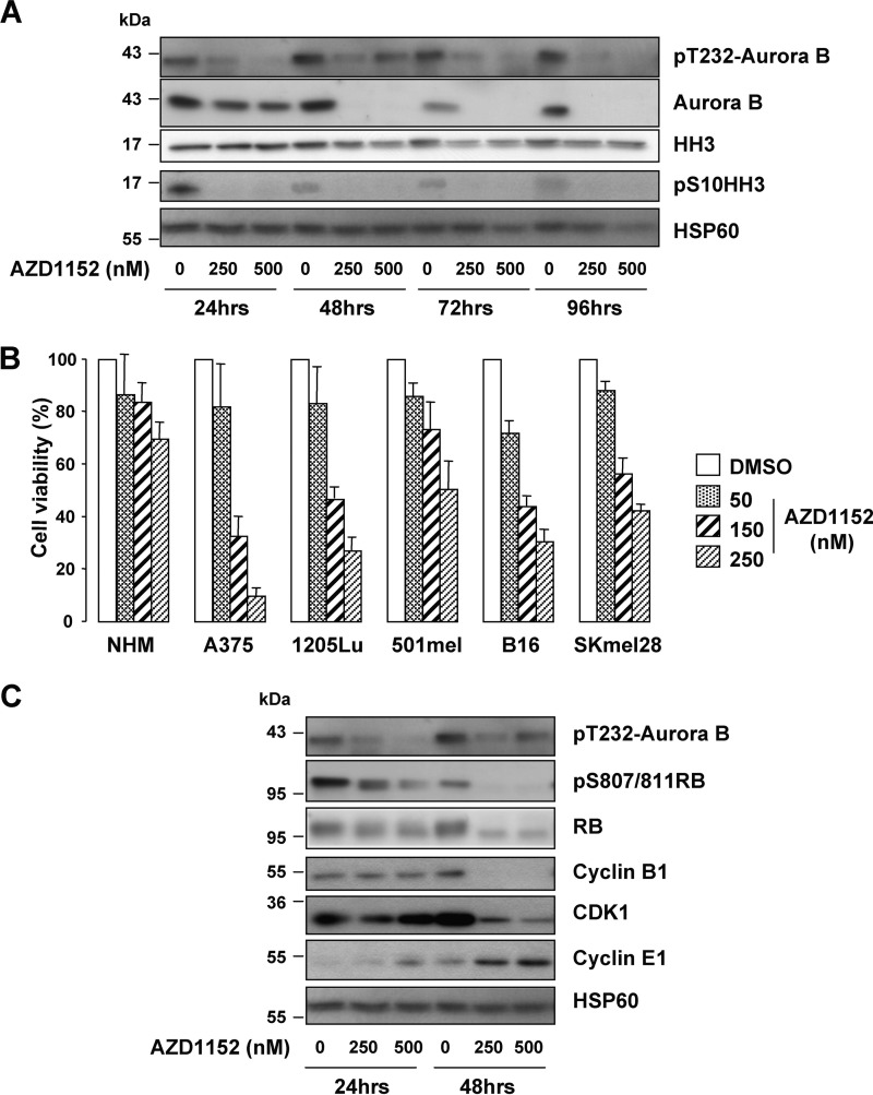
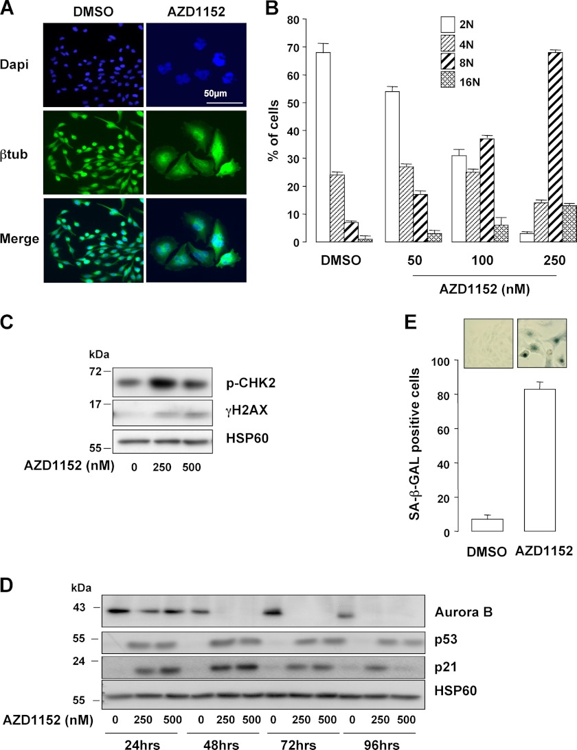
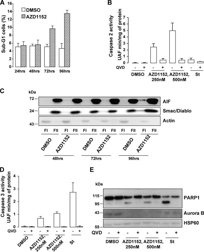
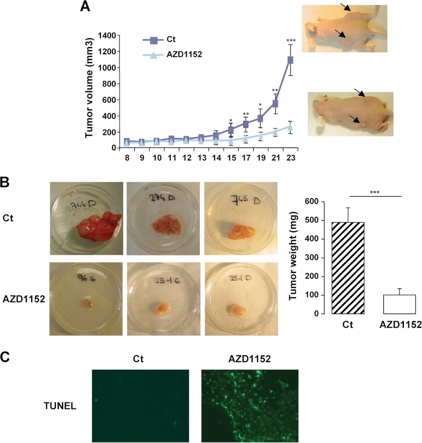
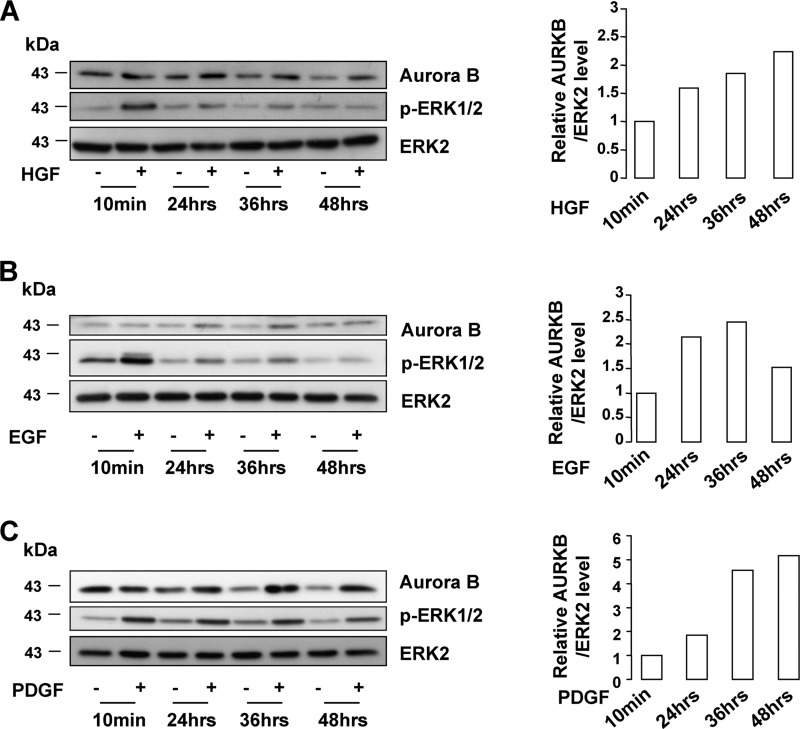
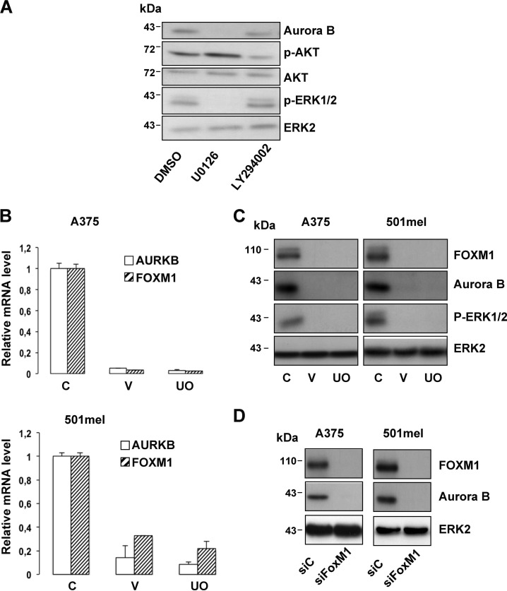
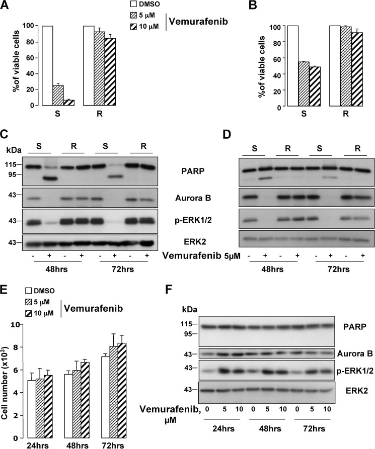
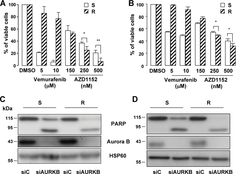
Metastatic melanoma is a deadly skin cancer and is resistant to almost all existing treatment. Vemurafenib, which targets the BRAFV600E mutation, is one of the drugs that improves patient outcome, but the patients next develop secondary resistance and a return to cancer. Thus, new therapeutic strategies are needed to treat melanomas and to increase the duration of v-Raf murine sarcoma viral oncogene homolog B1 (BRAF) inhibitor response. The ERK pathway controls cell proliferation, and Aurora B plays a pivotal role in cell division. Here, we confirm that Aurora B is highly expressed in metastatic melanoma cells and that Aurora B inhibition triggers both senescence-like phenotypes and cell death in melanoma cells. Furthermore, we show that the BRAF/ERK axis controls Aurora B expression at the transcriptional level, likely through the transcription factor FOXM1. Our results provide insight into the mechanism of Aurora B regulation and the first molecular basis of Aurora B regulation in melanoma cells. The inhibition of Aurora B expression that we observed in vemurafenib-sensitive melanoma cells was rescued in cells resistant to this drug. Consistently, these latter cells remain sensitive to the effect of the Aurora B inhibitor. Noteworthy, wild-type BRAF melanoma cells are also sensitive to Aurora B inhibition. Collectively, our findings, showing that Aurora B is a potential target in melanoma cells, particularly in those vemurafenib-resistant, may open new avenues to improve the treatment of metastatic melanoma.


Figures

FIGURE 1.
FIGURE 1.
Inhibition of metastatic melanoma cell proliferation by the Aurora B inhibitor AZD1152. A, human A375 melanoma cells were exposed to increasing concentration of AZD1152-HQPA (250 and 500 nm), and lysates were analyzed with the indicated antibodies. B, normal human melanocytes, human melanoma cells (A375, 1205LU, 501mel, and SkMel28), and mouse B16 melanoma cells were exposed to increasing concentrations of AZD1152-HQPA for 48 h, and then cell proliferation was assessed using the colorimetric XTT assay. C, lysates of A375 cells exposed to increasing concentrations of AZD1152 (250 and 500 nm) were analyzed by Western blotting with the indicated antibodies.
FIGURE 2.
FIGURE 2.
Aurora B inhibition induces polyploidization and senescence entry. A, immunofluorescence experiments of A375 cells exposed to 250 nm AZD1152-HQPA for 48 h were labeled with anti-β-tubulin antibody (btub). Nuclei were stained with DAPI. B, FACS analysis of A375 melanoma cells exposed to increasing concentrations of AZD1152-HQPA for 48 h. C, A375 cells treated with AZD1152-HQPA for 24 h were analyzed by immunoblotting with antibodies to phospho-Ser-387 CHK2 (pCHK2) and γH2AX. HSP60 ensures the even loading of each lane. D, human A375 melanoma cells were exposed to increasing concentrations of AZD1152-HQPA (250 and 500 nm), and lysates were analyzed with antibodies to p53 and p21. E, A375 cells were exposed to AZD1152-HQPA 250 nm for 96 h and cells remained adherent were analyzed for the SA-β-Galactosidase (SA-β-Gal) reactivity. The percentage of means ± S.D. was derived from counting 100 cells in duplicate plates.
FIGURE 3.
FIGURE 3.
Aurora B inhibition triggers cell death. A, sub-G1 FACS analysis of A375 melanoma cells exposed to 250 nm AZD1152-HQPA using propidium iodide. B, caspase-2 activity was assessed in lysates of A375 cells exposed to AZD1152-HQPA for 96 h. When indicated, the pan-caspase inhibitor QVD-OPH (QVD, 20 mm) was added to the reactions. The pro-apoptotic inducer staurosporine (1 μm) was used as a negative control. C, cytoplasmic (FI) and microsomal (FII) fractions of A375 cells exposed for the time indicated to 250 nm AZD1152-HQPA were analyzed by Western blotting with AIF, Smac/Diablo, and actin antibodies. D, caspase-3 activity was assessed in lysates of A375 cells exposed to AZD1152-HQPA for 96 h. When indicated, the pan-caspase inhibitor QVD-OPH (20 mm) was added to the reactions. The pro-apoptotic inducer staurosporine (1 μm) was used as a positive control. E, A375 cell lysates exposed for 96 h to 250 nm AZD1152-HQPA with or without 20 mm QVD-OPH were analyzed by Western blotting with total PARP1, Aurora B, and HSP60 antibodies.
FIGURE 4.
FIGURE 4.
In vivo anti-tumor effect of the Aurora B inhibitor AZD1152. A, A375 melanoma cells (2.5 × 10⁶) were subcutaneously engrafted in athymic nude mice. Mice were injected with the vehicle alone (Ct) or with AZD1152-HQPA. The growth tumor curves were determined by measuring the tumor volume using the equation V = (L × W2)/2. Results are presented as mean (±S.E.) tumor volumes (mm3), and p values are from Student's t test (*, p = 0.05; **, p = 0.01; ***, p = 0.001) comparing tumor size in AZD1152-HQPA-treated versus vehicle at each point. B, representative images and means of subcutaneous tumor weight from control (Ct) and AZD1152-HQPA treated mice are shown. C, TUNEL assay on 7-μm tumor sections.
FIGURE 5.
FIGURE 5.
Aurora B level is up-regulated upon growth factor ERK pathway. MeWo cells were treated with increasing duration of HGF (10 ng/ml) (A), EGF (10 ng/ml) (B), or PDGF (20 ng/ml) (C). Western blots were performed for Aurora B, p-ERK1/2, and ERK2. The densitometric scanning of the AURKB immunoblot bands was performed digitally using the ImageJ software program (National Institutes of Health) and was normalized to the total ERK expression levels.
FIGURE 6.
FIGURE 6.
Aurora B mRNA and protein levels are decreased in response to ERK inhibition. A, A375 melanoma cells were exposed to pharmacological inhibitors of the ERK (U0126, 10 μm) and PI3K (LY294002, 10 μm) signaling pathways for 48 h and were analyzed by Western blotting. B, quantitative PCR analysis of Aurora B and FOXM1 mRNA expression in A375 (upper panel) and 501mel (bottom panel) melanoma cells treated with vemurafenib (V, 10 μm) or UO126 (UO, 10 μm) for 48 h. C is control. C, A375 (left panel) or 501mel (right panel) melanoma cells were exposed to vemurafenib (V, 10 μm) or UO126 (UO, 10 μm) for 48 h. Cell lysates were analyzed by Western blotting. C is control. D, Western blot analysis of lysates from A375 (left panel) or 501mel (right panel) transfected with scrambled siRNA control (siC) or FOXM1 siRNA (siFOXM1).
FIGURE 7.
FIGURE 7.
Aurora B is re-expressed in vemurafenib-resistant melanoma cells. Freshly isolated human melanoma cells (#1) (left panel) (A) and WM9 cell line (right panel) (B) were rendered resistant to vemurafenib by continuous in vitro culture in increasing concentrations of the drug. Cell viability of the parental (vemurafenib-sensitive) (S) and of the resistant (R) melanoma cells treated with vemurafenib 5 and 10 μm for 48 h was assessed by XTT. C and D, vemurafenib-sensitive (S) and -resistant (R) melanoma cells (#1, left panel and WM9, right panel) were treated with 5 μm vemurafenib. Cell lysates were analyzed by Western blotting with the indicated antibodies. E, effect of vemurafenib 5 and 10 μm on the number of MeWo melanoma cells by cell count. F, MeWo melanoma cells were exposed to 5 and 10 μm vemurafenib for the time indicated on the figure and analyzed by Western blot with the indicated antibodies.
FIGURE 8.
FIGURE 8.
Aurora B is a potential therapeutic target in vemurafenib-resistant melanoma cells. A and B, XTT proliferation assay was performed on the vemurafenib-sensitive (S) and -resistant (R) melanoma cells (#1, left panel and WM9, right panel) treated with increasing concentrations of vemurafenib or AZD1152 for 48 h. Data are significantly different from the sensitive cells at **, p < 0.01, and *, p < 0.05. C and D, Western blot experiment of cell lysates from vemurafenib-sensitive (S) and -resistant (R) melanoma cells (#1, left panel, and WM9, right panel) transfected with Aurora B-specific inhibitor for 96 h.

Similar articles

Cited by

References

    1. Hodi F. S., O'Day S. J., McDermott D. F., Weber R. W., Sosman J. A., Haanen J. B., Gonzalez R., Robert C., Schadendorf D., Hassel J. C., Akerley W., van den Eertwegh A. J., Lutzky J., Lorigan P., Vaubel J. M., Linette G. P., Hogg D., Ottensmeier C. H., Lebbé C., Peschel C., Quirt I., Clark J. I., Wolchok J. D., Weber J. S., Tian J., Yellin M. J., Nichol G. M., Hoos A., Urba W. J. (2010) Improved survival with ipilimumab in patients with metastatic melanoma. N. Engl. J. Med. 363, 711–723 - PMC - PubMed
    1. Davies H., Bignell G. R., Cox C., Stephens P., Edkins S., Clegg S., Teague J., Woffendin H., Garnett M. J., Bottomley W., Davis N., Dicks E., Ewing R., Floyd Y., Gray K., Hall S., Hawes R., Hughes J., Kosmidou V., Menzies A., Mould C., Parker A., Stevens C., Watt S., Hooper S., Wilson R., Jayatilake H., Gusterson B. A., Cooper C., Shipley J., Hargrave D., Pritchard-Jones K., Maitland N., Chenevix-Trench G., Riggins G. J., Bigner D. D., Palmieri G., Cossu A., Flanagan A., Nicholson A., Ho J. W., Leung S. Y., Yuen S. T., Weber B. L., Seigler H. F., Darrow T. L., Paterson H., Marais R., Marshall C. J., Wooster R., Stratton M. R., Futreal P. A. (2002) Mutations of the BRAF gene in human cancer. Nature 417, 949–954 - PubMed
    1. Chapman P. B., Hauschild A., Robert C., Haanen J. B., Ascierto P., Larkin J., Dummer R., Garbe C., Testori A., Maio M., Hogg D., Lorigan P., Lebbe C., Jouary T., Schadendorf D., Ribas A., O'Day S. J., Sosman J. A., Kirkwood J. M., Eggermont A. M., Dreno B., Nolop K., Li J., Nelson B., Hou J., Lee R. J., Flaherty K. T., McArthur G. A. (2011) Improved survival with vemurafenib in melanoma with BRAF V600E mutation. N. Engl. J. Med. 364, 2507–2516 - PMC - PubMed
    1. Ruchaud S., Carmena M., Earnshaw W. C. (2007) Chromosomal passengers. Conducting cell division. Nat. Rev. Mol. Cell Biol. 8, 798–812 - PubMed
    1. Pirker C., Lötsch D., Spiegl-Kreinecker S., Jantscher F., Sutterlüty H., Micksche M., Grusch M., Berger W. (2010) Response of experimental malignant melanoma models to the pan-Aurora kinase inhibitor VE-465. Exp. Dermatol. 19, 1040–1047 - PubMed

Publication types

MeSH terms