Clinical Trial
. 2012 Aug 17;287(34):28378-85.
doi: 10.1074/jbc.M111.312884. Epub 2012 Jul 5.

Visfatin/pre-B-cell colony-enhancing factor (PBEF), a proinflammatory and cell motility-changing factor in rheumatoid arthritis

Affiliations
Clinical Trial

Visfatin/pre-B-cell colony-enhancing factor (PBEF), a proinflammatory and cell motility-changing factor in rheumatoid arthritis

Florian M P Meier et al. J Biol Chem. .

Abstract

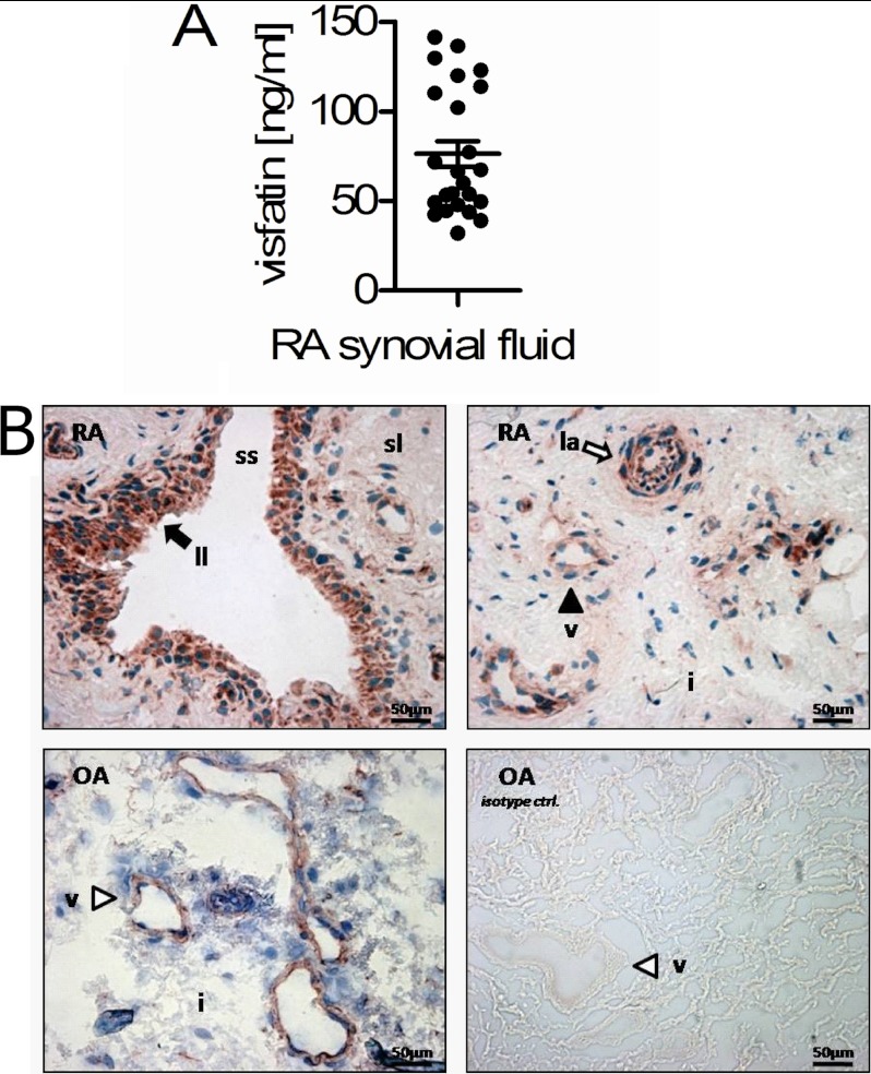
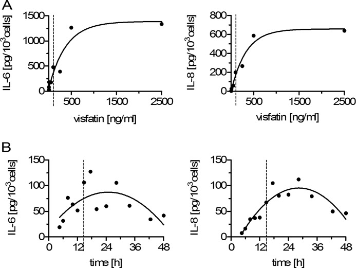
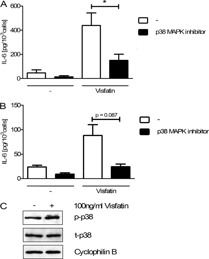
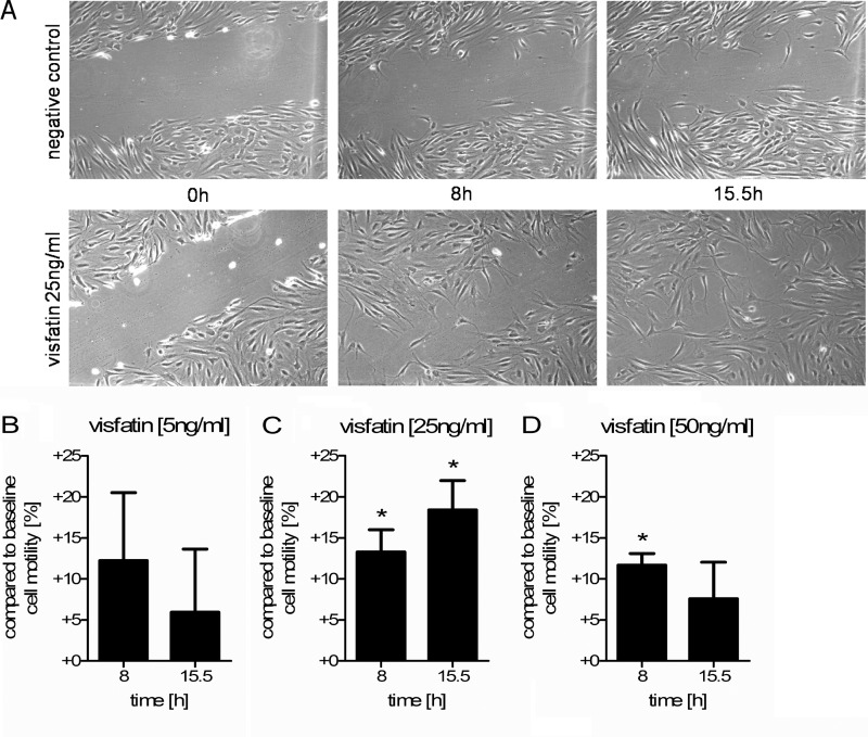
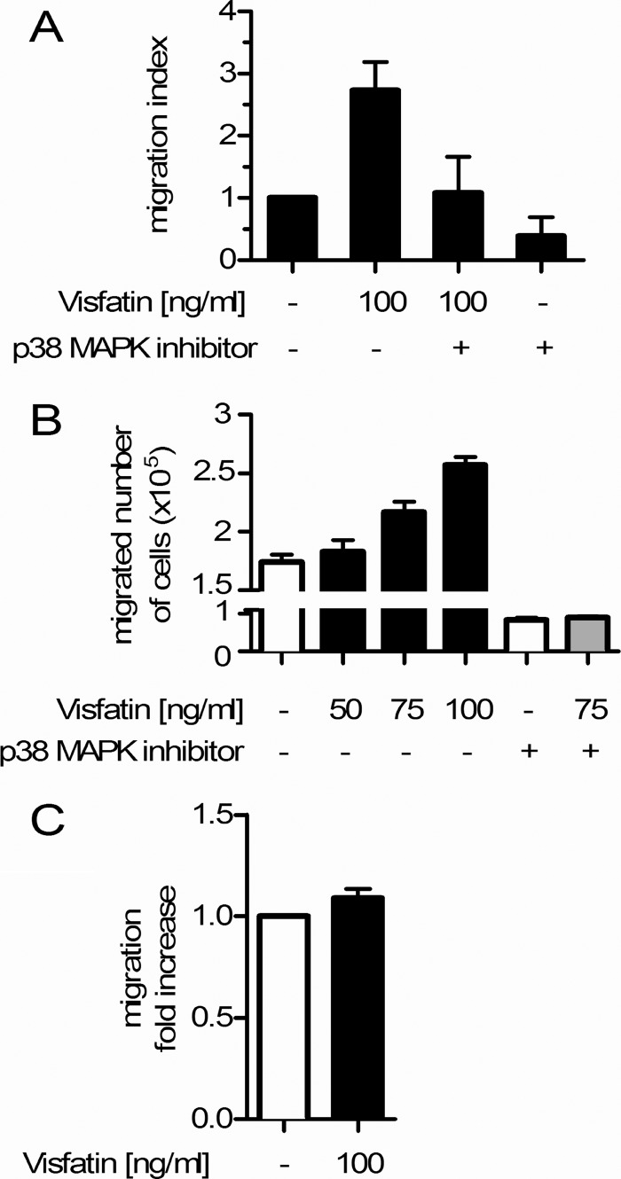
Adipokines such as adiponectin and visfatin/pre-B-cell colony-enhancing factor (PBEF) have been recently shown to contribute to synovial inflammation in rheumatoid arthritis (RA). In this study, we evaluated the pathophysiological implication of visfatin/PBEF in the molecular patterns of RA synovial tissue, focusing on RA synovial fibroblasts (RASFs), key players in RA synovium. Expression of visfatin/PBEF in synovial fluid and tissue of RA patients was detected by immunoassays and immunohistochemistry. RASFs were stimulated with different concentrations of visfatin/PBEF over varying time intervals, and changes in gene expression were evaluated at the RNA and protein levels using Affymetrix array, real-time PCR, and immunoassays. The signaling pathways involved were identified. The influence of visfatin/PBEF on fibroblast motility and migration was analyzed. In RA synovium, visfatin/PBEF was predominantly expressed in the lining layer, lymphoid aggregates, and interstitial vessels. In RASFs, visfatin/PBEF induced high amounts of chemokines such as IL-8 and MCP-1, proinflammatory cytokines such as IL-6, and matrix metalloproteinases such as MMP-3. Phosphorylation of p38 MAPK was observed after visfatin/PBEF stimulation, and inhibition of p38 MAPK showed strong reduction of visfatin-induced effects. Directed as well as general fibroblast motility was increased by visfatin/PBEF-induced factors. The results of this study indicate that visfatin/PBEF is involved in synovial fibroblast activation by triggering fibroblast motility and promoting cytokine synthesis at central sites in RA synovium.


Figures

FIGURE 1.
FIGURE 1.
Detection of visfatin/PBEF in RA synovial fluid and synovium. A, mean concentration of visfatin/PBEF in the synovial fluid of RA patients (n = 24; 66.1 ± 16 years of age; 83.3% female) measured by ELISA. B, Nucleus-counterstained immunohistochemical analysis for visfatin/PBEF protein in RA synovium (n = 3) and OA synovium (n = 2). Visfatin/PBEF protein is shown in red, and nuclei are shown in blue (magnification ×400). In RA synovium, synovial fibroblasts in the lining layer showed a strong expression of visfatin/PBEF (black arrow). Small interstitial vessels (black arrowhead) and lymphoid aggregates (white arrow) expressed visfatin/PBEF protein as well. In OA synovium, minor vessels of the interstitium showed visfatin/PBEF protein expression (white arrowhead). An isotype control experiment using OA synovium was performed. ss, synovial space; ll, lining layer; sl, sublining; v, vessel; la, lymphoid aggregate; i, interstitium.
FIGURE 2.
FIGURE 2.
Dose-dependent relationship and time-dependent response after visfatin/PBEF stimulation of RASFs. A, RASFs were incubated for 15 h with increasing concentrations of visfatin/PBEF (2.5–2500 ng/ml). IL-6 and IL-8 production was measured by ELISA. Data are expressed as the concentration of cytokine (pg produced per 103 cells) to stimulating concentrations of visfatin/PBEF. B, RASFs were stimulated with 100 ng/ml visfatin/PBEF for 4–48 h. IL-6 and IL-8 production was measured by ELISA. Data are presented as concentration of cytokine (pg produced per 103 cells) to stimulation time. Dashed lines display the stimulating concentration and time chosen for subsequent experiments.
FIGURE 3.
FIGURE 3.
Involvement of the p38 MAPK pathways in visfatin/PBEF-mediated production of IL-6 in RASFs using an intracellular signaling inhibitor. RASFs (n = 3) were stimulated with or without visfatin/PBEF (100 ng/ml) together with and without the chemical inhibitor of p38 MAPK. Values are expressed as means ± S.E. A, after 15 h. B, after 6 h. C, basal and visfatin/PBEF-induced activation of phospho-p38 (p-p38) in RASFs. *, p < 0.05. t-p38, total p38.
FIGURE 4.
FIGURE 4.
Visfatin/PBEF influences synovial fibroblast motility. A, scrape assay images represent the difference in cell motility over time between cell culture wells of the negative control and wells of the sample (25 ng/ml visfatin/PBEF). Images from the start of the experiment and after 8 and 15.5 h are shown. Scrape assays were performed as described under “Experimental Procedures” (n = 3). Results are expressed as comparison of samples to base line (percentages). The graphs show enhanced cell motility in the samples after 8 and 15.5 h for 5 ng/ml (B), 25 ng/ml (C), and 50 ng/ml (D) visfatin/PBEF. *, p < 0.05.
FIGURE 5.
FIGURE 5.
Visfatin/PBEF influences synovial fibroblast motility. A, RASF chemotaxis assays were performed (see “Experimental Procedures”), and the migration index was calculated (base line set to 1; ratio of sample to base line). Means ± S.E. are displayed as bars (n = 2). B, demonstration of lymphocyte migration to conditioned medium of RASFs (n = 2; the total number of migrated lymphocytes is shown). Results are expressed as means ± S.E. (see “Experimental Procedures”). C, incubation with 100 ng/ml visfatin/PBEF had only a slight effect on lymphocyte migration (n = 2). -Fold increase in migration is shown.

References

    1. Huber L. C., Distler O., Tarner I., Gay R. E., Gay S., Pap T. (2006) Synovial fibroblasts: key players in rheumatoid arthritis. Rheumatology 45, 669–675 - PubMed
    1. Lefèvre S., Knedla A., Tennie C., Kampmann A., Wunrau C., Dinser R., Korb A., Schnäker E. M., Tarner I. H., Robbins P. D., Evans C. H., Stürz H., Steinmeyer J., Gay S., Schölmerich J., Pap T., Müller-Ladner U., Neumann E. (2009) Synovial fibroblasts spread rheumatoid arthritis to unaffected joints. Nat. Med. 15, 1414–1420 - PMC - PubMed
    1. Knedla A., Neumann E., Müller-Ladner U. (2007) Developments in the synovial biology field 2006. Arthritis Res. Ther. 9, 209. - PMC - PubMed
    1. Müller-Ladner U., Ospelt C., Gay S., Distler O., Pap T. (2007) Cells of the synovium in rheumatoid arthritis. Synovial fibroblasts. Arthritis Res. Ther. 9, 223. - PMC - PubMed
    1. Kershaw E. E., Flier J. S. (2004) Adipose tissue as an endocrine organ. J. Clin. Endocrinol. Metab. 89, 2548–2556 - PubMed

Publication types

MeSH terms