. 2012;7(6):e39481.
doi: 10.1371/journal.pone.0039481. Epub 2012 Jun 29.

Small molecules targeted to a non-catalytic "RVxF" binding site of protein phosphatase-1 inhibit HIV-1

Affiliations

Small molecules targeted to a non-catalytic "RVxF" binding site of protein phosphatase-1 inhibit HIV-1

Tatiana Ammosova et al. PLoS One. 2012.

Abstract

HIV-1 Tat protein recruits host cell factors including CDK9/cyclin T1 to HIV-1 TAR RNA and thereby induces HIV-1 transcription. An interaction with host Ser/Thr protein phosphatase-1 (PP1) is critical for this function of Tat. PP1 binds to a Tat sequence, Q(35)VCF(38), which resembles the PP1-binding "RVxF" motif present on PP1-binding regulatory subunits. We showed that expression of PP1 binding peptide, a central domain of Nuclear Inhibitor of PP1, disrupted the interaction of HIV-1 Tat with PP1 and inhibited HIV-1 transcription and replication. Here, we report small molecule compounds that target the "RVxF"-binding cavity of PP1 to disrupt the interaction of PP1 with Tat and inhibit HIV-1 replication. Using the crystal structure of PP1, we virtually screened 300,000 compounds and identified 262 small molecules that were predicted to bind the "RVxF"-accommodating cavity of PP1. These compounds were then assayed for inhibition of HIV-1 transcription in CEM T cells. One of the compounds, 1H4, inhibited HIV-1 transcription and replication at non-cytotoxic concentrations. 1H4 prevented PP1-mediated dephosphorylation of a substrate peptide containing an RVxF sequence in vitro. 1H4 also disrupted the association of PP1 with Tat in cultured cells without having an effect on the interaction of PP1 with the cellular regulators, NIPP1 and PNUTS, or on the cellular proteome. Finally, 1H4 prevented the translocation of PP1 to the nucleus. Taken together, our study shows that HIV- inhibition can be achieved through using small molecules to target a non-catalytic site of PP1. This proof-of-principle study can serve as a starting point for the development of novel antiviral drugs that target the interface of HIV-1 viral proteins with their host partners.


Conflict of interest statement

Competing Interests: The authors have declared that no competing interests exist.

Figures

Figure 1
Figure 1. PP1 with RVxF peptide bound to its hydrophobic channel.
Peptide RRVSFA derived from Gm protein, a regulatory subunit of PP1 involved in glycogen metabolism, shown in ball-and-stick representation and colored after the CPK scheme, with carbon atoms colored in green for clarity. Solvent accessible surface area around the RVxF binding site is shown in transparent, and colored according to electrostatic potential.
Figure 2
Figure 2. Four binding modes for PP1 inhibitors.
Complexes with representative ligands, fulfilling each binding mode are shown. Representation scheme same as Figure 1. Hydrogen bonds are shown in yellow. Panel 1. Compounds are positioned toward Tyr255 were selected. Panel 2. Compounds reach Asp166. Panel 3. Compounds are span toward Gln262. Panel 4. Compounds form more than 4 hydrogen bonds with PP1.
Figure 3
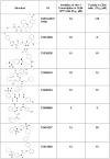
Figure 3. Selected compounds that inhibited HIV-1 transcription.
CEM-GFP cells were infected with Adeno-Tat and then treated with the indicated compounds at concentrations between 3 µM to 45 µM for 24 h. GFP fluorescence was measured in live cells. The cells were supplemented with propidium iodide (PI), and its fluorescence was measured to determine the toxicity of the compounds.
Figure 4
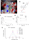
Figure 4. Inhibition of HIV-1 transcription and replication by 1H4.
A. The model of 1H4 interaction with PP1. 1H4 occupies hydrophobic sites of Phe68’ and Val 66′ side chains, interacts with Gln262 and forms a network of hydrogen bonds with Arg261 and Cys291. B. Chemical structure of 1H4. C. Inhibition of HIV-1 transcription in 293T cells. 293T cells were transfected with HIV-1 LTR-LacZ, CEM-GFP and Tat expressing vectors and treated with the indicated concentrations of 1H4 and 1G3 compounds. At 24 hours after the transfection, the cells were lysed and analyzed for green fluorescence and for β-galactosidase activity. D. Toxicity in CEM cells. CEM cells were treated with the indicated concentrations of 1H4, 1G3 and toxic A02 compound for 24 hours. The viability was determined using trypan exclusion assay and automated cell counter (Nexcelcom). E. HIV-1 replication is inhibited by 1H4. MT4 cells were infected with recombinant pNL4-3 HIV-1 and treated with different concentrations of 1H4 or inactive control 1G3. The RT activities were determined over the course of 8 days.
Figure 5
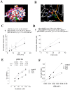
Figure 5. 1H4 compound competes with RVxF motif.
A. Superimposition of pRb-Tat peptide on the complex of PP1 with Spinophilin. PP1 surface is colored after the atom types. The spinophilin peptide is shown as magenta ribbon and the pRb-Tat peptide as orange ribbon. The Val25 and Phe27 residues of pRb-Tat and the Ile449 and Phe 451 residues of Spinophilin are shown as sticks. The 2-(N-morpholino)-ethanesulfonic acid bound in the active site of PP1 is shown in green spheres. The phosphorylated Ser6 residue of pRb-Tat peptide is shown as sticks. B. Phosphorylated Ser 6 residue binds to the active site of PP1. Comparative superimposition of pRb-Tat peptide over the crystal structure of MES bound in the active site of PP1. Catalytic resides are shown as sticks. MES is shown as green sticks, and the phosphorylated Ser6 residue of pRb-Tat peptide is shown as orange sticks. The pRb-Tat peptide is shown as orange ribbon. C. 1H4 inhibits kinetics of pRb-Tat peptide dephosphorylation by PP1α. Recombinant PP1α was assayed with pRb-Tat (WT or QACA mutant, 120 µM) in the absence or presence of 1H4 as indicated. The reactions were stopped at indicated time points and the phosphate release was quantified by malachite green assay. Initial velocity was calculated by linear regression in Prism. D. 1H4 inhibits kinetics of pRb-cdNIPP dephosphorylation by PP1α. Recombinant PP1α was assayed with pRb-cdNIPP1 in the absence or presence of 1H4 as indicated. Mutant pRb cdNIPP1 pA-RATA was used as negative control. The reactions were stopped at indicated time points and the phosphate release was quantified by malachite green assay. Initial velocity was calculated by linear regression in Prism. E and F. 1H4 competitively inhibits pRb-Tat peptide dephosphorylation by PP1α. Initial rates of pRb-Tat peptide dephosphorylation by PP1α were assayed at the indicated concentrations of the substrate in the absence or presence of 300 µM 1H4 or non-HIV-1 inhibitory 1G3. The amount of the released phosphate was quantified with malachite green. The VMAX and Km were calculated by non-linear regression analysis in Prism with the assumption that 25% of the substrate contained the phosphate group. Transformation of the data to Lineweaver-Burk plot (panel E) showed competitive inhibition of pRb-Tat dephosphorylation.
Figure 6
Figure 6. 1H4 has no effect on PP1 enzymatic activity.
A. 1H4 has no effect on the kinetics of KT(pT)IRR peptide dephosphorylation by PP1α. Recombinant PP1α (0.005 Units) was assayed with KT(pT)IRR peptide (3 µM) in the absence or presence of 1H4, and the reaction was stopped at indicated time points by the addition of malachite green solution. The amount of released phosphate was quantified by the absorbance and phosphate concentration was recalculated using standards. Initial velocity was calculated by linear regression in Prism. B and C. 1H4 has no effect on Km and VMAX of KT(pT)IRR peptide dephosphorylation by PP1α. Initial rates of KT(pT)IRR peptide dephosphorylation by PP1α were assayed at the indicated concentrations of the substrate in the absence or presence of 300 µM 1H4. The amount of released phosphate was quantified with malachite green. The VMAX and Km were calculated by non-linear regression analysis for Michaelis-Menten equation in Prism. The data were transformed to Lineweaver-Burk representation shown in panel C.
Figure 7
Figure 7. 1H4 prevents the interaction of Tat with PP1 in cultured cells.
A. Effect of 1H4 on PP1co-immunoprecipitation with Tat. 293T cells were transfected with Flag-tagged Tat and PP1α-EGFP. Flag-Tat was immunoprecipitated with anti-Flag antibodies from the cells extracts and probed with antibodies against EGFP to detect PP1and against Flag to detect Tat. Lane 1, untreated whole cell extract; lane 2, cells treated with 10 µM 1H4; lane 3, mock-transfected cells. B. 1H4 has no effect on PP1 association with NIPP1 and PNUTS. 293T cells were transfected with Flag-tagged Tat. PP1 was precipitated with microcystin agarose. The associated proteins were trypsinized and analyzed by nano-LC MS/MS. Liquid chromatography peak amplitudes for specific peptides derived from Tat (551.95 Da), PP1α (500.78 Da), NIPP1 (501.77 Da) and PNUTS (110.88 kDa) are shown, see details in text. The peptides were identified through MS/MS sequencing analysis by SEQUEST. C. Effect of 1H4 compound on a cell proteome. 293T cells were treated with 10 µM 1H4 for 18h or untreated and lysed as described in Materials and Methods. Lysates were trypsinized, fractionated by ion-exchange chromatography and then analyzed on by LC-MS-MS using C18 column. MS-MS data were analyzed by SEQUEST. The 28 major proteins having highest Score in SEQUEST are shown.
Figure 8
Figure 8. 1H4 compound disrupts the Tat-mediated translocation of PP1 into the nucleus.
HeLa cells were transfected with PP1α-EGFP (PP1α) (A), PP1α-EGFP and WT Flag-Tat (B, D and E) or PP1α-EGFP and Flag-Tat 35QACA38 mutant (C) and treated with 10 µM 1H4 (D) or control 1G3 compound (E) for 18 hours. The cells were photographed on Olympus IX51 using a blue filter for EGFP fluorescence or phase contrast with 600X magnification. F, 293T cells were transfected with PP1α-EGFP or PP1α-EGFP and WT Tat or Tat QACA mutant expression vectors. At 24 hrs posttransfection cells were lysed in low salt buffer and cytoplasmic extract was separated from the nuclear material by centrifugation. Fluorescence was measured in the nuclear and cytoplasmic fractions using Perkin-Elmer Luminoscan.

Similar articles

Cited by

References

    1. Berkhout B, Silverman RH, Jeang KT. Tat trans-activates the human immunodeficiency virus through a nascent RNA target. Cell. 1989;59:273–282. - PubMed
    1. Nekhai S, Jeang KT. Transcriptional and post-transcriptional regulation of HIV-1 gene expression: role of cellular factors for Tat and Rev. Future Microbiol. 2006;1:417–426. - PubMed
    1. Ammosova T, Jerebtsova M, Beullens M, Voloshin Y, Ray PE, et al. Nuclear protein phosphatase-1 regulates HIV-1 transcription. J Biol Chem. 2003;278:32189–32194. - PubMed
    1. Washington K, Ammosova T, Beullens M, Jerebtsova M, Kumar A, et al. Protein phosphatase-1 dephosphorylates the C-terminal domain of RNA polymerase-II. J Biol Chem. 2002;277:40442–40448. - PubMed
    1. Ammosova T, Yedavalli VR, Niu X, Jerebtsova M, Van Eynde A, et al. Expression of a protein phosphatase 1 inhibitor, cdNIPP1, increases CDK9 threonine 186 phosphorylation and inhibits HIV-1 transcription. J Biol Chem. 2011;286:3798–3804. - PMC - PubMed

Publication types

MeSH terms