. 2012;7(6):e39576.
doi: 10.1371/journal.pone.0039576. Epub 2012 Jun 29.

Activated human CD4+CD45RO+ memory T-cells indirectly inhibit NLRP3 inflammasome activation through downregulation of P2X7R signalling

Affiliations

Activated human CD4+CD45RO+ memory T-cells indirectly inhibit NLRP3 inflammasome activation through downregulation of P2X7R signalling

Vanessa Beynon et al. PLoS One. 2012.

Abstract

Inflammasomes are multi-protein complexes that control the production of pro-inflammatory cytokines such as IL-1β. Inflammasomes play an important role in the control of immunity to tumors and infections, and also in autoimmune diseases, but the mechanisms controlling the activation of human inflammasomes are largely unknown. We found that human activated CD4+CD45RO+ memory T-cells specifically suppress P2X7R-mediated NLRP3 inflammasome activation, without affecting P2X7R-independent NLRP3 or NLRP1 inflammasome activation. The concomitant increase in pro-IL-1β production induced by activated memory T-cells concealed this effect. Priming with IFNβ decreased pro-IL-1β production in addition to NLRP3 inflammasome inhibition and thus unmasked the inhibitory effect on NLRP3 inflammasome activation. IFNβ suppresses NLRP3 inflammasome activation through an indirect mechanism involving decreased P2X7R signaling. The inhibition of pro-IL-1β production and suppression of NLRP3 inflammasome activation by IFNβ-primed human CD4+CD45RO+ memory T-cells is partly mediated by soluble FasL and is associated with down-regulated P2X7R mRNA expression and reduced response to ATP in monocytes. CD4+CD45RO+ memory T-cells from multiple sclerosis (MS) patients showed a reduced ability to suppress NLRP3 inflammasome activation, however their suppressive ability was recovered following in vivo treatment with IFNβ. Thus, our data demonstrate that human P2X7R-mediated NLRP3 inflammasome activation is regulated by activated CD4+CD45RO+ memory T cells, and provide new information on the mechanisms mediating the therapeutic effects of IFNβ in MS.


Conflict of interest statement

Competing Interests: The authors have declared that no competing interests exist.

Figures

Figure 1
Figure 1. IFNβ inhibits IL-1β release in the presence of activated human CD4+CD45RO+ memory T-cells.
Monocytes were incubated overnight in the absence or presence of unstimulated or soluble αCD3 (0.5 ug/ml) activated CD4+CD45RO+CD45RA- memory T-cells with or without 1000 IU/ml IFNβ. The next day 100 ng/ml LPS was added to the co-culture for 4 h, followed by addition of 500 uM ATP for 45 min; a) IL-1β (n = 8) and b) TNFα levels (n = 5) were measured in the supernatant by ELISA. There was a significant reduction of IL-1β release in the presence of activated T-cells and IFNβ; TNFα secretion was not affected; c) memory T-cells were incubated with IFNβ either in the presence or absence of 2 ug/ml αCD3 and αCD28 for 18 h, washed twice with PBS and co-incubated with monocytes for 14 h. The release of IL-1β by monocytes after stimulation with LPS and ATP was suppressed by co-incubation with activated IFNβ-primed memory T-cells (n = 3) (Data are shown as means ± SD of duplicate cultures; *p<0.05; ***p<0.001 employing repeated measures ANOVA with post-hoc Bonferroni adjustment for multiple comparisons to avoid random correlations); d) immunoblot of pro-IL-1β cleavage and pro-caspase-1 levels in the cell lysate of monocytes.
Figure 2
Figure 2. Inhibition of NLRP3 inflammasome activation by IFNβ-primed human CD4+CD45RO+ memory T-cells is ATP-specific.
IL-1β release after incubation with a) Alum (400 ug/ml) (n = 3), b) MSU crystals (300 ug/ml) (n = 3) and c) MDP (10 ug/ml) (n = 3) was not inhibited by co-incubation of monocytes with IFNβ-primed CD4+CD45RO+ memory T-cells (Data are shown as means ± SD of duplicate cultures; ***p<0.01 employing repeated measures ANOVA with post-hoc Bonferroni adjustment for multiple comparisons to avoid random correlations).
Figure 3
Figure 3. Activated CD4+ CD45RO+ memory T-cells decrease secretion of soluble caspase-1 and P2X7R mRNA expression in monocytes leading to a reduced response upon ATP binding.
a) sCaspase-1 levels in the supernatant of monocytes was measured by ELISA (n = 8). A significant decrease of soluble Caspase-1 in the supernatant of cells co-cultured with activated memory T-cells was observed both in the presence and absence of IFNβ; b) intracellular levels of pro-IL-1β measured with an ELISA specific for the immature precursor of IL-1β showed a significant increase in pro-IL-1β levels in the presence of human activated CD4+CD45RO+ memory T-cells (n = 3), cell lysates from adherent monocytes were obtained after removal of non-adherent T-cells after addition of LPS and prior to incubation with ATP c) the ratio of intracellular pro-IL-1β to released soluble Caspase-1 is significantly increased in the presence of human activated CD4+CD45RO+ memory T-cells (n = 3). d) P2X7R mRNA expression in monocytes cultured overnight with activated CD4+CD45RO+ memory T-cells is decreased with and without addition of exogenous IFNβ (n = 3), mRNA was isolated from adherent monocytes after removal of non-adherent T-cells after addition of LPS and prior to incubation with ATP (Data are shown as means ± SD of duplicate cultures; *p<0.05; **p<0.01 employing repeated measures ANOVA with post-hoc Bonferroni adjustment for multiple comparisons to avoid random correlations); e) Ca2+-influx measurement in monocytes cultured with activated human CD4+CD45RO+ memory T-cells in the presence of IFNβ demonstrates the absence of slow sustained Ca2+-influx mediated by ATP binding to the purinergic P2X7-receptor (representative of 3 independent experiments); f) Annexin V staining of monocytes coincubated with T-cells and stimulated with LPS was measured by flow cytometry (representative flow cytometry histogram of n = 5).
Figure 4
Figure 4. IL10 does not mediate the observed suppression of active IL-1β release.
a) memory CD4+CD45RO+ T-cells cultured overnight in the presence of monocytes with or without αCD3 and/or IFNβ were restimulated with PMA/Ionomycin in the presence of Brefeldin A for 4 h and stained for IL-10 and IFNγ (representative of 3 independent experiments); b) upregulation of IL10 mRNA expression in resorted memory T-cells was confirmed by qPCR (n = 3; n.d. = not detected); c) inhibition of IL-10 (10 ug/ml) and/or IFNγ (10 ug/ml) by specific blocking antibodies did not affect the suppressive effect on IL-1β release by activated T-cells in the presence of IFNβ (n = 4) (% suppression is calculated by subtracting from 1 the ratio of IL-1β release from activated T-cells in the presence of IFNβ and non-activated T-cells in the absence of IFNβ and multiplying by 100; **p<0.01 employing repeated measures ANOVA with post-hoc Bonferroni adjustment for multiple comparisons to avoid random correlations).
Figure 5
Figure 5. Suppression of IL-1β release is cell contact independent and mediated by a soluble factor of 23–38 kDa MW.
a) Blocking OX40L and CD40L pathways by the use of inhibitory antibodies did not affect the suppressive effect of activated T-cells on monocytes in the presence of IFNβ (n = 3; % suppression calculated as described in Fig 4); b) CD39 mRNA expression by memory T-cells resorted after co-culture with monocytes was measured by qPCR (n = 3) (Data are shown as means ± SD of duplicate cultures; *p<0.01 employing repeated measures ANOVA with post-hoc Bonferroni adjustment for multiple comparisons to avoid random correlations); c) transfer of supernatant conditioned by a co-culture of monocytes and activated memory CD4+ T-cells in the presence of IFNβ onto fresh monocytes reproduced the suppressive effect on IL-1β release (n = 4); d) Kinetics of the inhibitory factor release and determination of molecular weight; supernatants of monocytes co-cultured with activated human memory T-cells in the presence of IFNβ were removed at the indicated time-points and fresh monocytes were cultured in the conditioned supernatants for 16 h before stimulation with LPS and activation with ATP (representative of 2 independent experiments); e) supernatants were filtered through molecular cut-off filters or f) SEC fractionated and molecular weight fractions in a 1∶1 ratio with fresh medium were used for the culture of fresh monocytes. IL-1β secretion was measured and inhibition of release was calculated according to Fig 4 (representative of 2 independent experiments).
Figure 6
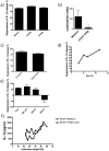
Figure 6. Identification of sFasL mediating the inhibition of IL-1β release.
a) Microarray analysis was performed in memory T-cells cultured in the presence of monocytes with and without IFNβ for 16 h and resorted by gating on CD3+CD4+7AAD- cells, upregulated candidates that fit the criteria of presence of a soluble form and molecular weight of 23–38 kDa are displayed; b) qPCR confirmation of upregulation of SSP1, LGALS9, APOL1 and FasL mRNA expression through incubation with IFNβ (n = 3); c) analysis of biological relevance of upregulated molecules by the use of blocking antibodies (10 ug/ml), only blockage of FasL significantly decreased the inhibitory effect on IL-1β release (n = 3) (% suppression calculated as described in Fig 4); d) soluble FasL levels were measured with a FasL specific ELISA in the supernatants of co-cultured activated CD4+CD45RO+ memory T-cells (n = 3); e) surface expression of FasL is measured after overnight co-culture by staining with FasL specific antibody and acquisition of cells by flow cytometry (representative of 3 independent experiments); f) IL-1β release is inhibited by recFasL in the presence of a crosslinking antibody (n = 3) (Data are shown as means ± SD of duplicate cultures; *p<0.05, **p<0.01, ***p<0.001 employing repeated measures ANOVA with post-hoc Bonferroni adjustment for multiple comparisons to avoid random correlations).
Figure 7
Figure 7. CD4+CD45RO+ memory T-cells of untreated MS patients are defective in the IFNβ-induced suppressive ability on active IL-1β release by monocytes.
This suppressive ability is rescued by in vivo treatment with IFNβ and is associated with increased serum levels of sFasL in IFNβ-treated MS patients; a) monocytes of HC (healthy controls) were cultured overnight with allogenic activated memory T-cells from HC (n = 13), untreated (n = 11) or IFNβ-treated MS patients (n = 10) and the suppressive effect of exogenous IFNβ on IL-1β release was measured as described in Fig 4; activated memory T-cells of MS patients showed a significantly decreased suppressive effect when compared to healthy controls, which was reversed by in vivo treatment with IFNβ; b) FasL levels in the serum of healthy controls (HC) (n = 24), untreated MS patients (MS) (n = 19) and IFNβ-treated MS patients (MS-IFNβ) (n = 16) were measured by ELISA; IFNβ treated MS patients showed an increased level of sFasL when compared to untreated MS patients and healthy controls; (*p<0.05; **p<0.01 employing repeated measures ANOVA with post-hoc Bonferroni adjustment for multiple comparisons to avoid random correlations).

References

    1. Dinarello CA. Immunological and inflammatory functions of the interleukin-1 family. Annu Rev Immunol. 2009;27:519–550. - PubMed
    1. Schroder K, Tschopp J. The inflammasomes. Cell. 2010. pp. 821–832. - PubMed
    1. Agostini L, Martinon F, Burns K, McDermott MF, Hawkins PN, et al. NALP3 forms an IL-1beta-processing inflammasome with increased activity in Muckle-Wells autoinflammatory disorder. Immunity. 2004. pp. 319–325. - PubMed
    1. Hoffman HM, Mueller JL, Broide DH, Wanderer AA, Kolodner RD. Mutation of a new gene encoding a putative pyrin-like protein causes familial cold autoinflammatory syndrome and Muckle-Wells syndrome. Nat Genet. 2001. pp. 301–305. - PMC - PubMed
    1. Latz E. The inflammasomes: mechanisms of activation and function. Current Opinion in Immunology. 2010. pp. 1–6. - PMC - PubMed

Publication types

MeSH terms