. 2012;7(6):e39613.
doi: 10.1371/journal.pone.0039613. Epub 2012 Jun 29.

Cell invasion by Neisseria meningitidis requires a functional interplay between the focal adhesion kinase, Src and cortactin

Affiliations

Cell invasion by Neisseria meningitidis requires a functional interplay between the focal adhesion kinase, Src and cortactin

Heiko Slanina et al. PLoS One. 2012.

Abstract

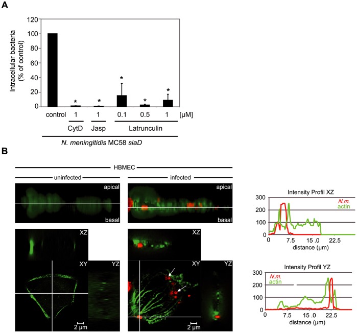
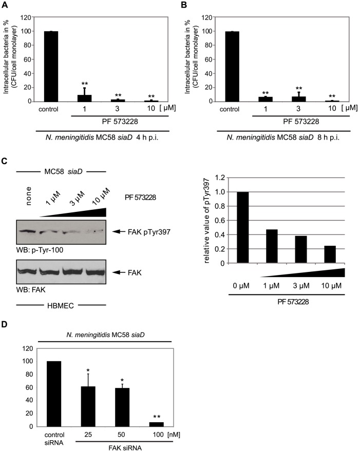
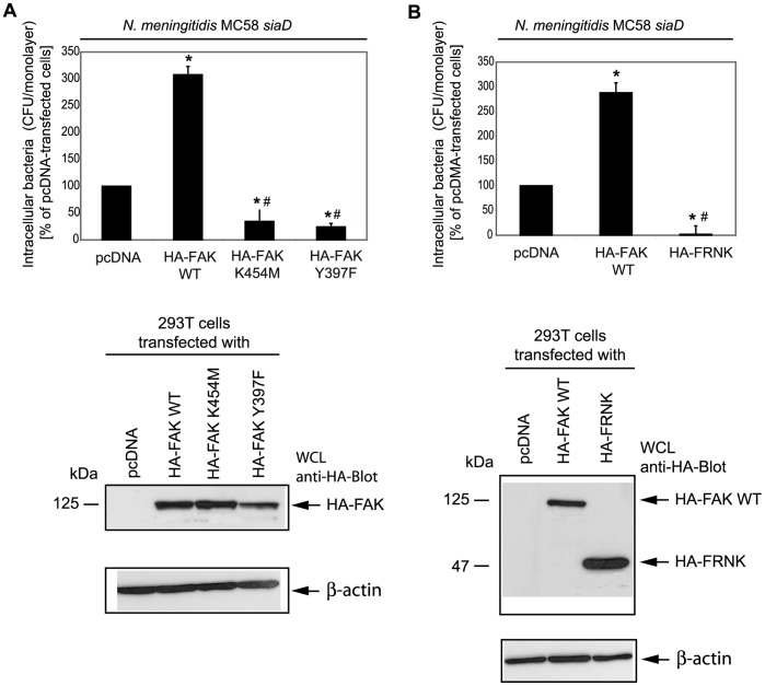
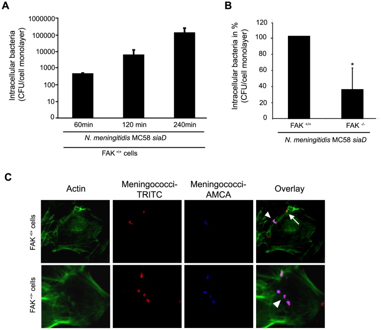
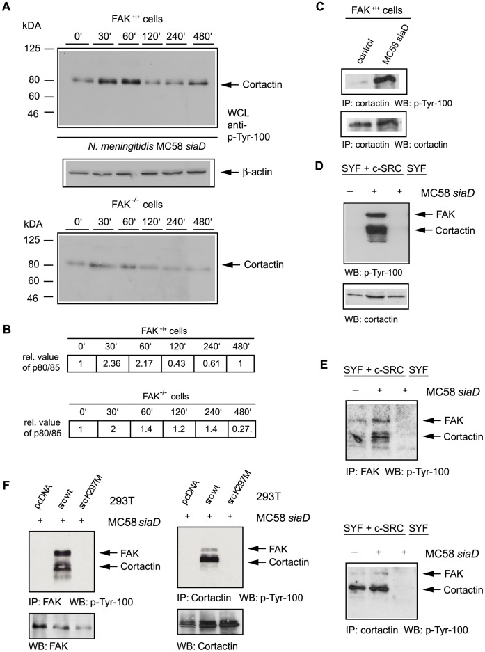
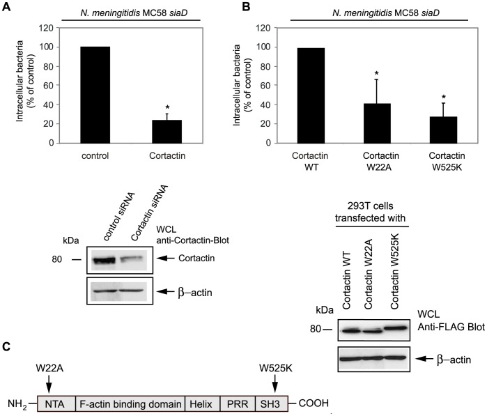
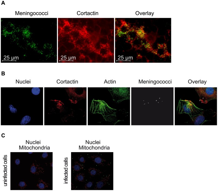
Entry of Neisseria meningitidis (the meningococcus) into human brain microvascular endothelial cells (HBMEC) is mediated by fibronectin or vitronectin bound to the surface protein Opc forming a bridge to the respective integrins. This interaction leads to cytoskeletal rearrangement and uptake of meningococci. In this study, we determined that the focal adhesion kinase (FAK), which directly associates with integrins, is involved in integrin-mediated internalization of N. meningitidis in HBMEC. Inhibition of FAK activity by the specific FAK inhibitor PF 573882 reduced Opc-mediated invasion of HBMEC more than 90%. Moreover, overexpression of FAK mutants that were either impaired in the kinase activity or were not capable of autophosphorylation or overexpression of the dominant-negative version of FAK (FRNK) blocked integrin-mediated internalization of N. meningitidis. Importantly, FAK-deficient fibroblasts were significantly less invaded by N. meningitidis. Furthermore, N. meningitidis induced tyrosine phosphorylation of several host proteins including the FAK/Src complex substrate cortactin. Inhibition of cortactin expression by siRNA silencing and mutation of critical amino acid residues within cortactin, that encompass Arp2/3 association and dynamin binding, significantly reduced meningococcal invasion into eukaryotic cells suggesting that both domains are critical for efficient uptake of N. meningitidis into eukaryotic cells. Together, these results indicate that N. meningitidis exploits the integrin signal pathway for its entry and that FAK mediates the transfer of signals from activated integrins to the cytoskeleton. A cooperative interplay between FAK, Src and cortactin then enables endocytosis of N. meningitidis into host cells.


Conflict of interest statement

Competing Interests: The authors have declared that no competing interests exist.

Figures

Figure 1
Figure 1. N. meningitidis internalization by HBMEC requires actin cytoskeleton dynamics.
(A) HBMEC were pre-incubated with indicated concentrations of inhibitors of the cytoskeleton, including cytochalasin D (Cyt D, 1 µM), jaspakinolide (Jasp), Latrunculin or solvent (DMSO) as a control and infected for 8 h with the unencapsulated invasive strain N. meningitidis strain MC58 siaD. Intracellular bacteria were defined by gentamicin protection assay. The graph shows mean values +/− S.D. of three independent experiments done in duplicate. * P<0.05, relative to cells infected without inhibitor. (B) Immunofluorescence analysis of HBMEC infected with N. meningitidis for 4 h. Uninfected and infected cells were fixed and labeled with Alexa Fluor® 488 phalloidin (green fluorescence). Bacteria were immunostained with a rabbit anti-meningococcal serum and secondary TRITC-labelled goat α-rabbit IgG (red fluorescence). The upper panels represent the z-stack projections, the white lane marks the level of the xy-planes shown in the lower panels. Meningococci attached to HBMEC induced an increase of actin concentration and caused the formation of actin stress fibers, consisting of long bundles of filaments traversing the cell, whereas uninfected control cells showed no formation of stress fibers. Internalized bacteria are marked with arrows. Fluorescence signals were separately quantified in the GFP and rhodamin channels and showed an increase in the local concentration of actin adjacent to the attached bacterium. Figure shows an XY plan view and the XZ and YZ sagittal cross-sections. Scale bar  = 2 µm.
Figure 2
Figure 2. N. meningitidis internalization by HBMEC requires focal adhesion kinase (FAK) activity.
(A and B) HBMEC were pre-incubated with indicated concentrations of the specific FAK inhibitor PF573228 and infected for 4 h and 8 h with the unencapsulated N. meningitidis strain MC58 siaD. Intracellular bacteria were defined by gentamicin protection assay. The graph shows mean values +/− S.D. of three independent experiments done in duplicate. ** P<0.01, relative to cells infected without inhibitor. (C) HBMEC were pre-incubated with the indicated concentrations of the FAK inhibitor PF 573228 for 1 h and infected with N. meningitidis MC58 siaD for 4 h. Cell lysates were resolved by SDS-PAGE and blotted with α-phospho-FAK Tyr397 demonstrating that PF 573228 blocked FAK Tyr397 phosphorylation in a dose-dependent manner. (D) 293T cells were transfected with indicated concentration of a commercial siRNA specific for FAK to limit FAK protein expression or transfected with unspecific control siRNA. siRNA transfected cells were infected with mutant strain MC58 siaD and internalized bacteria were measured by gentamicin protection assay at 4 h p.i. The graph represents mean values ± S.D. of three independent experiments done in duplicate. * P<0.05 and ** P<0.01, relative to cells transfected with the control siRNA.
Figure 3
Figure 3. Interference with FAK functions results in decreased uptake of N. meningitidis.
(A) 293T cells were transfected with a control plasmid (pcDNA) and a plasmid encoding wildtype FAK (HA-FAK WT) or a plasmid encoding a kinase inactive mutant (HA-FAK K454M) or a mutant that was not capable of autophosphorylation (HA-FAK Y397F). Transfected cell were infected with invasive strain MC58 siaD and intracellular bacteria were estimated 4 h post-infection by gentamicin protection assays. The graph represents mean values ± S.D. of three independent experiments done in duplicate. * P<0.05, relative to cells transfected with the control plasmid, # P ≤ 0.05, relative to cells transfected FAK wildtype were considered significant. In parallel, Western blotting of WCL extracts with anti-HA-tag antibody demonstrates expression of HA-tagged FAK constructs. (B) 293T cells were transfected with HA-FAK WT or a plasmid encoding the FAK related non-kinase (HA-FRNK) and infected with MC58 siaD as described above. The graph represents mean values ± S.D. of three independent experiments done in duplicate. * P<0.05. WCL were analyzed by Western blotting using an anti-HA-tag antibody and demonstrated overexpression of FRNK in transfected cells.
Figure 4
Figure 4. FAK-deficient cells are impaired in their ability to internalize N. meningitidis.
(A) FAK+/+ fibroblasts were infected with MC58 siaD at an MOI of 30 in presence of RPMI cell culture medium, supplemented with 10% human serum (HS). Intracellular bacteria were defined after gentamicin treatment at 60, 120 and 240 min post-infection (p.i.) demonstrating an invasion kinetic similar to 293T cells and HBMEC. The graphs represent mean value ± S.D. of three different independent experiments done in duplicate. * P<0.05. (B) FAK re-expressing (FAk+/+) and FAK-deficient (FAK−/−) fibroblasts were infected with invasive strain MC58 siaD in HS-supplemented RPMI cell culture medium. Intracellular bacteria were estimated at 4 h p.i. by gentamicin protection assays. The graph represents mean value ± S.D. of three different independent experiments done in duplicate. * P<0.05. (C) FAK+/+ and FAK−/− fibroblasts were infected with N. meningitidis strain MC58 siaD for 4 h and analyzed by immunofluorescence microscopy. Extracellular bacteria (arrowhead) stain positive with both TRITC (red fluorescence) and AMCA (blue fluorescence), whereas intracellular bacteria (arrow) are labeled with TRITC only. Cell actin was stained with Alexa Fluor® 488 phalloidin (green fluorescence).
Figure 5
Figure 5. N. meningitidis induced increased tyrosine phosphorylation of cellular proteins.
(A) FAK+/+ cells were serum starved and plated on poly-L-lysine coated dishes. Fibroblasts were left uninfected or infected with N. meningitidis MC58 siaD for a 8 h period and cell lysated were collected at from uninfected control cells and at 30 min, 60 min, 120 min, 240 min and 480 min post-infection and analyzed by Western blotting using the anti-phosphotyrosine antibody p-Tyr-100. (B) The increase of the phosphorylated protein was quantified by densitometric analysis and increase was estimated by comparison to the uninfected control. Densitometric analysis was performed as described in Experimental procedures. Staining of the samples with anti-actin antibody was used as loading control. (C) FAK+/+ cells were infected for 4 h as described above and samples were immunoprecipitated (IP) with an anti-cortactin antibody and immunoprecipitates were analyzed with α-p-Tyr-100 antibody (upper panel). Membranes were stripped and reprobed with polyclonal anti-cortactin antibody (lower panel). (D) Src re-expressing (SYF + c-Src) and c-Src-deficient (SYF) fibroblasts were infected with invasive strain MC58 siaD for 4 h as described above. Cell lysates were collected and analyzed by Western blotting using antibody p-Tyr-100. After stripping membranes were re-probed with polyclonal anti-cortactin antibody (lower panel). (E) SYF + c-Src and SYF cells were infected with strain MC58 siaD for 4 h and samples were immunoprecipitated either with an α-FAK antibody (Fig. 5E, upper panel) or α-cortactin antibody (Fig. 5E, lower panel) and immunoprecipitates were analyzed with α-p-tyr-100 antibody. (F) 293T cells were transfected with the empty control vector (pcDNA), a plasmid encoding wild-type c-Src and a vector encoding the inactive version of Src [Src K297M] to prove that Src kinase activity is required FAK/cortactin phosphorylation. Transfected cells were infected as described above, followed by an IP with an α-FAK antibody or α-cortactin antibody, respectively. Western blot analysis with α-p-tyr-100 demonstrated that Src kinase activity is required FAK/cortactin phosphorylation.
Figure 6
Figure 6. Interferences with cortactin expression results in diminished uptake of N. meningitidis into host cells.
(A) 293T cells were transfected with equal amounts (25 nM) of cortactin and control-siRNA. Invasion assays were performed 72 h post transfection with invasive strain MC58 siaD and intracellular bacteria were estimated 8 h post-infection by gentamicin protection assays. The graph represents mean values ± S.D. of three independent experiments done in duplicate. * P<0.05, relative to cells transfected with the control siRNA. In parallel whole cell lysates (WCL) extracts were prepared and subjected to Western blot analysis. Staining with polyclonal anti-cortactin antibody demonstrated genetic knock-down expression of cortactin in cortactin-siRNA-transfected cells compared to control siRNA-transfected cells. (B) 293T cells were transiently transfected with wildtype cortactin (cortactin WT), a plasmid encoding a mutant form of cortactin interfering with Arp2/3 association (W22A) or a plasmid encoding a mutant form of cortactin interfering with dynamin binding (W525K). Transfected cells were infected with strain MC58 siaD and the numbers of intracellular bacteria were determined after gentamicin treatment. The data in the graph are the mean values ± S.D. of three independent experiments done in duplicate. * P<0.05, relative to cells transfected with cortactin WT. Western blotting of WCL with anti-FLAG antibody demonstrated that there was equal overexpression of the wildtype cortactin and both cortactin mutant forms. The FLAG-tagged wildtype cortactin runs higher than endogenous cortactin, and the W525K mutant is anomalously high in SDS gels, as described elsewhere . (C) Schematic representation of the domain structure of cortactin. The cortactin point mutations are indicated. W22A, tryptophan to alanine point mutation in the NTA domain; and W525K, tryptophan to lysine point mutation in the SH3 domain. Domains include: NTA, N-terminal acidic (NTA) region; PRR, proline-rich region; and the SH3 domain that comprises the carboxy terminus.
Figure 7
Figure 7. N. meningitidis induces recruitment of cortactin.
(A) 293T cells were infected for 4 h with FITC-labeled meningococci (green fluorescence). Infected cells were fixed and stained with an anti-cortactin antibody followed by secondary α-rabbit TRITC-conjugated antibody (red fluorescence). Immunofluorescence demonstrated local recruitment of cortactin to cell-associated bacteria. Scale bars represent 25 µm. (B) For the close-up view, cells were infected with N. meningitidis for 4 h, fixed and incubated with DAPI (blue fluorescence), Alexa Fluor® 488 phalloidin (green fluorescence), cortactin goat α-rabbit/TRITC-conjugated goat α-rabbit (red fluorescence) and α-meningococcal serum/Alexa Fluor® 594 goat α-mouse (pseudocolored in gray). The immunofluorescence confirmed an accumulation of cortactin and the formation of stress fibers adjacent of bacterial attachment. (C) To exclude that bacterial attachment occurred solely on dead cells, cells were either infected with FITC-labeled bacteria or left uninfected and were incubated with MitoTracker (red fluorescence) and DAPI. No signs of apoptosis could be observed before or after 4 h of infection.

Similar articles

Cited by

References

    1. Schmitt C, Turner D, Boesl M, Abele M, Frosch M, et al. A functional two-partner secretion system contributes to adhesion of Neisseria meningitidis to epithelial cells. J Bacteriol. 2007;189:7968–7976. - PMC - PubMed
    1. Comanducci M, Bambini S, Brunelli B, Adu-Bobie J, Arico B, et al. NadA, a novel vaccine candidate of Neisseria meningitidis. J Exp Med. 2002;195:1445–1454. - PMC - PubMed
    1. Turner DP, Marietou AG, Johnston L, Ho KK, Rogers AJ, et al. Characterization of MspA, an immunogenic autotransporter protein that mediates adhesion to epithelial and endothelial cells in Neisseria meningitidis. Infect Immun. 2006;74:2957–2964. - PMC - PubMed
    1. Scarselli M, Serruto D, Montanari P, Capecchi B, Adu-Bobie J, et al. Neisseria meningitidis NhhA is a multifunctional trimeric autotransporter adhesin. Mol Microbiol. 2006;61:631–644. - PubMed
    1. Virji M. Pathogenic neisseriae: surface modulation, pathogenesis and infection control. Nat Rev Microbiol. 2009;7:274–286. - PubMed

Publication types

MeSH terms