. 2012;7(6):e39875.
doi: 10.1371/journal.pone.0039875. Epub 2012 Jun 29.

Herpes simplex virus type 1 infection facilitates invasion of Staphylococcus aureus into the nasal mucosa and nasal polyp tissue

Affiliations

Herpes simplex virus type 1 infection facilitates invasion of Staphylococcus aureus into the nasal mucosa and nasal polyp tissue

XiangDong Wang et al. PLoS One. 2012.

Abstract

Background: Staphylococcus aureus (S. aureus) plays an important role in the pathogenesis of severe chronic airway disease, such as nasal polyps. However the mechanisms underlying the initiation of damage and/or invasion of the nasal mucosa by S. aureus are not clearly understood. The aim of this study was to investigate the interaction between S. aureus and herpes simplex virus type 1 (HSV1) in the invasion of the nasal mucosa and nasal polyp tissue.

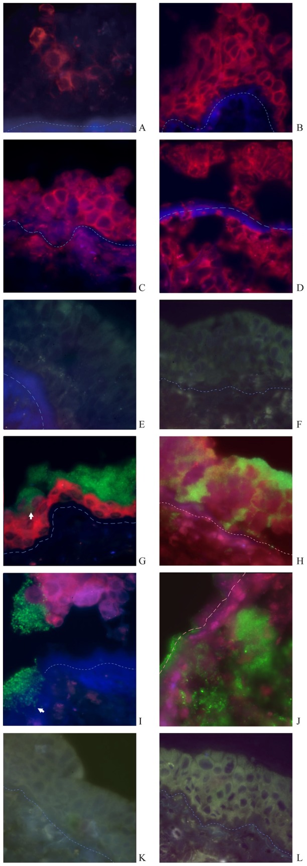
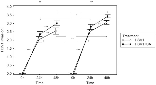
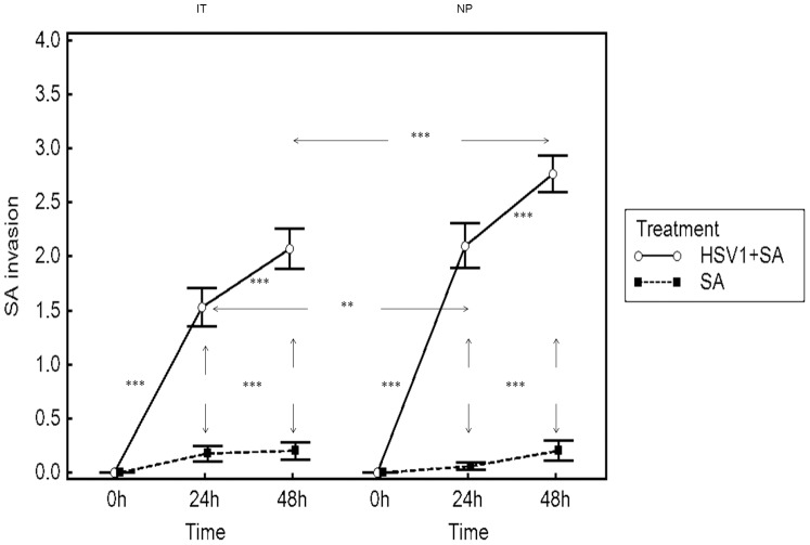
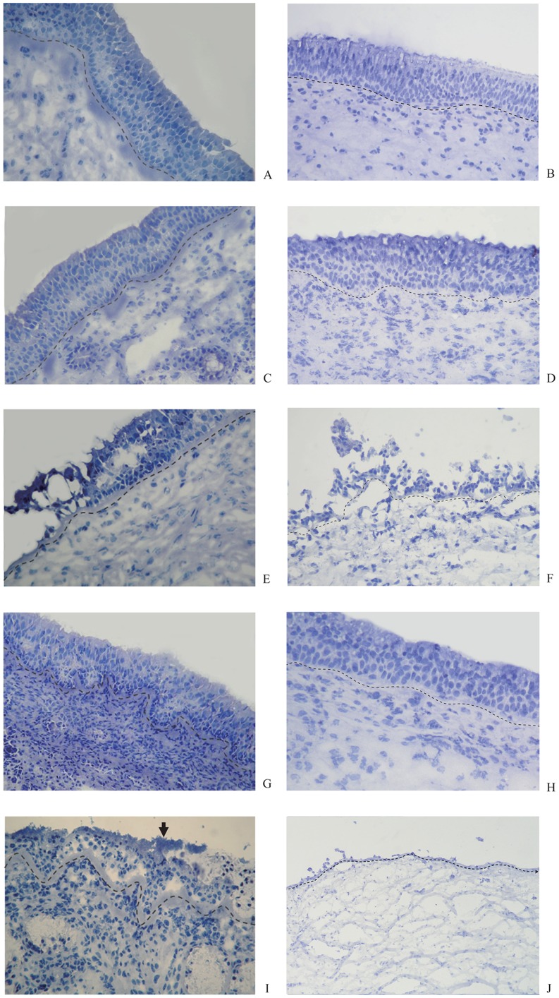
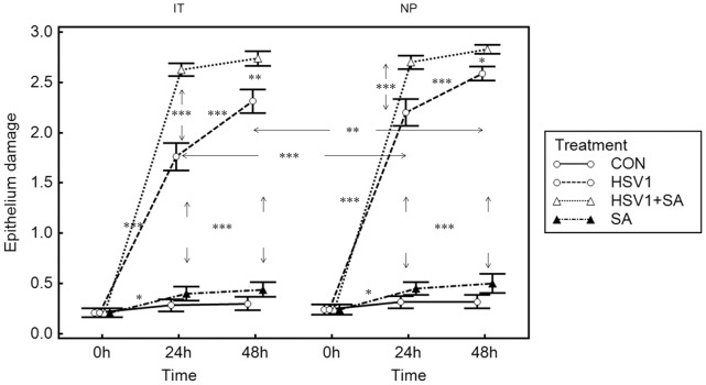
Methodology/principal findings: Inferior turbinate and nasal polyp samples were cultured and infected with either HSV1 alone, S. aureus alone or a combination of both. Both in turbinate mucosa and nasal polyp tissue, HSV1, with or without S. aureus incubation, led to focal infection of outer epithelial cells within 48 h, and loss or damage of the epithelium and invasion of HSV1 into the lamina propria within 72 h. After pre-infection with HSV1 for 24 h or 48 h, S. aureus was able to pass the basement membrane and invade the mucosa. Epithelial damage scores were significantly higher for HSV1 and S. aureus co-infected explants compared with control explants or S. aureus only-infected explants, and significantly correlated with HSV1-invasion scores. The epithelial damage scores of nasal polyp tissues were significantly higher than those of inferior turbinate tissues upon HSV1 infection. Consequently, invasion scores of HSV1 of nasal polyp tissues were significantly higher than those of inferior turbinate mucosa in the HSV1 and co-infection groups, and invasion scores of S. aureus of nasal polyp tissues were significantly higher than those of inferior turbinate tissues in the co-infection group.

Conclusions/significance: HSV1 may lead to a significant damage of the nasal epithelium and consequently may facilitate invasion of S. aureus into the nasal mucosa. Nasal polyp tissue is more susceptible to the invasion of HSV1 and epithelial damage by HSV1 compared with inferior turbinate mucosa.


Conflict of interest statement

Competing Interests: The authors have declared that no competing interests exist.

Figures

Figure 1
Figure 1. Immunofluorescence-stained sections of nasal mucosa of inferior turbinate (IT) and nasal polyp tissue samples (NP).
HSV1 infection: After 24 h HSV1+24 h medium incubation, HSV1 infects outer epithelial cells of IT focally, without reaching the basement membrane and results in loss of epithelial integrity (A). In contrast, HSV1infected the whole epithelium of NP after 24 h of HSV1 incubation (B). After 48 h HSV1+24 h medium incubation, HSV1 infects basal epithelial cells, and leads to loss of epithelial structure partly, penetration of the basement membrane and invasion of the virus into the lamina propria of IT (C). HSV1 infection leads to the loss of epithelial structures and invasion of the virus into the lamina propria of NP diffusively (D). S. aureus infection: After 24 h medium+24 h S. aureus incubation, S. aureus does not infect or invade the epithelium of IT and NP (E,F). HSV1+S. aureus infection: After 24 h HSV1+24 h S. aureus incubation, S. aureus forms clusters and attaches to the residual epithelial cells infected by HSV1, with S. aureus penetration of epithelium through inter-epithelial cell spaces (white arrow) (G). In contrast, cluster-forming S. aureus attached to epithelial cells of NP when infected by HSV1and penetrated the epithelium down to basal cells widely; planctonic bacteria invaded into the lamina propria after 24 h combined HSV1and S. aureus incubation (H). After 48 h HSV1+24h S. aureus incubation, clusters of S. aureus adhere to HSV1-infected epithelial cells and basement membrane of IT, with subsequent shedding of epithelial cells and penetration of the basement membrane (white arrow) (I). Whereas both HSV1 and S. aureus invade into the lamina propria of NP diffusively, with loss of epithelium(J). Control: The epithelium is intact and undamaged after 96 h totally in non-infected control tissues explants of IT and NP (K,L). (All slides viewed at ×630 magnification; red fluorescence demonstrates the presence of HSV1 and green fluorescence the presence of S. aureus in the nasal mucosa; dotted line indicates the basement membrane. The isotype control staining for HSV1 was completely negative and is not presented here.).
Figure 2
Figure 2. Invasion of nasal mucosa of inferior turbinate (IT) and nasal polyp tissue samples (NP) by HSV1.
Results are presented as mean scores ± SEM for IT from 10 patients and NP from 7 patients. Arrows represent differences between HSV1-infection and HSV1+S. aureus-infection groups or differences between IT group and NP groups. *P<0.05, **P<0.01 and ***P<0.001.
Figure 3
Figure 3. Invasion of nasal mucosa of inferior turbinate (IT) and nasal polyp tissue samples (NP) by S. aureus.
Results are presented as mean scores ±SEM for nasal turbinate samples from 10 patients and NP from 7 patients. Arrows represent differences between S. aureus-infection and HSV1+S. aureus-infection groups or differences between IT group and NP groups. ** P<0.01,*** P<0.001.
Figure 4
Figure 4. Hematoxylin-stained sections of inferior turbinate (IT) and nasal polyp tissue samples (NP).
The epithelium and basement membrane (dotted line) are intact and undamaged at baseline of IT (A) and NP (B) respectively and after 96 h totally incubation in uninfected control explants of IT (C) and NP (D), respectively. Extensive infection and damage to the epithelium is seen in the explants after 48 h HSV1+24h medium of IT (E) and NP (F) respectively, but not in the explants of IT (G) and NP (H) respectively after 48h medium+24 h S. aureus incubation. Combined HSV1 and S. aureus infection leads to adherence of S. aureus clusters to the epithelium (arrow) and partial damage of the epithelium of IT after 24 h HSV1+24 h S. aureus incubation (I), and complete loss of the epithelium of NP after 24 h HSV1+24 h S. aureus incubation (J). (All slides viewed at ×400 magnification.)
Figure 5
Figure 5. Epithelium damage in nasal mucosa.
Results are presented as mean±SEM scores for IT from 10 patients and NP from 7 patients; *P<0.05, **P<0.01, ***P<0.001 between time points. Control means no infection. Arrows indicate intergroup significance.

References

    1. Larkin EA, Carman RJ, Krakauer T, Stiles BG. Staphylococcus aureus: the toxic presence of a pathogen extraordinaire. Curr Med Chem. 2009;16:403–419. - PubMed
    1. Thomas D, Chou S, Dauwalder O, Lina G. Diversity in Staphylococcus aureus enterotoxins. Chem Immunol Allergy. 2007;93:24–41. - PubMed
    1. Patou J, Van Zele T, Gevaert P, Holtappels G, Van Cauwenberge P, et al. Staphylococcus aureus enterotoxin B, protein A and lipoteichoic acid stimulations in nasal polyps. J Allergy Clin Immunol. 2008;121:110–115. - PubMed
    1. Bachert C, Zhang N, Patou J, van Zele T, Gevaert P. Role of staphylococcal superantigens in upper airway disease. Curr Opin Allergy Clin Immunol. 2008;8:34–38. - PubMed
    1. Van Zele T, Gevaert P, Holtappels G, van Cauwenberge P, Bachert C. Local immunoglobulin production in nasal polyposis is modulated by superantigens. Clin Exp Allergy. 2007;37:1840–1847. - PubMed

Publication types