. 2012;7(6):e39885.
doi: 10.1371/journal.pone.0039885. Epub 2012 Jun 29.

Scaling-up of dental pulp stem cells isolated from multiple niches

Affiliations

Scaling-up of dental pulp stem cells isolated from multiple niches

Nelson F Lizier et al. PLoS One. 2012.

Abstract

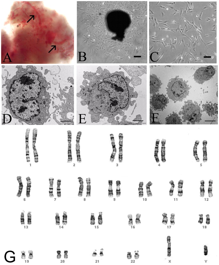
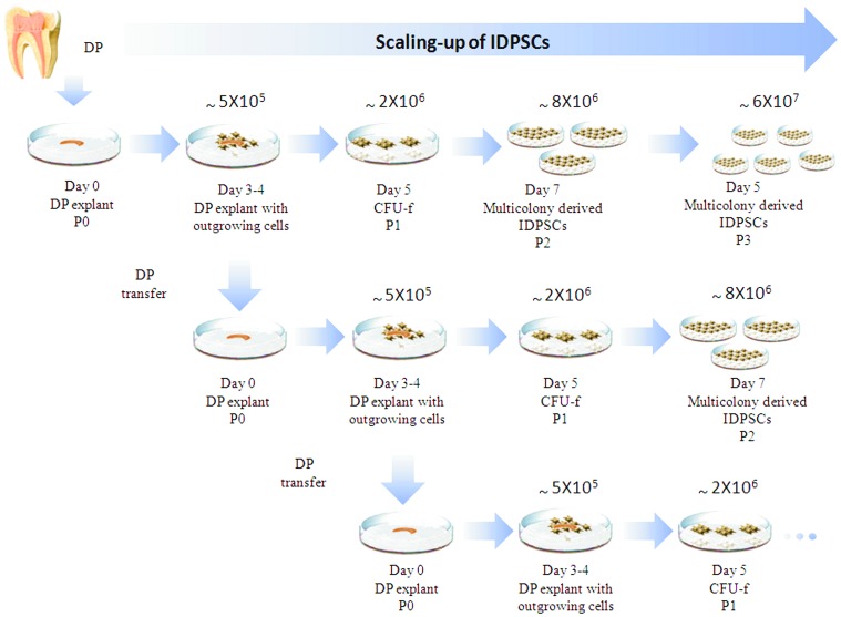
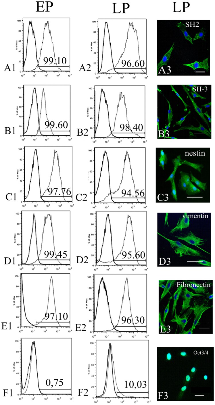
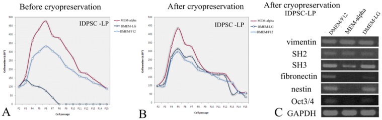
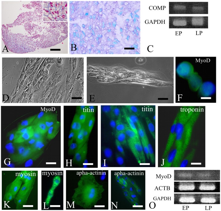
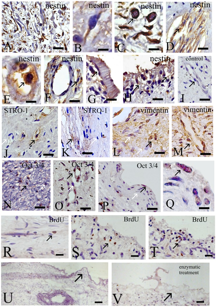
Dental pulp (DP) can be extracted from child's primary teeth (deciduous), whose loss occurs spontaneously by about 5 to 12 years. Thus, DP presents an easy accessible source of stem cells without ethical concerns. Substantial quantities of stem cells of an excellent quality and at early (2-5) passages are necessary for clinical use, which currently is a problem for use of adult stem cells. Herein, DPs were cultured generating stem cells at least during six months through multiple mechanical transfers into a new culture dish every 3-4 days. We compared stem cells isolated from the same DP before (early population, EP) and six months after several mechanical transfers (late population, LP). No changes, in both EP and LP, were observed in morphology, expression of stem cells markers (nestin, vimentin, fibronectin, SH2, SH3 and Oct3/4), chondrogenic and myogenic differentiation potential, even after cryopreservation. Six hours after DP extraction and in vitro plating, rare 5-bromo-2'-deoxyuridine (BrdU) positive cells were observed in pulp central part. After 72 hours, BrdU positive cells increased in number and were found in DP periphery, thus originating a multicellular population of stem cells of high purity. Multiple stem cell niches were identified in different zones of DP, because abundant expression of nestin, vimentin and Oct3/4 proteins was observed, while STRO-1 protein localization was restricted to perivascular niche. Our finding is of importance for the future of stem cell therapies, providing scaling-up of stem cells at early passages with minimum risk of losing their "stemness".


Conflict of interest statement

Competing Interests: The authors have declared that no competing interests exist.

Figures

Figure 1
Figure 1. Dental pulp and IDPSCs.
A) Highly vascularized (black arrows) DP just after extraction. B) Explant culture of DP with outgrowing IDPSCs. C) Culture of IDPSCs at 1st passage. D) IDPSCs showing ES-like cells morphology with a large nucleus. E) IDPSCs showing MSC-like morphology with several pseudopodes. F) IDPSCs showing uniform morphology resembling ES cells and MSCs. G) Karyotype of IDPSCs (LP): chromosomes in pairs, ordered by size and position did not reveal any numerical changes in chromosome number; G-banding analysis. A–C, G) Light Microscopy; D–F) Transmission Electron Microscopy; A = 20X, G = 63X; Scale bars: B = 20 µm; C, F = 10 µm; D, E = 3 µm.
Figure 2
Figure 2. Scaling-up of IDPSCs.
Horizontally, the process of DP in vitro plating (Day 0, P0) followed by DP adherence and cells outgrowth (Day 3–4). This process is followed by enzymatic treatment (P1) of the cells and formation of multiple colonies (CFU-f - Colony Forming Units-fibroblast). After 5 days, enzymatic treatment was performed to harvest multicolony-derived IDPSCs (P2) population. Next, in vitro expansion of IDPSCs (P3) has been performed. Upper numbers represent approximate quantity of harvested IDPSCs in each passage. Vertically, the same process is shown, albeit after multiple DP mechanical transfer.
Figure 3
Figure 3. Characterization of EP and LP of IDPSCs.
A1–F1) Flow cytometry showing EP of IDPSCs, which highly expressed such markers as SH2/CD105 (A1); SH3/CD73 (B1); nestin (C1); vimentin (D1); fibronectin (E1). F1) Low expression of Oct3/4 in EP; A2–F2) Flow cytometry showing LP of IDPSCs, which expressed same markers as EP. F2) Higher expression of Oct3/4 in LP, than in F1. A3–F3) Immunofluorescence of LP of IDPSCs using same markers as in (A2–E2). F3) Nuclear localization of Oct3/4 can be observed. A3–F3) Epi-fluorescence, nuclei stained with DAPI (blue). Scale bars: A3, B3, E3, F3 = 5 µm; C3, D3 = 10 µm.
Figure 4
Figure 4. Proliferation rate and gene expression of IDPSCs after cultivation in three distinct culture media. A
) Proliferation curve of LP before cryopreservation; B) Proliferation curve of LP after cryopreservation. C) Gene expression of LP after cryopreservation.
Figure 5
Figure 5. In vitro differentiation potential of IDPSCs.
A–C) Chondrogenic differentiation. A) Pellet culture: collagen fibers intensively stained by Massoǹs thrichrome. Inset: same as in (A) high magnification. B) The proteoglycans presence was revealed by Toloudine blue staining. C) RT-PCR shows the expression of COMP gene in EP and LP of IDPSCs. Housekeeping gene GAPDH is used as control. D–O) Myogenic differentiation. D, E) Morphological aspect showing stages of muscle fibers formation. F) Nuclear expression of MyoD1 protein in LP of IDPSCs-derived myocyte-like cells. G) Myosac composed by MyoD1 positive cells. H, I) Titin protein expression in LP of IDPSCs-derived myotubes. J) Expression of troponin I in Z-bands of myofibers. K) Myosin protein expression. L) Very small, satellite-like cells, showing positive myosin immunostaining. M) Binuclear cell positive for alpha-actinin (spot-like labeling). N) Fused myotubes, which deferentially express alpha-actinin protein. O) RT-PCR shows the expression of MyoD1 and ACTB genes in EP and LP of IDPSCs. Housekeeping gene GAPDH is used as control. A, B, D, E) Light Microscopy; F-N) Epi-fluorescence, nuclei stained with DAPI (blue). Scale bars: A = 200 µm; B = 20 µm; D = 50 µm; E, N = 10 µm; F–M = 5 µm.
Figure 6
Figure 6. Expression of nestin, STRO-1, vimentin, Oct3/4 and BrdU in DP.
A–H) Nestin expression. A–C) Cell rich zone. A) Multiple nestin positive cells can be observed. Here and below black arrows indicate immunopositive, while white arrows - immunonegative cells. B) Supposedly undifferentiated MSC shows nestin cytoplasm localization. C) Nestin positive cells with two distinct morphologies round epithelial-like (ES-like) and fibroblast-like cells. D–F) Cell free zone. D) Nestin showing intermediate filament staining in nerve plexus. E) Small capillary with two intensively stained nestin positive cells. F) Same as in (E) with nestin positive cells in lateral of capillary (arrow). G, H) Odontoblastic layer. Nestin positive obontoblasts (G, H) can be observed. I) Negative control: only secondary antibody was used. J, K) Cell free zone. J) STRO-1 positive cells within capillaries (perivascular niche). K) Very poor STRO-1 immunostaining was observed within nerve plexus. L, M) Vimentin positive (black arrows) cells localization in cell rich (L) and cell free (M) zones. N–Q) Oct3/4 positive cells localization in cell rich (N) and cell free (O–Q) zones. R–T) BrdU immunostaining of DP. R) DP just after plating in culture medium. S) 48 hours after in vitro cultivation. T) 72 hours after in vitro cultivation. U–V) DP without (U) and with (V) enzymatic treatment. V) External cell layer of DP is destroyed by such treatment. A–V) Light Microscopy. Scale bars: A, D, F–P, R–T = 20 µm; B, C, Q = 5 µm; U, V = 50 µm.

References

    1. Gronthos S, Mankani M, Brahim J, Robey PG, Shi S. Postnatal human DP stem cells (DPSCs) in vitro and in vivo. Proc Natl Acad Sci U S A. 2000;97:13625–13630. - PMC - PubMed
    1. Miura M, Gronthos S, Zhao M, Lu B, Fisher LW, et al. SHED: Stem cells from human exfoliated deciduous teeth. Proc Natl Acad Sci U S A. 2003;100:5807–5812. - PMC - PubMed
    1. Kerkis I, Caplan AI. Stem cells in dental pulp of deciduous teeth. Tissue Eng Part B Rev Dec 28 [Epub ahead of print] 2011. - PMC - PubMed
    1. Kerkis I, Kerkis A, Dozortsev D, Stukart-Parsons GC, Gomes Massironi SM, et al. Isolation and characterization of a population of immature dental pulp stem cells expressing OCT-4 and other embryonic stem cell markers. Cells Tissues Organs. 2006;184:105–116. - PubMed
    1. Fonseca SAS, Abdelmassih S, de Mello Cintra Lavagnolli T, Serafim RC, Clemente Santos EJ, et al. Human immature dental pulp stem cells’ contribution to developing mouse embryos: production of human/mouse preterm chimaeras. Cell Prolif. 2009;42:132–140. - PMC - PubMed

Publication types

MeSH terms