

Quantitative analysis of viral load per haploid genome revealed the different biological features of Merkel cell polyomavirus infection in skin tumor
- PMID: 22768181
- PMCID: PMC3386999
- DOI: 10.1371/journal.pone.0039954
Quantitative analysis of viral load per haploid genome revealed the different biological features of Merkel cell polyomavirus infection in skin tumor
Abstract
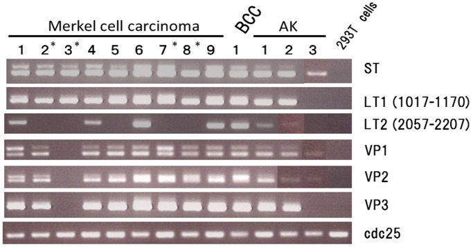
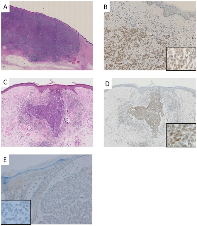
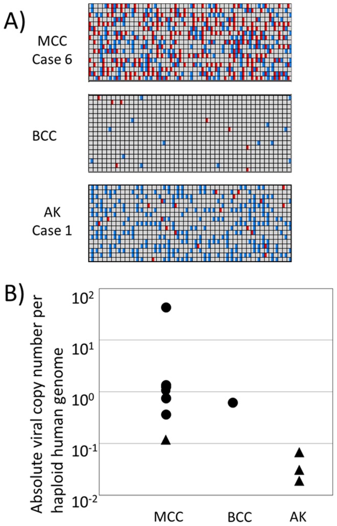
Merkel cell polyomavirus (MCPyV) has recently been identified in Merkel cell carcinoma (MCC), an aggressive cancer that occurs in sun-exposed skin. Conventional technologies, such as polymerase chain reaction (PCR) and immunohistochemistry, have produced conflicting results for MCPyV infections in non-MCC tumors. Therefore, we performed quantitative analyses of the MCPyV copy number in various skin tumor tissues, including MCC (n = 9) and other sun exposure-related skin tumors (basal cell carcinoma [BCC, n = 45], actinic keratosis [AK, n = 52], Bowen's disease [n = 34], seborrheic keratosis [n = 5], primary cutaneous anaplastic large-cell lymphoma [n = 5], malignant melanoma [n = 5], and melanocytic nevus [n = 6]). In a conventional PCR analysis, MCPyV DNA was detected in MCC (9 cases; 100%), BCC (1 case; 2%), and AK (3 cases; 6%). We then used digital PCR technology to estimate the absolute viral copy number per haploid human genome in these tissues. The viral copy number per haploid genome was estimated to be around 1 in most MCC tissues, and there were marked differences between the MCC (0.119-42.8) and AK (0.02-0.07) groups. PCR-positive BCC tissue showed a similar viral load as MCC tissue (0.662). Immunohistochemistry with a monoclonal antibody against the MCPyV T antigen (CM2B4) demonstrated positive nuclear localization in most of the high-viral-load tumor groups (8 of 9 MCC and 1 BCC), but not in the low-viral-load or PCR-negative tumor groups. These results demonstrated that MCPyV infection is possibly involved in a minority of sun-exposed skin tumors, including BCC and AK, and that these tumors display different modes of infection.
Conflict of interest statement
Figures
Similar articles
-
The prevalence of Merkel cell polyomavirus in Japanese patients with Merkel cell carcinoma.J Dermatol Sci. 2013 May;70(2):99-107. doi: 10.1016/j.jdermsci.2013.02.010. Epub 2013 Mar 6. J Dermatol Sci. 2013. PMID: 23517683
-
Merkel cell carcinoma: histopathologic and prognostic features according to the immunohistochemical expression of Merkel cell polyomavirus large T antigen correlated with viral load.Hum Pathol. 2015 Mar;46(3):443-53. doi: 10.1016/j.humpath.2014.12.001. Epub 2014 Dec 23. Hum Pathol. 2015. PMID: 25623078
-
No evidence for a causal role of Merkel cell polyomavirus in keratoacanthoma.J Am Acad Dermatol. 2012 Jul;67(1):41-6. doi: 10.1016/j.jaad.2011.07.026. Epub 2011 Oct 11. J Am Acad Dermatol. 2012. PMID: 21996295
-
Merkel cell polyomavirus and non-Merkel cell carcinomas: guilty or circumstantial evidence?APMIS. 2020 Feb;128(2):104-120. doi: 10.1111/apm.13019. Epub 2020 Jan 28. APMIS. 2020. PMID: 31990105 Review.
-
Merkel cell carcinoma: The first human cancer shown to be associated with a polyomavirus.Presse Med. 2014 Dec;43(12 Pt 2):e405-11. doi: 10.1016/j.lpm.2014.09.008. Epub 2014 Nov 6. Presse Med. 2014. PMID: 25455636 Review.
Cited by
-
The Future of Digital Polymerase Chain Reaction in Virology.Mol Diagn Ther. 2016 Oct;20(5):437-47. doi: 10.1007/s40291-016-0224-1. Mol Diagn Ther. 2016. PMID: 27351921 Review.
-
Prevalence of Merkel Cell Polyomavirus in Normal and Lesional Skin: A Systematic Review and Meta-Analysis.Front Oncol. 2022 Mar 22;12:868781. doi: 10.3389/fonc.2022.868781. eCollection 2022. Front Oncol. 2022. PMID: 35392226 Free PMC article.
-
Tumor Content Chart-Assisted HER2/CEP17 Digital PCR Analysis of Gastric Cancer Biopsy Specimens.PLoS One. 2016 Apr 27;11(4):e0154430. doi: 10.1371/journal.pone.0154430. eCollection 2016. PLoS One. 2016. PMID: 27119558 Free PMC article.
-
Quantitative analysis of Merkel cell polyomavirus (MCPyV) genome in non-melanoma skin cancer and normal tumor margins.Braz J Microbiol. 2022 Dec;53(4):1987-1994. doi: 10.1007/s42770-022-00850-x. Epub 2022 Oct 24. Braz J Microbiol. 2022. PMID: 36279096 Free PMC article.
-
Frequent detection of Merkel cell polyomavirus DNA in sera of HIV-1-positive patients.Virol J. 2013 Mar 13;10:84. doi: 10.1186/1743-422X-10-84. Virol J. 2013. PMID: 23496956 Free PMC article.
References
-
- LeBoit PE, Burg G, Weedon D, Sarasin A. World Health Organization Classification of Tumours Pathology and Genetics Skin Tumours. IARC Press. 2006.
-
- Kassem A, Schopflin A, Diaz C, Weyers W, Stickeler E, et al. Frequent detection of Merkel cell polyomavirus in human Merkel cell carcinomas and identification of a unique deletion in the VP1 gene. Cancer Res. 2008;68:5009–5013. - PubMed
-
- Becker JC, Houben R, Ugurel S, Trefzer U, Pfohler C, et al. MC polyomavirus is frequently present in Merkel cell carcinoma of European patients. J. Invest. Dermatol. 2009;129:248–250. - PubMed
Publication types
MeSH terms
Substances
LinkOut - more resources
Full Text Sources
Medical
