. 2012;7(6):e40110.
doi: 10.1371/journal.pone.0040110. Epub 2012 Jun 29.

Cardiac kallikrein-kinin system is upregulated in chronic volume overload and mediates an inflammatory induced collagen loss

Affiliations

Cardiac kallikrein-kinin system is upregulated in chronic volume overload and mediates an inflammatory induced collagen loss

Chih-Chang Wei et al. PLoS One. 2012.

Abstract

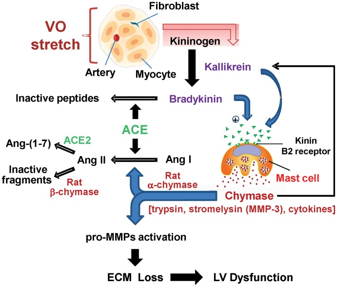
Background: The clinical problem of a "pure volume overload" as in isolated mitral or aortic regurgitation currently has no documented medical therapy that attenuates collagen loss and the resultant left ventricular (LV) dilatation and failure. Here, we identify a potential mechanism related to upregulation of the kallikrein-kinin system in the volume overload of aortocaval fistula (ACF) in the rat.

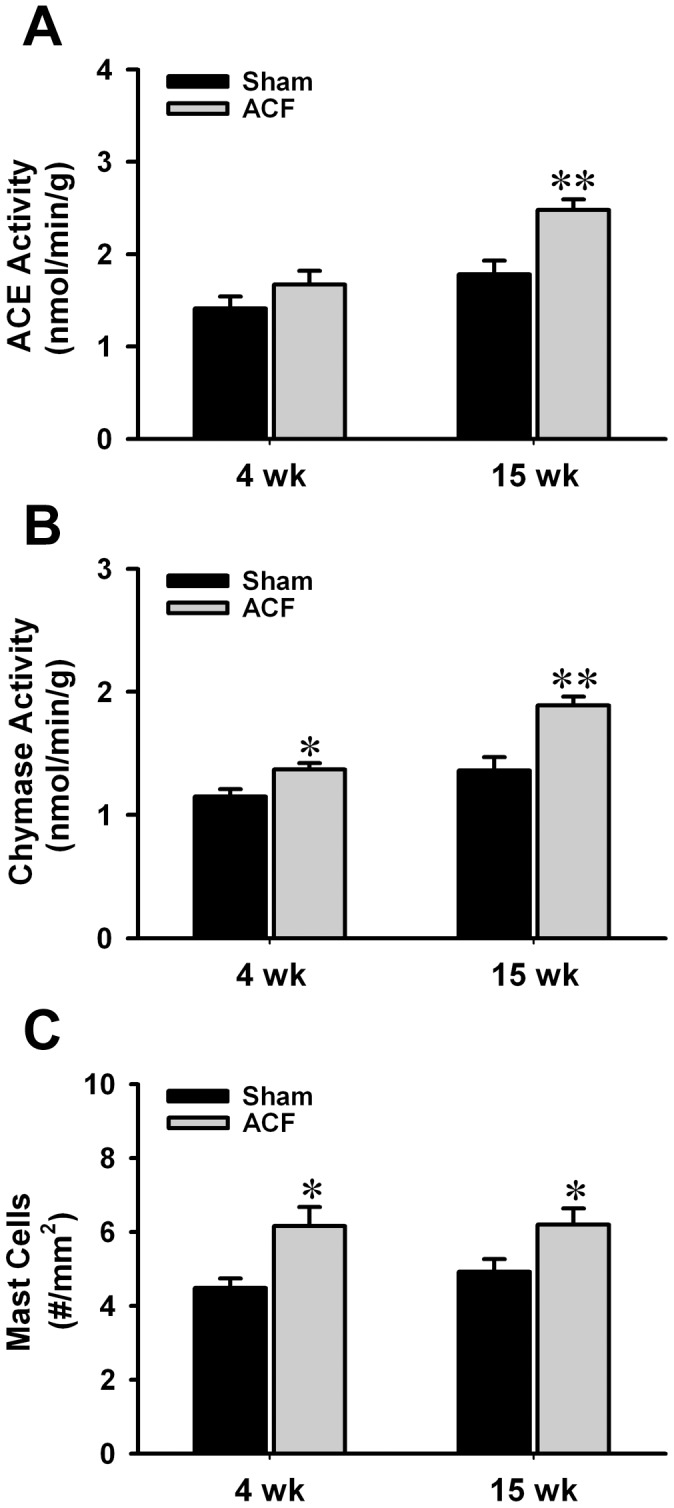
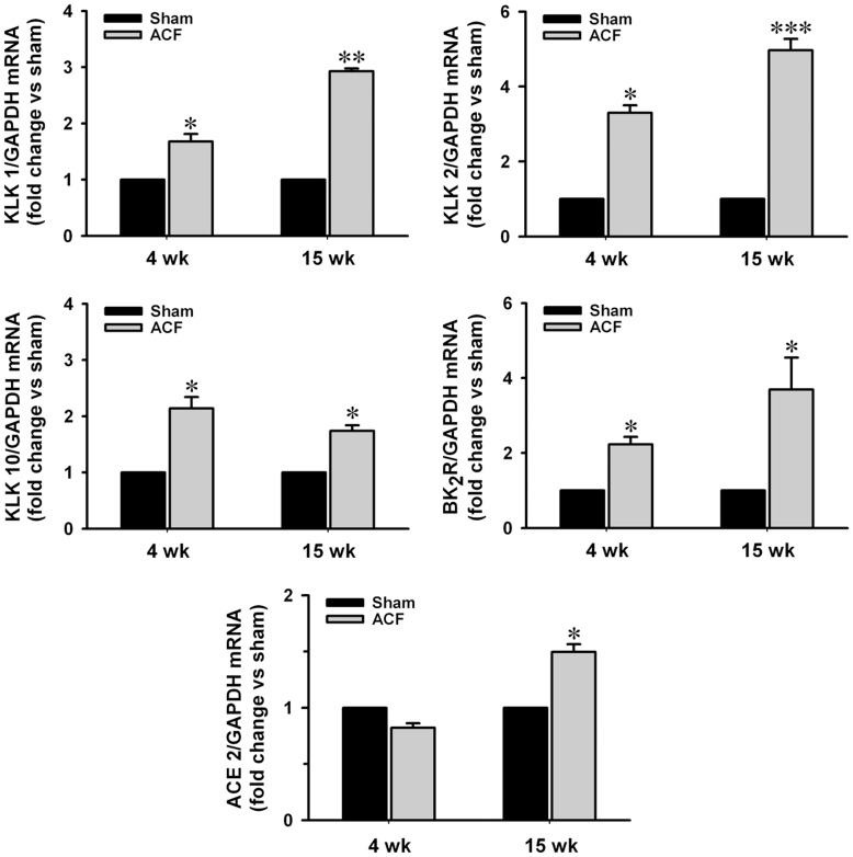
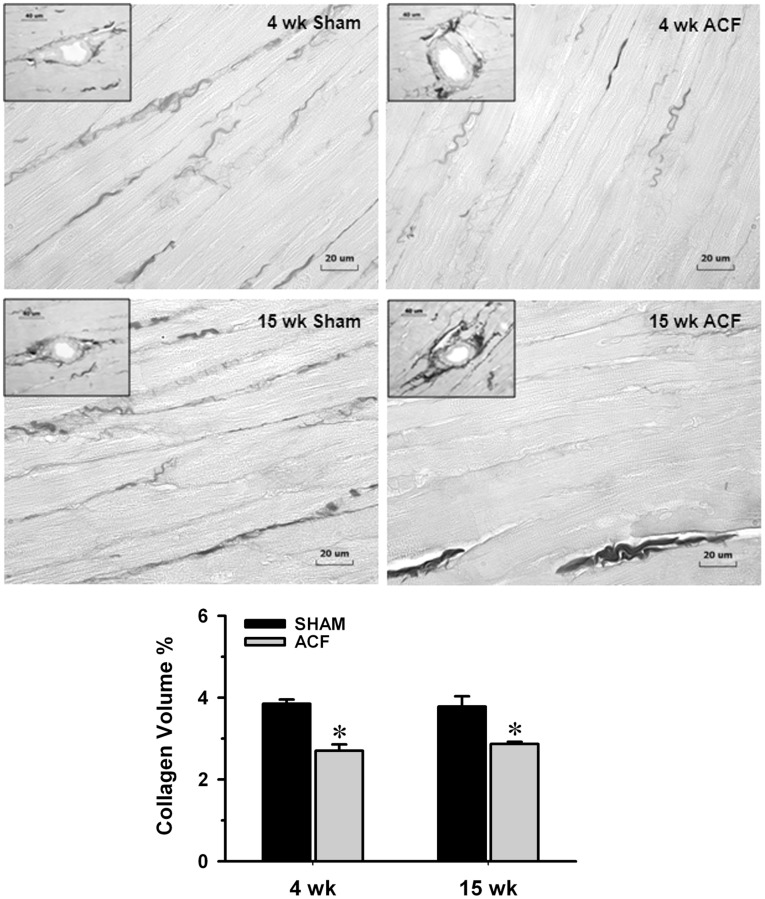
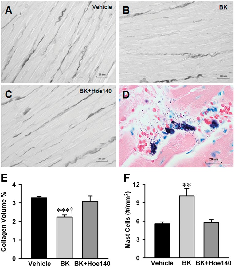
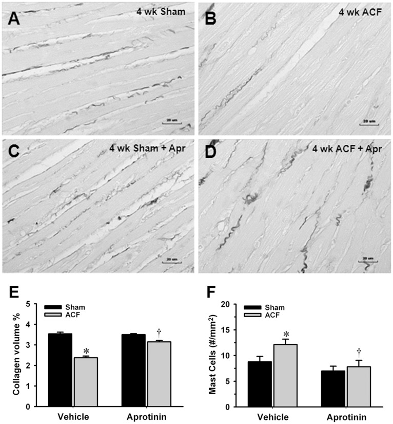
Methodology/principal findings: LV interstitial fluid (ISF) collection, hemodynamics, and echocardiography were performed in age-matched shams and 4 and 15 wk ACF rats. ACF rats had LV dilatation and a 2-fold increase in LV end-diastolic pressure, along with increases in LV ISF bradykinin, myocardial kallikrein and bradykinin type-2 receptor (BK(2)R) mRNA expression. Mast cell numbers were increased and interstitial collagen was decreased at 4 and 15 wk ACF, despite increases in LV ACE and chymase activities. Treatment with the kallikrein inhibitor aprotinin preserved interstitial collagen, prevented the increase in mast cells, and improved LV systolic function at 4 wk ACF. To establish a cause and effect between ISF bradykinin and mast cell-mediated collagen loss, direct LV interstitial bradykinin infusion in vivo for 24 hrs produced a 2-fold increase in mast cell numbers and a 30% decrease in interstitial collagen, which were prevented by BK(2)R antagonist. To further connect myocardial stretch with cellular kallikrein-kinin system upregulation, 24 hrs cyclic stretch of adult cardiomyocytes and fibroblasts produced increased kallikrein, BK(2)R mRNA expressions, bradykinin protein and gelatinase activity, which were all decreased by the kallikrein inhibitor-aprotinin.

Conclusions/significance: A pure volume overload is associated with upregulation of the kallikrein-kinin system and ISF bradykinin, which mediates mast cell infiltration, extracellular matrix loss, and LV dysfunction-all of which are improved by kallikrein inhibition. The current investigation provides important new insights into future potential medical therapies for the volume overload of aortic and mitral regurgitation.


Conflict of interest statement

Competing Interests: The authors have declared that no competing interests exist.

Figures

Figure 1
Figure 1. Increasing LV ACE and chymase activity and mast cell numbers in ACF.
(A) By using ACE-specific substrate hippuryl-his-leu, LV myocardium ACE activity was defined as captopril-inhibitable hippuric acid (HA) formation expressed as nmoles of HA formed/g/min of tissue. (B) By using Ang I as substrate, chymase-like activity was defined as chymostatin-inhibitable Ang II formation expressed as nmoles of Ang II formed/min/g of tissue. Both activities were measured at 4 and 15 wk age-matched shams and ACF rats. (C) The number of mast cells was quantitatively determined for the entire LV wall using Giemsa-stained paraffin sections. LV mast cell numbers were increased at the 4 and 15 wk ACF rats vs. age-matched shams. Values are mean±SEM. n = 8–10 in each group. *P<0.05, **P<0.01 vs. age-matched shams.
Figure 2
Figure 2. Increasing expressions of kallikreins (KLKs), BK2 receptor and ACE 2 mRNA in ACF.
Total RNA was extracted from LV tissue with TRIzol reagent. Expressions of kallikrein 1, 2, 10 and BK2 receptor and ACE 2 mRNA in normalized to GAPDH at 4 and 15 wk ACF rats compared to age-matched shams. Values are mean±SEM. n = 6–8 in each group. *P<0.05, **P<0.01 and ***P<0.001 vs. age-matched shams.
Figure 3
Figure 3. Immunohistochemistry of tissue kallikrein in LV myocardium of sham and ACF rats.
Immunohistochemistry were performed on the formalin fixed paraffin-embedded LV. Image acquisition (100x objective, 4000x video-screen magnification) was performed on a Leica DM6000 epifluorescence microscope with SimplePCI software. LV tissues were stained with anti-tissue kallikrein (red), anti-desmin (green), and DAPI (blue). Representative images of the sections: (A) negative control (cross section), (B) 4 wk sham (cross section), (C) 4 wk ACF (cross section), (D) 15 wk sham (longitudinal section), (E) 15 wk ACF (longitudinal section). Arrows demonstrate tissue kallikrein in the LV myocytes or interstitium of each group. Bar scale: 10 µm.
Figure 4
Figure 4. Determination of interstitial collagen volume percent in sham and ACF rats.
Representative images of LV interstitial collagen stained with picric acid sirius red F3BA (PASR) were measured at 4 and 15 wk ACF and their age-matched sham rats. The loss of interstitial collagen (shown as dark collagen fibers excluding perivascular areas) between cardiomyocytes at 4 and 15 wk ACF was reflected by the decrease in collagen volume percent (%). However from insets, when comparing the 4 and 15 wk ACF, there is an obvious increase in perivascular collagen compared to sham in the 15 wk ACF. Panel in the bottom displays quantification of the interstitial collagen at 4 and 15 wk ACF and their age-matched shams. Values are mean±SEM. n = 8–10 in each group. *P<0.05 vs. age-matched shams. Bar scale: 20 µm and 40 µm in insets.
Figure 5
Figure 5. Effect of LV interstitial bradykinin infusion on mast cell infiltration, degranulation and collagen loss.
Interstitial collagen volume percent was decreased 30% by bradykinin infusion group (B) compared to saline infused vehicle control (A) and prevented by Hoe-140 (C). Infusion of bradykinin into LV myocardium increased mast cell number by 2-fold compared to vehicle group and mast cell increases were inhibited by Hoe-140 (F). Representative image (D) demonstrates mast cells and mast cell granules show degranulation after interstitial bradykinin infusion. The number of mast cell was quantitatively determined for the entire LV wall using Giemsa-stained paraffin sections. It produces intense staining (purple) specific for mast cell granules. Panel (E) displays quantification of the interstitial collagen of normal rat with interstitial saline or bradykinin (5 ng/ml) infusion for 24 hrs with and without BK2R antagonist (Hoe-140, 0.5 mg/kg/d given by osmotic infusion pump). Values are mean±SEM. n = 6 in each group. ***P<0.001 vs. Vehicle. P<0.05 vs. bradykinin infusion plus Hoe 140. **P<0.01 vs. Vehicle and bradykinin infusion plus Hoe-140. Bar scale: 20 ∝m.
Figure 6
Figure 6. Effect of aprotinin on interstitial collagen volume percent and mast cells in 4 wk ACF.
Interstitial collagen volume percent was decreased >30% at 4 wk ACF (B) compared to age-matched sham rats (A) and prevented by aprotinin (D). Panel (C) represents sham with aprotinin treatment which displays no change in the interstitial collagen volume % compared to Vehicle treated sham. Quantification of the interstitial collagen volume % and mast cells were shown in Panels E and F. Mast cell numbers were increased in the 4 wk ACF rats and decreased by aprotinin (F). Values are mean±SEM. n = 7–8 in each group. *P<0.05 vs. Vehicle treated age-matched shams. P<0.05 vs. Vehicle treated ACF rats.
Figure 7
Figure 7. A diagram demonstrates the hypothesized pathway of volume overload stress effect on LV remodeling and functions.
The pure stretch of volume overload induces cardiac kallikrein-kinin upregulation from cardiomyocytes and fibroblasts that result in increased ISF bradykinin and activate kinin B2 receptor. Kallikrein and kinin cause mast cell degranulation. Mast cell degranulation causes the release of proteolytic enzymes like tryptase and chymase, as well as other cytokines that activate MMPs. Chymase also can activate kallikreins, MMPs and degrade collagen and non-collagen extracellular matrix components, such as fibronectin. The increase in bradykinin can further decrease extracellular matrix synthesis. Furthermore, tissue kallikreins can activate the kinin B2 receptor in the absence of kininogen. Because the kininogen substrate is abundant in plasma and tissues, the expression and availability of tissue kallikrein are the rate-limiting factors in kinin production.

References

    1. Borer JS, Bonow RO. Contemporary approach to aortic and mitral regurgitation. Circulation. 2003;108:2432–2438. - PubMed
    1. Chandrashekhar Y. Embracing the diversity of remodeling. J Am Coll Cardol. 2007;49:822–825. - PubMed
    1. Bonow RO, Carabello BA, Chatterjee K, de Leon AC, Jr, Faxon DP, et al. ACC/AHA 2006 guidelines for the management of patients with valvular heart disease: a report of the American College of Cardiology/American Heart Association Task Force on practice guidelines (writing committee to revise the 1998 guidelines for the management of patients with valvular heart disease) developed in collaboration with the Society of Cardiovascular Anesthesiologists endorsed by the Society for Cardiovascular Angiography and Interventions and the Society of Thoracic Surgeons. J Am Coll Cardiol. 2006;48:e1–e148. - PubMed
    1. Ryan TD, Rothstein EC, Aban I, Tallaj JA, Husain A, et al. Left ventricular eccentric remodeling and matrix loss are mediated by bradykinin and precede cardiomyocyte elongation in rats with volume overload. J Am Coll Cardiol. 2007;49:811–821. - PubMed
    1. Wei CC, Lucchesi PA, Tallaj J, Bradley WE, Powell PC, et al. Cardiac interstitial bradykinin and mast cells modulate pattern of LV remodeling in volume overload in rats. Am J Physiol Heart Circ Physiol. 2003;285:H784–H792. - PubMed

Publication types

MeSH terms