. 2012;7(6):e40185.
doi: 10.1371/journal.pone.0040185. Epub 2012 Jun 29.

First human model of in vitro Candida albicans persistence within granuloma for the reliable study of host-fungi interactions

Affiliations

First human model of in vitro Candida albicans persistence within granuloma for the reliable study of host-fungi interactions

Nidia Alvarez-Rueda et al. PLoS One. 2012.

Abstract

Background: The balance between human innate immune system and Candida albicans virulence signaling mechanisms ultimately dictates the outcome of fungal invasiveness and its pathology. To better understand the pathophysiology and to identify fungal virulence-associated factors in the context of persistence in humans, complex models are indispensable. Although fungal virulence factors have been extensively studied in vitro and in vivo using different immune cell subsets and cell lines, it is unclear how C. albicans survives inside complex tissue granulomas.

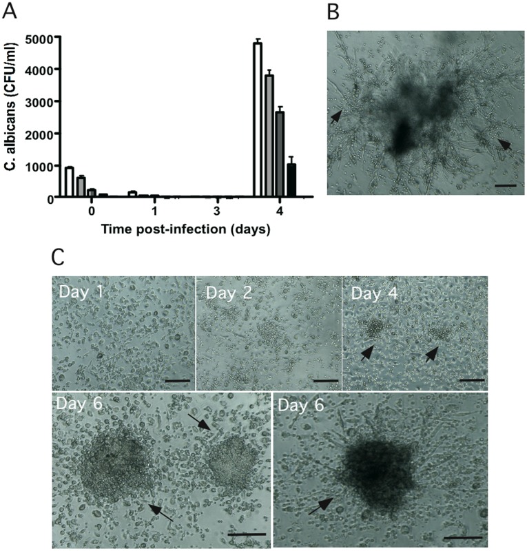
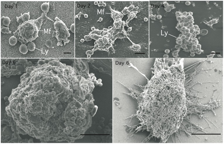
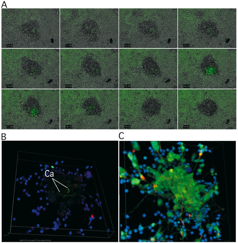
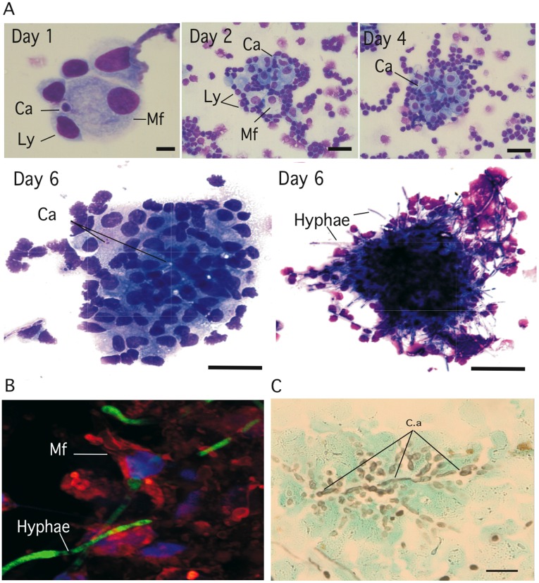
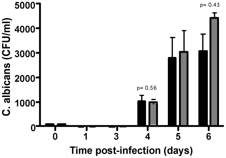
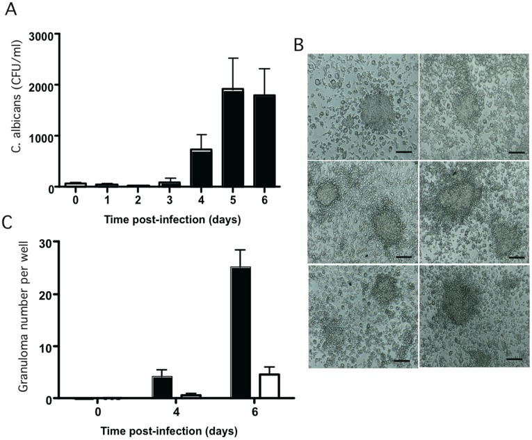
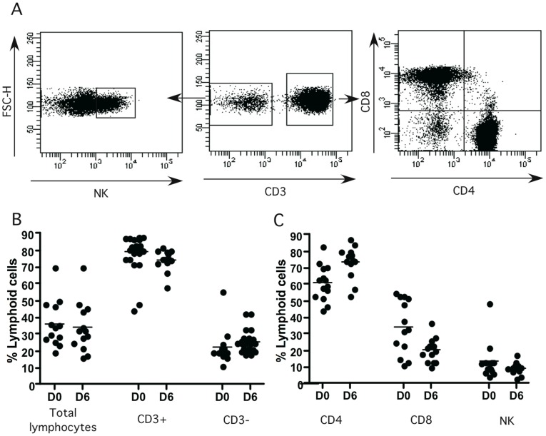
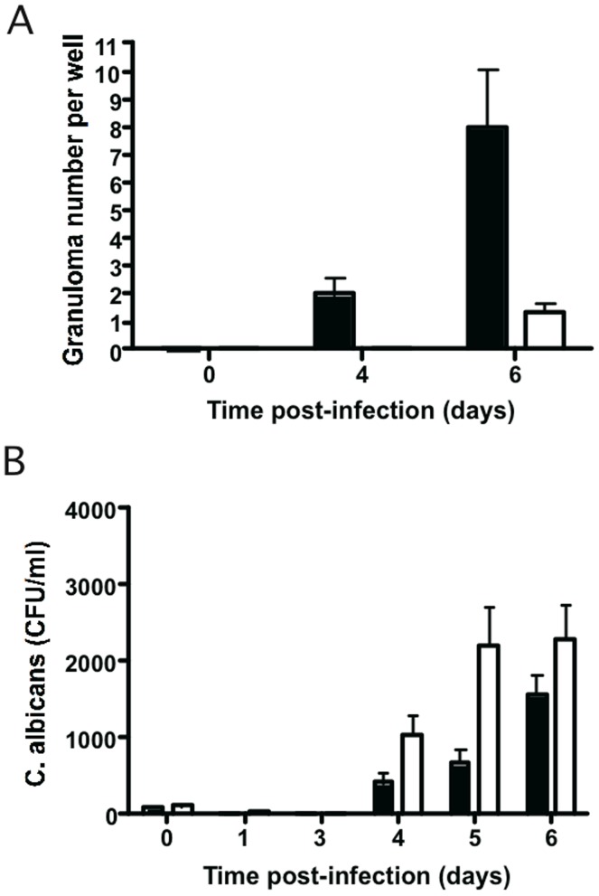
Methodology/principal finding: We developed an original model of in vitro human granuloma, reproducing the natural granulomatous response to C. albicans. Persistent granulomas were obtained when the ratio of phagocytes to fungi was high. This in vitro fungal granuloma mimics natural granulomas, with infected macrophages surrounded by helper and cytotoxic T lymphocytes. A small proportion of granulomas exhibited C. albicans hyphae. Histological and time-lapse analysis showed that C. albicans blastoconidia were located within the granulomas before hyphae formation. Using staining techniques, fungal load calculations, as well as confocal and scanning electron microscopy, we describe the kinetics of fungal granuloma formation. We provide the first direct evidence that C. albicans are not eliminated by immunocompetent cells inside in vitro human granulomas. In fact, after an initial candicidal period, the remaining yeast proliferate and persist under very complex immune responses.

Conclusions/significance: Using an original in vitro model of human fungal granuloma, we herein present the evidence that C. albicans persist and grow into immunocompetent granulomatous structures. These results will guide us towards a better understanding of fungal invasiveness and, henceforth, will also help in the development of better strategies for its control in human physiological conditions.


Conflict of interest statement

Competing Interests: The authors have declared that no competing interests exist.

Figures

Figure 1
Figure 1. Recruitment of local human PBMCs around living C. albicans.
(A): Multiplicity of infection calculations (MOIs). 106 cells/ml PBMC were infected with different concentrations of yeast cells and incubated at 37°C, 5% CO2. Cellular aggregation was followed every day under light microscopy. Uninfected PBMCs were used as a negative control of aggregation. The amount of surviving yeasts in granuloma structures at different time points was expressed as colony-forming units per ml (CFU/ml). The data are presented as the mean ± SD for three independent experiments, each in triplicate. MOIs tested were: 200∶1 (white column), 400∶1 (light grey column), 800∶1 (grey column) and 2000∶1 (black column). (B): Light-microscopic observation of a representative granuloma six days after C. albicans infection (MOI of 400∶1). Large hyphae envading from granuloma (arrows). Bar represents 50 µm. (C): Light microscopy observation of time course evolution of the granulomatous reaction after infection with C. albicans (MOI of 2000∶1). Bars represent 50 µm.
Figure 2
Figure 2. Fungal granuloma progression by scanning electron microscopy.
Kinetics of granuloma formation after infection with C. albicans (MOI of 2000∶1). The granuloma structures were collected at different time points from co-culture plates, and fixed in 2.5% glutaraldehyde, O.1 M sucrose in cacodylate buffer for 1 h. After dehydratation through a gradient ethanol series and alcohol-freon substitution, specimens were coated with 100 Å of gold-palladium mix in an ion sputter and photographed under a scanning electron microscope. Bars represent 5 µm (Days 1 post-infection), 10 µm (days 2 and 4) and 50 µm (day 6). Activated macrophages (Mf), lymphocytes (Ly).
Figure 3
Figure 3. Distribution of C. albicans cells in the granuloma.
(A): For co-culture analysis by video-imaging, PBMCs were infected with C. albicans cells containing the pACT1-GFP fusion protein at a MOI of 200∶1. The cells were illuminated at day 0 and every 10 min over 72 h of incubation with a 300 W xenon lamp fitted with 488 nm excitation filter. Emission at 515 nm was used for analysis of C. albicans fluorescence with a Leica DMI6000B camera cool Snap HQ2 and processed with Metamorph imaging software version 7.7.4.0. (B and C): For confocal microscopy imaging granulomas were generated by infecting PBMCs at an MOI of 2000∶1. Cells were stained with rhodamine-phalloidin and Hoechst six days after infection. Confocal images showed GFP- tagged C. abicans inside granulomas (B). Large green-fluorescent hyphae of C. abicans emerging from granulomas (C). Image scan was 1024 pixels × 1024 pixels. Image size was 1212 µm.
Figure 4
Figure 4. Histological analysis of human cell subsets surrounding fungal granulomas.
Granuloma structures were collected at different time points of incubation, plated on glass slides with a cytospin and stained. (A): May-Grünwald-Giemsa (MGG, Sigma) staining of granulomas at different time points. Bars represent 5 µm (Day 1), 15µm (day 2 and 4) and 50 µm (day 6). (B): Confocal images showing the interaction between mature macrophages and green-fluorescent C. abicans hyphae inside granulomas. (C): Grocott-Gomori methenamine silver nitrate staining of a granuloma six days post-infection (bar represent 50 µm). Mature macrophages (Mf), lymphocytes (Ly), C. albicans (C.a).
Figure 5
Figure 5. Capability to C. albicans clinical isolates to generate granulomas.
Granuloma formation by different C. albicans clinical isolates. Human PBMCs were infected with CAAL93 (black columns) and DSY735 (grey columns) clinical isolates. Granuloma formation was followed by light microscopy. The fungal load was expressed as CFU/ml at different incubation times. The data are presented as the mean ± SD for three independent experiments, each in triplicate.
Figure 6
Figure 6. Fungal granuloma-response variability between donors.
(A): The fungal granuloma-response variability between healthy individuals was analyzed by infecting the PBMCs from 23 donors with C. albicans (MOI of 2000∶1). The granuloma-response was followed by calculating the colony-forming units (CFU/ml) at different time points and by light microscopy observations. The data are presented as the mean ± SD for 23 independent experiments. (B): Light-microscopic observation of representative granulomas from donors after six days of infection with C. albicans. Bar represents 50 µm. (C): Number of specific granulomas per culture-well at different times of infection with C. albicans. Intact granulomas (black columns), granulomas with C. albicans hyphae (white columns).
Figure 7
Figure 7. Recruitment of cell subsets in fungal granuloma.
The granuloma structures were collected at different times points from co-culture plates under light microscopy, and stained with a cocktail of fluorescent-conjugated antibodies specific to CD3, CD4, CD8, and NKp46. (A): The cells were analyzed by flow cytometry after gating on CD3 lymphocytes: the CD3+ population was separated into conventional CD4+ and CD8+ T cells. Natural killer cells were gated from the CD3- population. (B): Total lymphocytes, CD3+ and CD3- subsets percentages into granulomas for each donor on day 0 (D0) and day 6 (D6) (n = 13). (C): CD4+, CD8+ and NK lymphocyte cells into granulomas (n = 13 donors). All data were acquired using a FACS Canto II instrument (BD Biosciences) and analyzed with FlowJo software version 9.4.10 (Tree Star Inc) and DIVA software version 6.2 (BD).
Figure 8
Figure 8. Effect of PMNs on granuloma formation.
PBMCs alone or PBMCs with PMNs from healthy individuals were used to induce in vitro granulomas after C. albicans (CAAL93) infection. Host cells were infected with yeasts at an MOI of 2000∶1 and incubated at 37°C, 5% CO2. Cellular aggregation was followed every day under light microscopy. Uninfected PBMCs and PMNs were used as a negative control of aggregation. (A): The number of granulomas per well was followed at different time points. Intact granulomas (black columns), granulomas with C. albicans hyphae (white columns). (B): The amount of surviving yeasts in granuloma structures at different time points was expressed as colony-forming units per ml (CFU/ml). PBMCs with PMNs co-cultures (black columns), PBMCs co-cultures (white columns). The data are presented as the mean ± SD, each experiment in triplicate (n = 5).

References

    1. Calderone RA, Fonzi WA. Virulence factors of Candida albicans. Trends Microbiol. 2001;9:327–335. - PubMed
    1. Nantel A, Dignard D, Bachewich C, Harcus D, Marcil A, et al. Transcription profiling of Candida albicans cells undergoing the yeast-to-hyphal transition. Mol Biol Cell. 2002;13:3452–3465. - PMC - PubMed
    1. Calderone R, Suzuki S, Cannon R, Cho T, Boyd D, et al. Candida albicans: adherence, signaling and virulence. Med Mycol. 2000;38:125–137. - PubMed
    1. Calera JA, Herman D, Calderone R. Identification of YPD1, a gene of Candida albicans which encodes a two-component phosphohistidine intermediate protein. Yeast. 2000;16:1053–1059. - PubMed
    1. Pankhurst CL. Candidiasis (oropharyngeal). Clin Evid (Online) 2009. 2009. - PMC - PubMed