. 2012;7(6):e40293.
doi: 10.1371/journal.pone.0040293. Epub 2012 Jun 29.

Ell3 enhances differentiation of mouse embryonic stem cells by regulating epithelial-mesenchymal transition and apoptosis

Affiliations

Ell3 enhances differentiation of mouse embryonic stem cells by regulating epithelial-mesenchymal transition and apoptosis

Hee-Jin Ahn et al. PLoS One. 2012.

Abstract

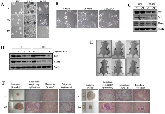
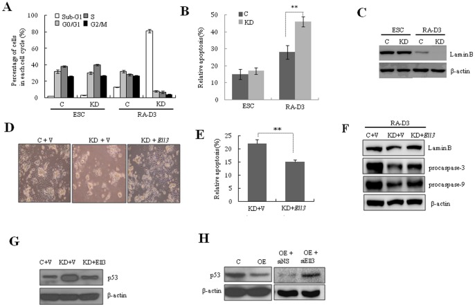
Ell3 is a testis-specific RNA polymerase II elongation factor whose cellular function is not clear. The present study shows that Ell3 is activated during the differentiation of mouse embryonic stem cells (mESCs). Furthermore, Ell3 plays a critical role in stimulating lineage differentiation of mESCs by promoting epithelial-mesenchymal transition (EMT) and suppressing apoptosis. Mouse ESCs engineered to stably express Ell3 were rapidly differentiated compared with control cells either under spontaneous differentiation or neural lineage-specific differentiation conditions. Gene expression profile and quantitative RT-PCR analysis showed that the expression of EMT markers, such as Zeb1 and Zeb2, two major genes that regulate EMT, was upregulated in Ell3-overexpressing mESCs. Remarkably, knockdown of Zeb1 attenuated the enhanced differentiation capacity of Ell3-overexpressing mESCs, which indicates that Ell3 plays a role in the induction of mESC differentiation by inducing EMT. In contrast to Ell3-overexpressing mESCs, Ell3-knock down mESCs could not differentiate under differentiation conditions and, instead, underwent caspase-dependent apoptosis. In addition, apoptosis of differentiating Ell3-knock out mESCs was associated with enhanced expression of p53. The present results suggest that Ell3 promotes the differentiation of mESCs by activating the expression of EMT-related genes and by suppressing p53 expression.


Conflict of interest statement

Competing Interests: The authors have declared that no competing interests exist.

Figures

Figure 1
Figure 1. Ell3 is specifically expressed in mESCs.
(A) RT-PCR and real-time RT-PCR analysis shows that Ell3 is expressed in mESCs but not in MEF and NIH3T3 cells. (B) Quantitative RT-PCR analysis of Ell3 in ESCs, EBs, and differentiated cell stages (RA-D4, RA-D8, RA-D12 and RA-D16). Ell3 transcripts increased at the EB and RA-D4 stages, and subsequently decreased as differentiation progressed (RA-D8, RA-D12 and RA-D16). (C) Ell3 transcript levels in Ell3-OE and KD cells at passage 10 were compared with those in control cells. Passage was counted after Ell3-OE and KD stable cell lines were established. (D) Expression of Stat3, p-Stat3, and Oct4 in Ell3-OE and KD cells was compared with that in control mESCs. β-actin was used as a loading control for immunoblot analysis. (E) Primary EBs of Ell3-OE, Ell3-KD, and control mESCs were dissociated into single cells and re-seeded at a density of 1×10⁶ cells/mL in the same medium. The number of secondary EBs was counted under a bright microscope (n>3). All experiments were performed at least in triplicate, and all values represent the mean ± s.d. from at least triplicate experiments. *Indicates significant (P<0.05) results (Student's t-test).
Figure 2
Figure 2. Ell3 expression level in mESCs affects the differentiation capacity of the cells both in vitro and in vivo.
(A) Five-day-old EBs (EB-D5) or differentiated Ell3-OE, Ell3-KD, and control mESCs were examined under a bright microscope. Differentiation was induced for 3 days (RA-D3) by removing LIF and adding retinoic acid (RA) to the mESCs culture media. (B) Nonspecific siRNA (siNS) or siRNAs targeting Ell3 (siEll3) were transfected into Ell3-OE cells in the ESCs state, and transfected cells were spontaneously differentiated for 2 days. Differentiating cells were examined under the microscope. (C) Expression of Oct4, Sox2, and Nanog in Ell3-OE cells was compared with that in control cells both in the ESC and differentiated states (RA-D3). β-actin was used as the loading control for immunoblot analysis. (D) Expression levels of Stat3 and phospho-Stat3 were compared between Ell3-OE and control cells as differentiation progressed. β-actin was used as the loading control for immunoblot analysis. (E) Ell3-OE, Ell3-KD, and control mESCs were injected into Balb/c nude mice and teratoma development was monitored. Teratomas of Ell3-OE and KD cell-injected mice were compared with those of control animals at 4 and 7 weeks after injection, respectively. (F) Morphology of mESCs teratomas obtained from Balb/c nude mice injected with either control, Ell3-OE, or Ell3-KD cells. MESC-induced teratomas were stained with H&E. Respiratory epithelium, muscle, cartilage, and epidermis were examined. All experiments were performed at least in triplicate.
Figure 3
Figure 3. Ell3 expression promotes neural differentiation of mESCs.
(A) Ell3-OE and control mESCs were differentiated into neural lineage, and differentiating cells were examined under a bright microscope 3 days (ND-D3) and 5 days (ND-D5) after differentiation. (B) Nestin expression in Ell3-OE cells was compared with that in control cells by qRT-PCR 0, 3, 5, and 7 days after neural differentiation (ND0, ND3, ND5, and ND7, respectively). (C) Nestin expression in Ell3-OE cells was compared with that of control cells by immunostaining 7 days after neural differentiation. (D) Nestin+ cells in differentiating Ell3-OE and control cells were analyzed by FACS. MESCs were cultured in neural differentiation media for 7 days and 1×10⁴ cells were analyzed for nestin expression by FACS. (E) The number of adherent cells was counted at the indicated days after 1×10⁴ cells of Ell3-OE and control mESCs were differentiated into the neural lineage. All experiments were performed at least in triplicate and all values represent the mean ± s.d. from at least triplicate experiments. * Indicates significant (P<0.05) results (Student's t-test).
Figure 4
Figure 4. Suppression of Zeb1 attenuates the enhanced differentiation of Ell3-OE cells.
(A) The expression of Zeb1 and Zeb2 in the ESC state or RA-induced differentiating state of control or Ell3-OE cells was analyzed by real-time RT-PCR. (B) The expression of E-cadherin in the ESC state or RA-induced differentiating state of ESC or Ell3-OE cells was analyzed by immunoblot analysis. β-actin was used as a loading control. (C) The expression of EMT markers during RA-induced differentiation of mESCs was compared between control and Ell3-OE cells by real-time RT-PCR. (D) Control or Ell3-OE cells were transfected with siRNAs targeting Zeb1 or Zeb2 and spontaneously differentiated 1 day after transfection. Cell morphology was examined 3 days after RA-induced differentiation (RA-D3). Nonspecific siRNA was transfected as a control. (E) The expression of Nestin was analyzed by real-time RT-PCR 3 days after RA-induced differentiation of siZeb1- or siZeb2-transfected control or Ell3-OE cells. All experiments were performed at least in triplicate and all values represent the mean ± s.d. from at least triplicate experiments. * Indicates significant (P<0.05) and ** highly significant (p<0.01) results (Student's t-test).
Figure 5
Figure 5. Differentiating Ell3-KD cells undergo apoptosis, which is associated with enhanced p53 expression and activated caspase pathway.
(A) Cell cycle distribution of control and Ell3-KD cells stained with propidium iodide (PI). Cells in the ESC state or differentiated for 3 days (RA-D3) by removing LIF and adding retinoic acid (RA) were analyzed by flow cytometry. (B) Apoptosis of control and Ell3-KD cells was quantitatively analyzed either in the ESC or differentiated state by determining the number of Annexin V-positive cells. Cells were spontaneously differentiated for 3 days (RA-D3). (C) The amounts of Lamin B in control or Ell3-KD cells were determined either in the ESC state or in spontaneously differentiated cells for 3 days (RA-D3) by immunoblot analysis. β-actin was used as a loading control. Control (V) or Ell3-expressing vectors (Ell3) were transfected into Ell3-KD cells in the ESC state, and transfected cells were spontaneously differentiated for 3 days. Cells were examined under the microscope (D) and apoptosis was quantitatively analyzed by determining the number of Annexin V-positive cells (E). (F) The amounts of Lamin B, procaspase-3, and procaspase-9 in Ell3-KD cells transfected with control or Ell3-expressing plasmids were determined either in the ESC state or in spontaneously differentiated cells after 3 days by immunoblot analysis. β-actin was used as a loading control. (G) Ell3-KD cells were transfected with control (V) or Ell3-expressing plasmids (Ell3). Transfected cells were spontaneously differentiated for 3 days, and p53 levels were examined by immunoblot analysis. β-actin was used as a loading control. (H) p53 in control or Ell3-OE cells was determined after 3 days of spontaneous differentiation by immunoblot analysis (left panel). Ell3-OE cells were transfected with nonspecific siRNA (siNS) or Ell3-targeting siRNA (siEll3). Transfected cells were spontaneously differentiated for 3 days, and p53 was examined by immunoblot analysis (right panel). β-actin was used as a loading control. All experiments were performed at least in triplicate and all values represent the mean ± s.d. from at least triplicate experiments. * Indicates significant (P<0.05) and ** highly significant (p<0.01) results (Student's t-test).

References

    1. Evans MJ, Kaufman MH. Establishment in culture of pluripotential cells from mouse embryos. Nature. 1981;292:154–156. - PubMed
    1. Martin GR. Isolation of a pluripotent cell line from early mouse embryos cultured in medium conditioned by teratocarcinoma stem cells. Proc Natl Acad Sci U S A. 1981;78:7634–7638. - PMC - PubMed
    1. Bernstein BE, Mikkelsen TS, Xie X, Kamal M, Huebert DJ, et al. A bivalent chromatin structure marks key developmental genes in embryonic stem cells. Cell. 2006;125:315–326. - PubMed
    1. Boyer LA, Lee TI, Cole MF, Johnstone SE, Levine SS, et al. Core transcriptional regulatory circuitry in human embryonic stem cells. Cell. 2005;122:947–956. - PMC - PubMed
    1. Boyer LA, Plath K, Zeitlinger J, Brambrink T, Medeiros LA, et al. Polycomb complexes repress developmental regulators in murine embryonic stem cells. Nature. 2006;441:349–353. - PubMed

Publication types

MeSH terms