. 2012;7(7):e40029.
doi: 10.1371/journal.pone.0040029. Epub 2012 Jul 2.

Probing of exosites leads to novel inhibitor scaffolds of HCV NS3/4A proteinase

Affiliations

Probing of exosites leads to novel inhibitor scaffolds of HCV NS3/4A proteinase

Sergey A Shiryaev et al. PLoS One. 2012.

Abstract

Background: Hepatitis C is a treatment-resistant disease affecting millions of people worldwide. The hepatitis C virus (HCV) genome is a single-stranded RNA molecule. After infection of the host cell, viral RNA is translated into a polyprotein that is cleaved by host and viral proteinases into functional, structural and non-structural, viral proteins. Cleavage of the polyprotein involves the viral NS3/4A proteinase, a proven drug target. HCV mutates as it replicates and, as a result, multiple emerging quasispecies become rapidly resistant to anti-virals, including NS3/4A inhibitors.

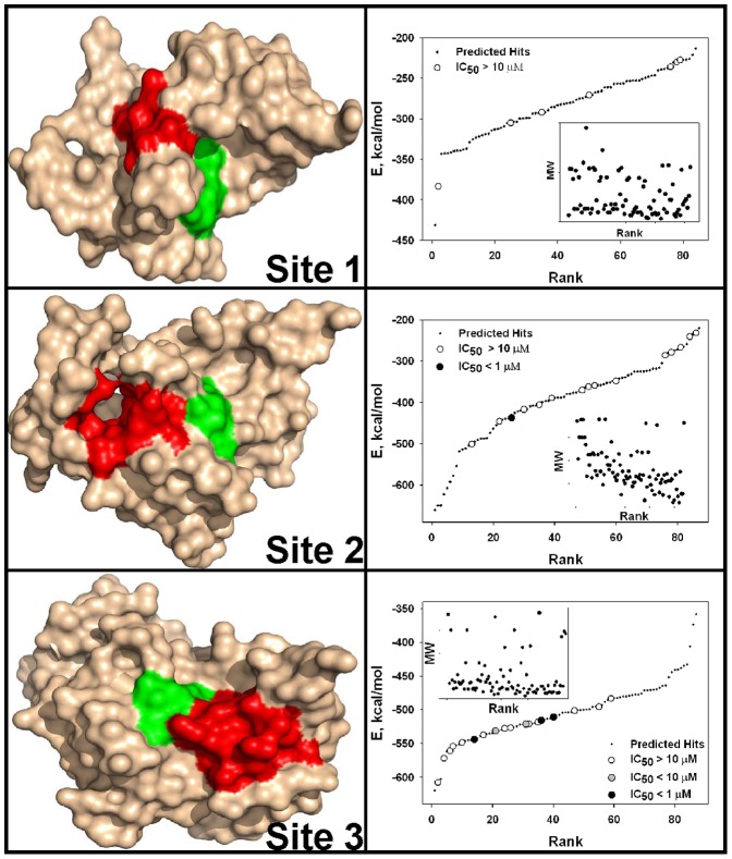
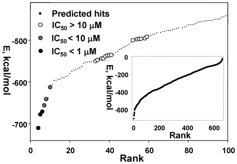
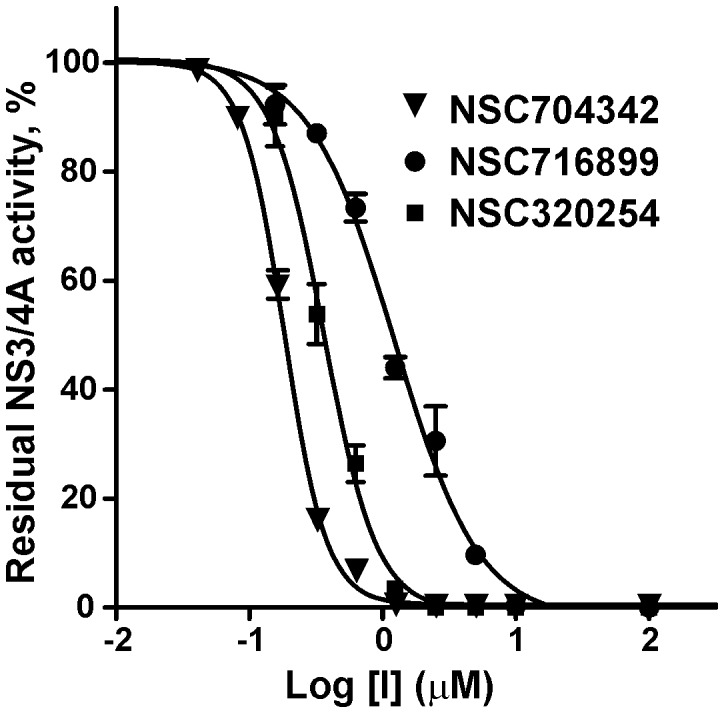
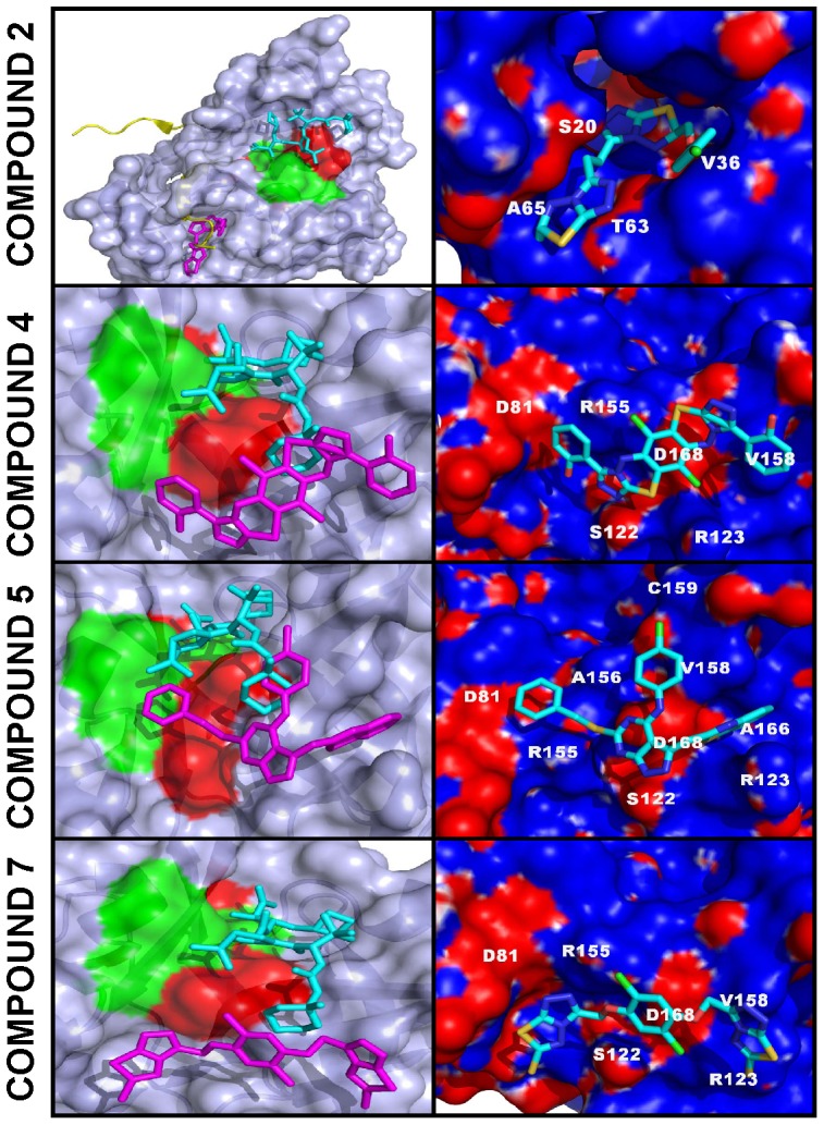
Methodology/principal findings: To circumvent drug resistance and complement the existing anti-virals, NS3/4A inhibitors, which are additional and distinct from the FDA-approved telaprevir and boceprevir α-ketoamide inhibitors, are required. To test potential new avenues for inhibitor development, we have probed several distinct exosites of NS3/4A which are either outside of or partially overlapping with the active site groove of the proteinase. For this purpose, we employed virtual ligand screening using the 275,000 compound library of the Developmental Therapeutics Program (NCI/NIH) and the X-ray crystal structure of NS3/4A as a ligand source and a target, respectively. As a result, we identified several novel, previously uncharacterized, nanomolar range inhibitory scaffolds, which suppressed of the NS3/4A activity in vitro and replication of a sub-genomic HCV RNA replicon with a luciferase reporter in human hepatocarcinoma cells. The binding sites of these novel inhibitors do not significantly overlap with those of α-ketoamides. As a result, the most common resistant mutations, including V36M, R155K, A156T, D168A and V170A, did not considerably diminish the inhibitory potency of certain novel inhibitor scaffolds we identified.

Conclusions/significance: Overall, the further optimization of both the in silico strategy and software platform we developed and lead compounds we identified may lead to advances in novel anti-virals.


Conflict of interest statement

Competing Interests: The authors have the following competing interests: The affiliation of Dr. Anton Cheltsov with a commercial company (Q-MOL L.L.C., San Diego, CA) does not alter the authors' adherence to all the PLoS ONE policies on sharing data and materials.

Figures

Figure 1
Figure 1. Three docking sites in NS3/4A and VLS of the NCI/DTP compound library.
Left panels, positions of docking sites 1, 2 and 3 in the PDB 3EYD X-ray structure of NS3/4A, surface model. The catalytic triad (His-57, Asp-81, and Ser-139) is green. Docking sites 1, 2 and 3 are red. Right panels, VLS of the 275,000-compound NCI library against docking sites 1, 2 and 3. VLS led to identification of the top 84, 87 and 88 hits, from which 7, 15 and 18 available compounds for sites 1, 2 and 3, respectively, were tested in the NS3/4A inhibitory assays. Compounds were ranked according to their relative binding energy. Black, grey and open circles correspond to the tested compounds with the IC50 values below 1 µM, below 10 µM and above 10 µM, respectively. Predicted (but untested) hits are shown as small back dots. E, relative binding energy. Inset, relations between the molecular weight (MW) and ranking of the ligands.
Figure 2
Figure 2. Structural similarity of the flaviviral NS3 proteinases.
(A) Sequence alignment of NS3 proteinases. Asterisks mark the catalytic triad. Identical and homologous residue positions are shaded gray. (B) Sequence alignment of the flaviviral WNV and DV2 NS2B and HCV NS4A (PDB 3EYD) co-factors. Dots indicate identical residues. Secondary structure elements above and below the sequences are for WNV NS2B-NS3 proteinase (PDB 2IJO) and HCV NS3/4A (PDB 3EYD), respectively. The secondary structure of the minimal, 14-residue, NS4A co-factor required for activation of the NS3 proteinase in vitro is shown. WNV, West Nile virus. DV2, Dengue virus type 2.
Figure 3
Figure 3. VLS and SAR optimization of the focused compound sub-library.
The 665-compound sub-library was docked into docking site 3 of NS3/4A. Compounds were ranked according to their relative binding energy (inset, a complete ranking curve, each dot represents a single compound). The screening led to the identification of the top 100 compounds with the lowest binding energy. Compounds were visually inspected and the 20 available compounds were ordered from the NCI/DTP for the follow-up in vitro activity tests. Black, grey and open circles correspond to the tested compounds with the IC50 values below 1 µM, below 10 µM and above 10 µM, respectively. Predicted (but untested) hits are shown as small back dots. E, relative binding energy.
Figure 4
Figure 4. Selected compounds efficiently inhibit the catalytic activity of NS3/4A: representative dose-response curves.
Before the addition of the Ac-DE-D(Edans)-EE-Abu-ψ-[COO]-AS-K(Dabsyl)-NH2 substrate (10 µM), NS3/4A (10 nM) was co-incubated for 30 min at ambient temperature with increasing concentrations of compounds 1, 4 and 7. The residual activity was then monitored continuously at λex = 355 nm and λem = 500 nm to determine the initial velocity of the reactions. The initial velocity was calculated as a percentage of residual activity versus the untreated proteinase (control). Refer to Table S1 for the compound structures.
Figure 5
Figure 5. Models of NS3/4A-inhibitor complexes.
Left panels, NS3/4A is shown as a surface model. Docked compounds 2, 4, 5 and 7 are shown as stick models (magenta). Mutant residue positions, which confer resistance to telaprevir and boceprevir , , , , , , are red. Catalytic triad residues (His-57, Asp-81, and Ser-139) are green. The superimposed co-crystallized boceprevir derivative (1R,2S,5S)-N-[(1S)-3-amino-1-(cyclobutylmethyl)-2,3-dioxopropyl]-6,6-dimethyl-3-{3-methyl-N-[(1-methylcyclohexyl)carbamoyl]-L-valyl}-3-azabicyclo[3.1.0]hexane-2-carboxamide (PDB 3LOX) is shown as a stick model (cyan). In the compound 2 panel the NS2B cofactor is yellow. Right panels. Close-up of the binding modes of compounds 2, 4, 5 and 7. NS3/4A is shown as a molecular surface model colored by electrostatic potential. The latter was corrected for solvation using the Poisson-Boltzmann equation , . Docked compounds 2, 4, 5 and 7 are shown as stick models colored by the chemical element type. Amino acid numbering corresponds to that of PDB 3EYD. The figures were created using Pymol.

References

    1. Chevaliez S, Pawlotsky JM. HCV genome and life cycle. In: Tan SL, editor. Chapter 1 Hepatitis C viruses: genomes and molecular biology. Norfolk (UK): Horizon Bioscience; 2006. pp. 5–47. - PubMed
    1. Penin F, Dubuisson J, Rey FA, Moradpour D, Pawlotsky JM. Structural biology of hepatitis C virus. Hepatology. 2004;39:5–19. - PubMed
    1. Kim JL, Morgenstern KA, Lin C, Fox T, Dwyer MD, et al. Crystal structure of the hepatitis C virus NS3 protease domain complexed with a synthetic NS4A cofactor peptide. Cell. 1996;87:343–355. - PubMed
    1. Stempniak M, Hostomska Z, Nodes BR, Hostomsky Z. The NS3 proteinase domain of hepatitis C virus is a zinc-containing enzyme. J Virol. 1997;71:2881–2886. - PMC - PubMed
    1. Kwong AD, Kim JL, Rao G, Lipovsek D, Raybuck SA. Hepatitis C virus NS3/4A protease. Antiviral Res. 1999;41:67–84. - PubMed

Publication types

MeSH terms