. 2012;7(7):e40056.
doi: 10.1371/journal.pone.0040056. Epub 2012 Jul 2.

BVT.2733, a selective 11β-hydroxysteroid dehydrogenase type 1 inhibitor, attenuates obesity and inflammation in diet-induced obese mice

Affiliations

BVT.2733, a selective 11β-hydroxysteroid dehydrogenase type 1 inhibitor, attenuates obesity and inflammation in diet-induced obese mice

Long Wang et al. PLoS One. 2012.

Abstract

Background: Inhibition of 11β-hydroxysteroid dehydrogenase type 1 (11β-HSD1) is being pursued as a new therapeutic approach for the treatment of obesity and metabolic syndrome. Therefore, there is an urgent need to determine the effect of 11β-HSD1 inhibitor, which suppresses glucocorticoid action, on adipose tissue inflammation. The purpose of the present study was to examine the effect of BVT.2733, a selective 11β-HSD1 inhibitor, on expression of pro-inflammatory mediators and macrophage infiltration in adipose tissue in C57BL/6J mice.

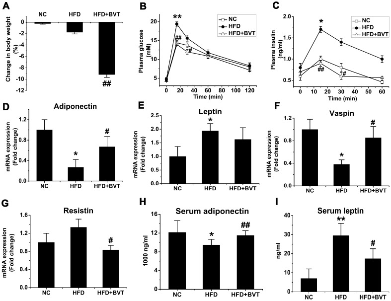
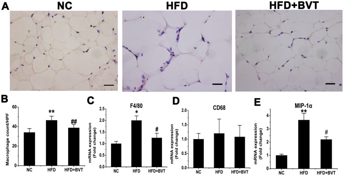
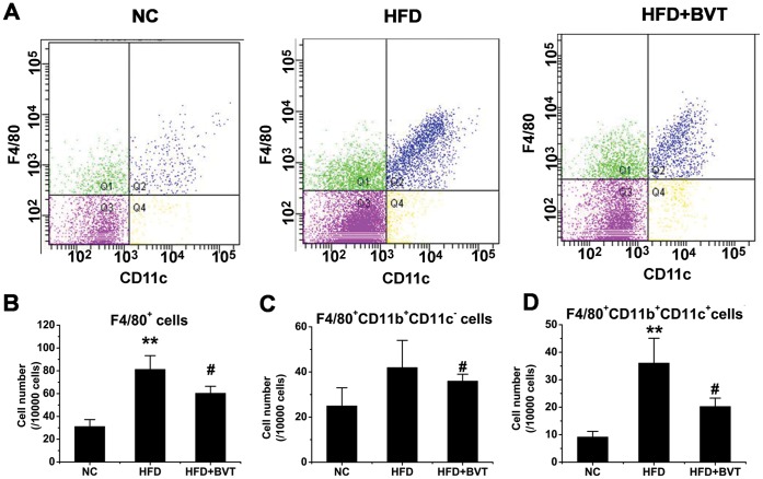
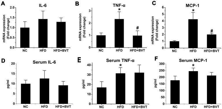
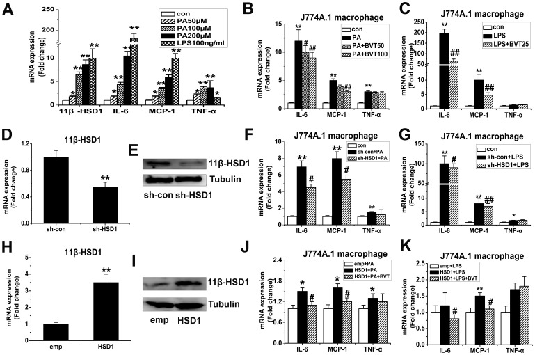
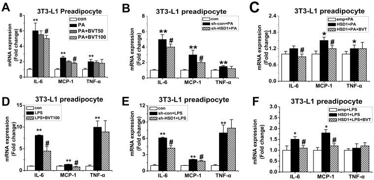
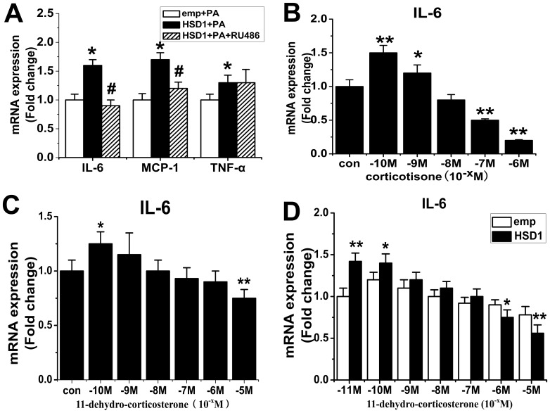
Methodology/principal findings: C57BL/6J mice were fed with a normal chow diet (NC) or high fat diet (HFD). HFD treated mice were then administrated with BVT.2733 (HFD+BVT) or vehicle (HFD) for four weeks. Mice receiving BVT.2733 treatment exhibited decreased body weight and enhanced glucose tolerance and insulin sensitivity compared to control mice. BVT.2733 also down-regulated the expression of inflammation-related genes including monocyte chemoattractant protein 1 (MCP-1), tumor necrosis factor alpha (TNF-α) and the number of infiltrated macrophages within the adipose tissue in vivo. Pharmacological inhibition of 11β-HSD1 and RNA interference against 11β-HSD1 reduced the mRNA levels of MCP-1 and interleukin-6 (IL-6) in cultured J774A.1 macrophages and 3T3-L1 preadipocyte in vitro.

Conclusions/significance: These results suggest that BVT.2733 treatment could not only decrease body weight and improve metabolic homeostasis, but also suppress the inflammation of adipose tissue in diet-induced obese mice. 11β-HSD1 may be a very promising therapeutic target for obesity and associated disease.


Conflict of interest statement

Competing Interests: The authors have declared that no competing interests exist.

Figures

Figure 1
Figure 1. Effect of HDF and BVT.2733 on adiposity and metabolic parameters in C57BL/6J mice.
A, Percentage change in body weight. B–C, Glucose tolerance and plasma insulin level. D–G, Changes in adipose gene mRNA expression. H–I, Serum adiponectin and leptin concentration. The results are shown as the means ± SEM. *, P<0.05; **, P<0.01 compared with NC group; #, P<0.05; ##, P<0.01 compared with HFD group. n = 5−10 animals per group.
Figure 2
Figure 2. Effect of HFD and BVT.2733 treatment on the abundance of macrophage in adipose tissue.
A–B, Representative immunohistochemical staining of white adipose tissue using the specific macrophage marker F4/80 and quantification of the of macrophages present in adipose tissue. C–E, Changes in the expression of macrophage marker genes determined by real time PCR. The results are shown as the means ± SEM. *, P< 0.05; **, P< 0.01 compared with NC group; #, P<0.05; ##, P< 0.01 compared with HFD group. n = 6−8 animals per group. Bar  = 50 µm.
Figure 3
Figure 3. Effect of HFD and BVT.2733 treatment on the number of macrophages in SVF of epididymal fat tissue.
Cells in the SVF of epididymal fat tissue from three groups of mice were analyzed using flow cytometry as described in METHODS section. A, Representative flow cytometric profiles of cells in the SVF of epdidymal fat tissue derived from NC, HFD, and HFD+BVT mice individually. B, The cell number in F4/80-positive fraction. C, F4/80-positive/CD11b-positive/CD11c- negetive fraction. D, F4/80-positive/CD11b-positive/CD11c- positive fraction. The results are shown as the means ± SEM. *, P<0.05; **, P<0.01 compared with NC group; #, P<0.05; ##, P<0.01 compared with HFD group. n = 3−4 animals per group.
Figure 4
Figure 4. Effect of HFD and BVT.2733 treatment on the inflammation gene expression in the adipose tissue and the level of circulating inflammation markers.
A–C, The mRNA expression of IL-6, TNF-α and MCP-1 in adipose tissue. D–E, Plasma concentrations of IL-6, TNF-α and MCP-1. The results are shown as the means ± SEM. *, P<0.05; **, P< 0.01 compared with NC group; #, P<0.05; ##, P<0.01 compared with HFD group. n = 5−6 animals per group.
Figure 5
Figure 5. Effect of 11β-HSD1 on the inflammation gene expression in PA or LPS-activated J774A.1 macrophages in vitro.
A, J774.1 macrophages were treated with palmitic acid (PA) (50−200 µmol/L) or LPS (100 ng/ml) for 24 h. B–C, J774.1 macrophages were activated by PA (200 µmol/L) or LPS (100 ng/ml) and co-treated with 11β-HSD1 inhibitor BVT.2733 (25−100 µmol/L) for 24 h. D–G, J774.1 macrophages were transfected with either sh-RNA for mouse 11β-HSD1 (sh-HSD1) or a negative control (sh-con) by Lentivirus. After 72 h incubation, cells were treated with PA (200 µmol/L) or LPS (100 ng/ml) for 24 h. Efficiency of 11β-HSD1 knockdown on mRNA level (D) and protein level (E). Effects of knockdown of 11β-HSD1 on MCP-1, IL-6, and TNF-α expression in PA (F) or LPS (G) treated macrophages. H–K, J774.1 macrophages were transfected with the expression vector for 11β-HSD1 (HSD1) or a corresponding empty vector (emp) using Lentivirus. After 72 h incubation, cells were treated with PA (100 µmol/L) or LPS (50 ng/ml) and co-treated with 11β-HSD1 inhibitor BVT.2733 for 24 h. Efficiency of 11β-HSD1 overexpression on mRNA level (H) and protein level (I). Effects of overexpression of 11β-HSD1 on MCP-1, IL-6, and TNF-α expression in PA (J) or LPS (K) treated macrophages. mRNA for IL-6, MCP-1 and TNF-α were determined by real-time PCR, protein of 11β-HSD1 were determined by Western blot. The results are shown as the means ± SEM of three individual experiments. *P<0.05; **P<0.01 vs con (B, C, F, G) or sh-con (D) or emp (H) or emp+PA (J) or emp+LPS (K). # P<0.05; ## P<0.01 vs PA (B) or LPS (C) or sh-con+PA (F) or sh-con+LPS (G) or HSD1+PA (J) or HSD1+LPS (K).
Figure 6
Figure 6. Effect of 11β-HSD1 on the inflammation gene expression in PA or LPS-activated 3T3-L1 preadipocytes in vitro.
3T3-L1 preadipocytes were activated by PA (200 µmol/L) (A) or LPS (200 ng/ml) (D) and co-treated with 11β-HSD1 inhibitor BVT.2733 (50−100 µmol/L) for 24 h. 3T3-L1 preadipocytes were transfected with either sh-RNA for mouse 11β-HSD1 (sh-HSD1) or a negative control (sh-con) by Lentivirus. Cells were treated with PA (200 µmol/L) (B) or LPS (200 ng/ml) (E) for 24 h. 3T3-L1 preadipocytes were transfected with the expression vector for 11β-HSD1 (HSD1) or a corresponding empty vector (emp) using Lentivirus. Cells were treated with PA (200 µmol/L) (C) or LPS (200 ng/ml) (F) for 24 h. mRNA for IL-6, MCP-1 and TNF-α were determined by real-time PCR. The results are shown as the means ± SEM of three individual experiments. *P<0.05; **P<0.01 vs con (A, B, D, E) or or emp+PA (C) or emp+LPS (F). # P<0.05 vs PA (A) or LPS (D) or sh-con+PA (B) or sh-con+LPS (E) or HSD1+PA (C) or HSD1+LPS (F).
Figure 7
Figure 7. Effect of glucocorticoid receptor (GR) on proinflammatory properties of 11β-HSD1 in J774A.1 macrophages.
A, J774A.1 macrophages were transfected with the expression vector for 11β-HSD1 (HSD1) or a corresponding empty vector (emp) using Lentivirus. Cells were treated with PA (100 µmol/L) or co-treated with glucocorticoid antagonist RU486 for 24 h. B, J774A.1 macrophages were activated by LPS(100 ng/ml) in the absence (con) or presence of increasing amounts of corticosterone (10−10 M to 10−6 M) for 24 h in steroid hormones free media. C, J774A.1 macrophages were activated by LPS (100 ng/ml) in the absence (con) or presence of increasing amounts of 11-dehydro corticosterone (10−10 M to 10−5 M) for 24 h in Charcoal Dextran Stripped Serum media. D, J774A.1 macrophages were transfected with the expression vector for 11β-HSD1 (HSD1) or a corresponding empty vector (emp) using Lentivirus. Cells were activated by LPS(100 ng/ml) in the absence (con) or presence of increasing amounts of 11-dehydro corticosterone (10−11 M to 10−5 M) for 24 h in Charcoal Dextran Stripped Serum media. mRNA for IL-6, MCP-1 and TNF-α were determined by real-time PCR. The results are shown as the means ± SEM of three individual experiments. *P<0.05; **P<0.01 vs emp+PA (A) or con (B–C) or emp (D), # P<0.05 vs HSD1+PA (A).

References

    1. Greenberg AS, Obin MS. Obesity and the role of adipose tissue in inflammation and metabolism. Am J Clin Nutr. 2006;83:461S–465S. - PubMed
    1. Lenz A, Diamond FB., Jr Obesity: the hormonal milieu. Curr Opin Endocrinol Diabetes Obes. 2008;15:9–20. - PubMed
    1. Stienstra R, Duval C, Keshtkar S, van der Laak J, Kersten S, et al. Peroxisome proliferator-activated receptor gamma activation promotes infiltration of alternatively activated macrophages into adipose tissue. J Biol Chem. 2008;283:22620–22627. - PubMed
    1. Hara Y, Wakino S, Tanabe Y, Saito M, Tokuyama H, et al. Rho and Rho-kinase activity in adipocytes contributes to a vicious cycle in obesity that may involve mechanical stretch. Sci Signal. 2011;4:ra3. - PubMed
    1. Rask E, Olsson T, Soderberg S, Andrew R, Livingstone DE, et al. Tissue-specific dysregulation of cortisol metabolism in human obesity. J Clin Endocrinol Metab. 2001;86:1418–1421. - PubMed

Publication types

MeSH terms

Substances