. 2012 Jul 7:9:163.
doi: 10.1186/1742-2094-9-163.

Effects of dimethyl fumarate on neuroprotection and immunomodulation

Affiliations

Effects of dimethyl fumarate on neuroprotection and immunomodulation

Philipp Albrecht et al. J Neuroinflammation. .

Abstract

Background: Neuronal degeneration in multiple sclerosis has been linked to oxidative stress. Dimethyl fumarate is a promising novel oral therapeutic option shown to reduce disease activity and progression in patients with relapsing-remitting multiple sclerosis. These effects are presumed to originate from a combination of immunomodulatory and neuroprotective mechanisms. We aimed to clarify whether neuroprotective concentrations of dimethyl fumarate have immunomodulatory effects.

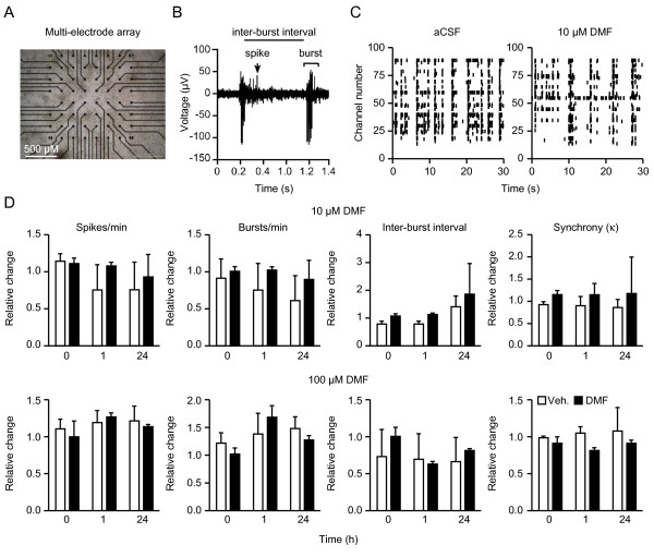
Findings: We determined time- and concentration-dependent effects of dimethyl fumarate and its metabolite monomethyl fumarate on viability in a model of endogenous neuronal oxidative stress and clarified the mechanism of action by quantitating cellular glutathione content and recycling, nuclear translocation of transcription factors, and the expression of antioxidant genes. We compared this with changes in the cytokine profiles released by stimulated splenocytes measured by ELISPOT technology and analyzed the interactions between neuronal and immune cells and neuronal function and viability in cell death assays and multi-electrode arrays. Our observations show that dimethyl fumarate causes short-lived oxidative stress, which leads to increased levels and nuclear localization of the transcription factor nuclear factor erythroid 2-related factor 2 and a subsequent increase in glutathione synthesis and recycling in neuronal cells. Concentrations that were cytoprotective in neuronal cells had no negative effects on viability of splenocytes but suppressed the production of proinflammatory cytokines in cultures from C57BL/6 and SJL mice and had no effects on neuronal activity in multi-electrode arrays.

Conclusions: These results suggest that immunomodulatory concentrations of dimethyl fumarate can reduce oxidative stress without altering neuronal network activity.


Figures

Figure 1
Figure 1
Dimethyl fumarate (DMF) protects from oxidative stress by enhancing nuclear factor erythroid 2-related factor 2 (Nrf2) abundance and translocation to the nucleus in a time- and concentration-dependent manner.A) DMF protects from oxidative glutamate toxicity, a model of endogenous oxidative stress where extracellular glutamate blocks the cystine import ultimately causing glutathione (GSH) depletion and cell death. Primary cortical cultures or hippocampal HT22 cells were preincubated with 10 μM DMF, monomethylfumarate (MMF) or vehicle for 24 h and exposed to the indicated concentrations of glutamate for 24 h before cell viability was measured by the Cell Titer Blue (CTB) assay. B) DMF and MMF increase cellular GSH concentrations. HT22 cells were treated with 10 μM DMF, MMF or vehicle for 24 h and exposed to the indicated concentrations of glutamate for 8 h before intracellular glutathione was measured enzymatically. C-F) Dose and time-course of DMF and MMF effect on oxidative glutamate toxicity. HT22 cells were treated for the indicated times and concentrations with DMF, MMF or vehicle before addition of glutamate. Viability was quantitated 24 h later as described above. G) Time course of DMF effects on glutathione content. HT22 cells were incubated with 10 μM DMF for the indicated periods of time before intracellular glutathione was measured enzymatically. H) DMF enhances Nrf2 abundance quantitated by immunoblots done on nuclear fractions from HT22 cells treated with the indicated concentrations of DMF for 4 h. I) and J) DMF induces nuclear localization of Nrf2 but has no effect on the nuclear translocation of NF-κB as shown by high content imaging. I) HT22 cells were treated with vehicle (n = 9,561 cells), 10 μM DMF for 24 h (n = 8,170 cells) or with 25 μM TBHQ (n = 3,281 cells) as positive control for 4 h. J) HT22 cells were treated with vehicle or 10 ng/ml TNFα in the presence or absence of 10 μM DMF (vehicle n = 1,048 cells, DMF n = 943 cells, TNFα n = 1,410 cells, DMF + TNFα n = 1,085 cells). Cells were fixed, stained and nuclear localization analyzed by immunocytochemistry. K) DMF has no effect on GSH levels in fibroblasts derived from Nrf2-deficient mice. Cells were treated with 10 μM DMF (black bars) or vehicle (white bars) for 24 h before GSH was measured enzymatically. Graphs of all experiments represent the means ± standard error of the mean (SEM) of three independent experiments performed in triplicate. *P < 0.05, two-way ANOVA with Bonferroni post hoc test in A), B), G), I), J), and paired Student’s t-test in K).
Figure 2
Figure 2
Dimethyl fumarate (DMF)-mediated protection in neuronal cells involves glutathione recycling.A) DMF treatment induces mRNA expression of transcripts involved in the antioxidant response. Cells were treated for 24 h with 10 μM DMF or vehicle and mRNA quantitated by real-time PCR using β-actin and hprt as endogenous controls. B) DMF protects from inhibition of GCLC by BSO and inhibition of system Χc − by (S)-4- carboxyphenylglycine (s-4-CPG). HT22 cells were treated for 24 h with 10 μM DMF or vehicle and exposed to the indicated concentrations of s-4-CPG or BSO for another 24 h before cell viability was measured by the CTB assay. C) DMF still elevates cellular GSH when GSH synthesis is blocked by GCLC inhibition by BSO or system Χc − inhibition by incubation in cystine-free medium. HT22 cells were treated for 24 h with 10 μM DMF (black bars) or vehicle (white bars) and then exposed to the indicated concentrations of BSO or to cystine-free medium for another 24 or 4 h respectively before intracellular GSH was measured enzymatically. Graphs of all experiments represent the means ± standard error of the mean (SEM) of three independent experiments performed in triplicate.*P < 0.05, paired Student’s t-test.
Figure 3
Figure 3
Neuroprotective concentrations of dimethyl fumarate (DMF) suppress cytokine production in activated splenocytes from two different mouse strains without altering viability. 10 μM DMF does not significantly affect cell viability of splenocytes from C57BL/6 mice (A) or SJL mice (A’) while 100 μM is toxic (A) and (A’). Cells were treated for 24 h with the indicated concentrations of DMF or vehicle. Cell viability was measured by flow cytometry quantitating 7-AAD and Annexin V-positive cells. B) and B’) DMF concentration-dependently reduced TNFα production from anti-CD3-stimulated splenocytes from C57BL/6 (B) and SJL mice (B’). Primary splenocytes from seven mice were treated with the indicated concentrations of DMF for 48 h and co-stimulated with 1 μg/ml anti-CD3 for the same time before TNFα was measured in the supernatants by ELISA. C) and C’) DMF decreases anti-CD3-induced production of IL-17 and IL-2 in splenocytes from C57BL/6 mice (C) and of the production of IF-γ, IL-2, IL-4, IL-5, IL-6 and IL-17 in splenocytes from SJL mice (C’). Primary splenocytes from seven mice were treated with 10 μM of DMF for 24 h and costimulated with 0.5 μg/ml anti-CD3 for the same time before interferon-gamma, IL-2, IL-4, IL-5, IL-6 and IL-17 were measured using ELISPOT technology. Graphs of all experiments represent the means ± standard error of the mean (SEM) of three independent experiments performed in triplicate.*P < 0.05, paired Student’s t-test in (C) and (C’) and two-way analysis of variance (ANOVA) with Bonferroni post hoc test in (A), (A), (B), and (B’).
Figure 4
Figure 4
Dimethyl fumarate (DMF)-treated neuronal cells but not splenocytes secrete neuroprotective glutathione (GSH).A) HT22 cells and splenocytes show increased intracellular glutathione after 24 h pretreatment with 10 μM DMF, but only HT22 cells release glutathione into the extracellular space. GSH was quantitated enzymatically and normalized to cellular protein content and vehicle-treated cells, respectively. Released GSH in the medium was quantitated after 4 h incubation in cystine-free medium following a 24 h incubation in medium supplemented with 10 μM DMF (black bars) or vehicle (white bars). B) HT22 cells treated with conditioned medium (CM) from HT22 cells but not splenocytes take up released GSH and C) are protected from glutamate toxicity. HT22 cells were treated for 24 h with conditioned medium before addition of the indicated concentrations of glutamate for another 24 h. Viability was quantitated by the CTB assay. (D) Viability of unstimulated splenocytes quantitated by flow cytometry using Annexin V and 7AAD staining is unaffected by HT22 conditioned medium. Graphs of all experiments represent the means ± standard error of the mean (SEM) of three independent experiments performed in triplicate. *P < 0.05, paired Student’s t-test for all assays except two-way analysis of variance (ANOVA) with Bonferroni post hoc test for (C).
Figure 5
Figure 5
No effect of dimethyl fumarate (DMF) on the network activity of primary dissociated cortical cultures grown on multi-electrode arrays (MEAs).A) Dissociated cortical cultures were grown on multi-electrode arrays and visualized by phase-contrast microscopy. Black discs correspond to electrodes. B) Typical trace recorded from one MEA electrode showing the parameters used for statistical analysis. C) Spike raster plot of spontaneously active cortical neurons on one MEA in artificial cerebrospinal fluid (aCSF) or 10 μM DMF. Each bar represents one spike; electrode numbers are displayed on the vertical axis. The network exhibits correlated burst activity across several electrodes, which is not altered from baseline by DMF. D) Treatment with 10 (top) or 100 (bottom) μM DMF for the indicated times does not change the number of spikes and bursts per minute, the inter-burst interval, or the network activity using Cohen’s kappa as measure of synchrony of the activity on all 64 electrodes on the chip. Bar graphs of all experiments represent the means ± standard error of the mean (SEM) of three independent experiments performed in triplicate. *P < 0.05, two-tailed Student’s t-test.

References

    1. Compston A, Coles A. Multiple sclerosis. Lancet. 2008. pp. 1502–1517. - DOI - PubMed
    1. Gonsette RE. Neurodegeneration in multiple sclerosis: the role of oxidative stress and excitotoxicity. J Neurol Sci. 2008;274:48–53. doi: 10.1016/j.jns.2008.06.029. - DOI - PubMed
    1. Nave K-A, Trapp BD. Axon-glial signaling and the glial support of axon function. Annu Rev Neurosci. 2008;31:535–561. doi: 10.1146/annurev.neuro.30.051606.094309. - DOI - PubMed
    1. Kappos L, Gold R, Miller DH, MacManus DG, Havrdova E, Limmroth V, Polman CH, Schmierer K, Yoursry TA, Yang M, Eraksoy M, Meluzinova E, Rektor I, Dawson KT, Sandrock AW, O’Neill GN. bg-12 Phase IIb Study Investigators. Efficacy and safety of oral fumarate in patients with relapsing-remitting multiple sclerosis: a multicentre, randomised, double-blind, placebo-controlled phase IIb study. Lancet. 2008;372:1463–1472. doi: 10.1016/S0140-6736(08)61619-0. - DOI - PubMed
    1. Gold R, Kappos L, Bar-Or D, Arnold D, Giovannoni G, Selmaj K, Yang M, Dawson K. Clinical efficacy of BG-12, an oral therapy, in relapsing-remitting multiple sclerosis: data from the phase 3 DEFINE trial. , Amsterdam; October 19-22 2011. 17:S9-S52.

LinkOut - more resources